584 Endocr<strong>in</strong>e Reviews, October 2003, 24(5):571–599 Thomas et al. • <strong>Gastro<strong>in</strong>test<strong>in</strong>al</strong> <strong>Hormones</strong> <strong>and</strong> <strong>Proliferation</strong>Townsend et al. (209) demonstrated that growth <strong>of</strong> <strong>the</strong> hamsterpancreatic cancer cell l<strong>in</strong>e, H2T, was stimulated by cerule<strong>in</strong><strong>and</strong> that growth could be fur<strong>the</strong>r enhanced by concomitanttreatment with secret<strong>in</strong>. Both gastr<strong>in</strong> <strong>and</strong> Gly-G canstimulate <strong>the</strong> proliferation <strong>of</strong> AR42J rat pancreatic tumorcells (210, 211); <strong>the</strong>se growth-promot<strong>in</strong>g effects can beblocked by L-365,260, a CCK-B receptor antagonist (210–212). Therefore, <strong>the</strong>se studies provide strong support for amodulatory effect <strong>of</strong> CCK, secret<strong>in</strong>, <strong>and</strong> gastr<strong>in</strong> on certa<strong>in</strong>pancreatic cancers. In contrast, Liehr et al. (213) found thatCCK, at dosages <strong>of</strong> 10 12 to 10 6 m, had no effect on <strong>the</strong>growth <strong>of</strong> ei<strong>the</strong>r MIA PaCa-2 or PANC-1 cells. Moreover,stable transfection <strong>of</strong> MIA PaCa-2 <strong>and</strong> PANC-1 cells withCCK-A or CCK-B receptors resulted <strong>in</strong> growth-<strong>in</strong>hibitoryresponses after activation <strong>of</strong> both receptor subtypes by <strong>the</strong>irlig<strong>and</strong>s. The reason for <strong>the</strong>se discrepant results is not readilyapparent.In vivo studies support a role for CCK <strong>in</strong> <strong>the</strong> stimulation<strong>of</strong> pancreatic cancer growth. Pancreatic tumors <strong>in</strong> rats can be<strong>in</strong>duced by endogenous CCK us<strong>in</strong>g CCK-releas<strong>in</strong>g agents,such as tryps<strong>in</strong> <strong>in</strong>hibitors, by biliary diversion or us<strong>in</strong>g bilesalt-b<strong>in</strong>d<strong>in</strong>g drugs (214–216). For example, feed<strong>in</strong>g rats asoybean diet, a natural tryps<strong>in</strong> <strong>in</strong>hibitor, <strong>in</strong>duced <strong>the</strong> formation<strong>of</strong> preneoplastic lesions <strong>in</strong> <strong>the</strong> pancreas <strong>and</strong>, whentreatment was prolonged for years, pancreatic cancer developed(217–219).Evidence exists that CCK can enhance chemical carc<strong>in</strong>ogen-<strong>in</strong>ducedpancreatic cancers, as well. When comb<strong>in</strong>edwith azaser<strong>in</strong>e (a carc<strong>in</strong>ogenic DNA alkylat<strong>in</strong>g agent), <strong>the</strong>long-term adm<strong>in</strong>istration <strong>of</strong> exogenous CCK or elevation <strong>of</strong>endogenous CCK levels (220–222) enhanced <strong>the</strong> developmentor shortened <strong>the</strong> latency period <strong>of</strong> <strong>the</strong> preneoplasticac<strong>in</strong>ar lesions. These effects could be blocked by CCK-Areceptor antagonists (223–225). Consistent with <strong>the</strong>se results,Satake et al. (107) found that adm<strong>in</strong>istration <strong>of</strong> cerule<strong>in</strong>enhanced <strong>the</strong> carc<strong>in</strong>ogenic effect <strong>of</strong> N-nitrosobis (2-hydroxypropyl) am<strong>in</strong>e <strong>in</strong> hamsters. Fur<strong>the</strong>rmore, Howatson<strong>and</strong> Carter (226) found that both CCK <strong>and</strong> secret<strong>in</strong> enhancedpancreatic carc<strong>in</strong>ogenesis <strong>in</strong>duced by N-nitrosobis (2-oxypropyl) am<strong>in</strong>e (BOP) <strong>in</strong> hamsters. In contrast, Johnsonet al. (227) reported that CCK-8 <strong>in</strong>hibited development <strong>of</strong>nitrosam<strong>in</strong>e-<strong>in</strong>duced pancreatic cancer <strong>in</strong> hamsters. The differencesnoted <strong>in</strong> <strong>the</strong>se studies may relate to differences <strong>in</strong> <strong>the</strong>animal models. With <strong>the</strong> exception <strong>of</strong> this study, <strong>the</strong> majority<strong>of</strong> studies suggest that agents capable <strong>of</strong> <strong>in</strong>creas<strong>in</strong>g endogenousCCK <strong>and</strong> stimulat<strong>in</strong>g <strong>the</strong> growth <strong>of</strong> normal pancreascan also stimulate pancreatic carc<strong>in</strong>ogenesis.Similar to CCK, BBS/GRP, ano<strong>the</strong>r pancreatic trophic factor,has been associated with pancreatic carc<strong>in</strong>ogenesis.Douglas et al. (223) demonstrated that rats treated with azaser<strong>in</strong>e(30 mg/kg) <strong>and</strong> BBS (10 mg/kg) for 16 wk developedpreneoplastic lesions at a higher rate than rats treated withazaser<strong>in</strong>e alone. Similar results were noted by Meijers et al.(228) <strong>and</strong> Lhoste <strong>and</strong> Longhecker (208). This effect was notmediated solely by <strong>in</strong>duction <strong>of</strong> CCK because <strong>the</strong> development<strong>of</strong> preneoplastic lesions was not blocked by <strong>the</strong> CCKreceptor antagonist CR-1409 (223). In contrast, ano<strong>the</strong>r studydemonstrated that adm<strong>in</strong>istration <strong>of</strong> BBS was accompaniedby a decrease <strong>in</strong> <strong>the</strong> number <strong>of</strong> preneoplastic lesions <strong>in</strong> BOPtreatedhamsters. The differential effects <strong>of</strong> BBS <strong>in</strong> <strong>the</strong>sestudies may be attributed to species differences <strong>and</strong> also to<strong>the</strong> differences <strong>in</strong> <strong>the</strong> carc<strong>in</strong>ogenic agents.In ano<strong>the</strong>r study, Szepeshazi et al. (229) demonstratedthat adm<strong>in</strong>istration <strong>of</strong> <strong>the</strong> BBS receptor antagonist, RC-3095, decreased BOP-<strong>in</strong>duced pancreatic cancers <strong>in</strong> hamsters.RC-3095 significantly decreased EGF b<strong>in</strong>d<strong>in</strong>g capacity,suggest<strong>in</strong>g that <strong>in</strong>direct effects might be largelyresponsible for <strong>the</strong> reduction identified <strong>in</strong> BOP-associatedhamster cancer. In a subsequent study (230), <strong>the</strong>se <strong>in</strong>vestigatorsshowed that <strong>the</strong> comb<strong>in</strong>ation <strong>of</strong> GRP or BBS withRC-3095 was not able to overcome <strong>the</strong> effects <strong>of</strong> RC-3095but, conversely, augmented <strong>the</strong> <strong>in</strong>hibitory effects <strong>of</strong> RC-3095. These data are more consistent with <strong>the</strong> observations<strong>of</strong> Meijer <strong>and</strong> Baak (231) <strong>in</strong> hamsters <strong>and</strong> show <strong>the</strong> complexity<strong>of</strong> BBS/GRP regulation <strong>in</strong> pancreatic cancer, whichmay be highly species-dependent.More recently, <strong>the</strong> effects <strong>of</strong> a potent <strong>and</strong> specific GRPreceptor antagonist, BIM 26226 [[d-F5 Phe 6, d-Ala 11] BBS(6–13) OMe], were evaluated <strong>in</strong> pancreatic cancers <strong>in</strong> vivo<strong>and</strong> <strong>in</strong> vitro (232). Chronic BIM 26226 adm<strong>in</strong>istration significantlyreduced tumor volume, prote<strong>in</strong>, RNA, amylase, <strong>and</strong>chymotryps<strong>in</strong> content <strong>in</strong> both GRP-responsive <strong>and</strong> GRPunresponsivepancreatic cancers. These f<strong>in</strong>d<strong>in</strong>gs suggest thatGRP receptor antagonists may work through ei<strong>the</strong>r a director <strong>in</strong>direct effect to <strong>in</strong>hibit tumor growth. Fur<strong>the</strong>rmore, arecent study by Burghardt et al. (233) demonstrated that BBS<strong>in</strong>creases expression <strong>of</strong> three transcription factors (c-fos,c-myc, <strong>and</strong> high-mobility group prote<strong>in</strong> IY) that are associatedwith proliferation <strong>in</strong> <strong>the</strong> human pancreatic cell l<strong>in</strong>eHPAF. These results provide potential novel mechanisms fortarget<strong>in</strong>g <strong>the</strong> trophic effects <strong>of</strong> BBS/GRP <strong>in</strong> pancreaticcancers.Similar to CCK, NT also stimulates pancreatic cancer proliferation.Our laboratory has shown that <strong>the</strong> human pancreaticcancer cell l<strong>in</strong>e MIA PaCa-2 possesses NTRs <strong>and</strong> mobilizes<strong>in</strong>tracellular calcium (234). Iwase et al. (181) haveshown that <strong>the</strong> NTR antagonist SR48692 <strong>in</strong>hibits <strong>in</strong>tracellularcalcium mobilization, IP-3 turnover, <strong>and</strong> MIA PaCa-2 <strong>in</strong> vitrocell growth <strong>in</strong>duced by NT <strong>in</strong> a dose-dependent fashion.Fur<strong>the</strong>rmore, <strong>in</strong> vivo experiments demonstrated that NT significantly<strong>in</strong>creased <strong>the</strong> size, weight, <strong>and</strong> DNA <strong>and</strong> prote<strong>in</strong>contents <strong>of</strong> xenografted MIA PaCa-2 tumors; SR48692blocked this effect. Recent f<strong>in</strong>d<strong>in</strong>gs by Reubi et al. (235) haveshown that approximately 75% <strong>of</strong> pancreatic adenocarc<strong>in</strong>omasexpress NTRs. Consistent with <strong>the</strong>se reports, Ehlers et al.(236) have shown NTR expression <strong>in</strong> approximately 90% <strong>of</strong>resected pancreatic cancers.The signal<strong>in</strong>g mechanisms regulat<strong>in</strong>g NT-mediated proliferation<strong>in</strong> NTR-positive cancers have been assessed. NTstimulates ERK <strong>and</strong> JNK activity <strong>in</strong> MIA PaCa-2 cells <strong>and</strong><strong>in</strong>creases AP-1 b<strong>in</strong>d<strong>in</strong>g activity (237). Consistent with <strong>the</strong>seresults, Ryder et al. (238) reported that NT stimulated Ca 2mobilization <strong>and</strong> activated ERK1 <strong>and</strong> ERK2 <strong>and</strong> DNA syn<strong>the</strong>sis<strong>in</strong> <strong>the</strong> human pancreatic cancer cell l<strong>in</strong>e, PANC-1.Recently, Guha et al. (239) reported that NT <strong>in</strong>duced a rapidactivation <strong>of</strong> <strong>the</strong> PKC is<strong>of</strong>orm, PKD, <strong>in</strong> PANC-1 cells, whichwas l<strong>in</strong>ked to <strong>the</strong> mitogenic effect <strong>of</strong> NT.The effects <strong>of</strong> PYY on pancreatic cancer growth have beenexam<strong>in</strong>ed. Treatment <strong>of</strong> <strong>the</strong> human pancreatic cancer celll<strong>in</strong>es, MIA PaCa-2 <strong>and</strong> PANC-1, with <strong>the</strong> syn<strong>the</strong>tic analog <strong>of</strong>Downloaded from edrv.endojournals.org by on July 16, 2007
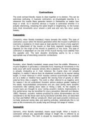
Thomas et al. • <strong>Gastro<strong>in</strong>test<strong>in</strong>al</strong> <strong>Hormones</strong> <strong>and</strong> <strong>Proliferation</strong> Endocr<strong>in</strong>e Reviews, October 2003, 24(5):571–599 585PYY, PYY 22–36, <strong>in</strong>hibited <strong>the</strong> growth <strong>of</strong> <strong>the</strong>se cells <strong>in</strong> vitro(240). A significant decrease <strong>in</strong> <strong>the</strong> growth <strong>of</strong> human pancreaticcancer xenografts was demonstrated <strong>in</strong> nude micegiven PYY 22–36 for 2 wk (241). Similarly, <strong>the</strong>re was a decrease<strong>in</strong> cyclic adenos<strong>in</strong>e monophosphatase expression <strong>and</strong>augmentation <strong>of</strong> <strong>the</strong> tumor-suppress<strong>in</strong>g capability <strong>of</strong> 5-fluorouracil <strong>and</strong> leucovor<strong>in</strong> (241, 242). More recently, Liuet al. (243) reported that a biot<strong>in</strong>ylated PYY analog 14–36,l<strong>in</strong>ked to a fluorescent dye attached to streptavid<strong>in</strong>, significantlybound to human pancreatic cancer cells MIA PaCa-2<strong>and</strong> PANC-1 <strong>and</strong> <strong>in</strong>hibited <strong>the</strong> growth <strong>of</strong> <strong>the</strong>se pancreaticcancer cells. It was postulated that biot<strong>in</strong>ylated PYY 14–36could be used to selectively deliver toxic agents to <strong>the</strong> cancersor, conversely, as a diagnostic tool tagged with a fluorescentagent to identify <strong>the</strong> extent <strong>of</strong> disease. The results <strong>of</strong> <strong>the</strong>sestudies suggest that PYY can <strong>in</strong>hibit pancreatic cancergrowth. However, it has not been determ<strong>in</strong>ed whe<strong>the</strong>r amajority <strong>of</strong> pancreatic cancers express receptors for PYY.Also, <strong>the</strong> cellular mechanisms for <strong>the</strong> <strong>in</strong>hibitory effect <strong>of</strong> PYYneed to be better del<strong>in</strong>eated.Similar to PYY, somatostat<strong>in</strong> <strong>and</strong> its analogs have beenevaluated as antiproliferative agents <strong>in</strong> <strong>the</strong> treatment <strong>of</strong> pancreaticcancer. Many pancreatic cancers have high-aff<strong>in</strong>ityb<strong>in</strong>d<strong>in</strong>g sites for somatostat<strong>in</strong> (244, 245); however, <strong>the</strong> precisemechanisms regulat<strong>in</strong>g <strong>the</strong> antiproliferative effect <strong>of</strong> somatostat<strong>in</strong>have not been entirely elucidated. Potential mechanisms<strong>in</strong>clude a direct receptor-mediated effect <strong>and</strong> possiblyan <strong>in</strong>direct effect that could <strong>in</strong>clude <strong>in</strong>hibition <strong>of</strong>angiogenesis <strong>and</strong>/or <strong>the</strong> suppression <strong>of</strong> GH <strong>and</strong> IGF secretion(244, 245). Redd<strong>in</strong>g <strong>and</strong> Schally (246) reported significantreductions <strong>in</strong> tumor weight <strong>and</strong> volume <strong>in</strong> Wistar-Lewisrats bear<strong>in</strong>g <strong>the</strong> pancreatic ac<strong>in</strong>ar tumor DNC-322 after a21-d adm<strong>in</strong>istration <strong>of</strong> somatostat<strong>in</strong> analog [l-5-Br-Trp 8 ]somatostat<strong>in</strong>-14.Upp et al. (64) demonstrated <strong>in</strong>hibition <strong>of</strong> twohuman pancreatic cancer xenografts (SKI <strong>and</strong> CAV) <strong>in</strong> nudemice by <strong>the</strong> adm<strong>in</strong>istration <strong>of</strong> <strong>the</strong> long-act<strong>in</strong>g somatostat<strong>in</strong>analog SMS 201-995 (octreotide). O<strong>the</strong>r growth factors (e.g.,EGF) may modulate <strong>the</strong> growth <strong>of</strong> pancreatic cancers. In vitrostudies by Hierowski et al. (247) <strong>and</strong> Liebow et al. (248)demonstrated receptors for EGF <strong>and</strong> somatostat<strong>in</strong> <strong>in</strong> <strong>the</strong> MIAPaCa-2 cancer cell l<strong>in</strong>e. The effects <strong>of</strong> somatostat<strong>in</strong> may bethrough its actions on o<strong>the</strong>r growth factors <strong>and</strong> <strong>the</strong>ir receptors.For example, <strong>the</strong> long-act<strong>in</strong>g analog RC-160 <strong>in</strong>hibitsproliferation <strong>of</strong> <strong>the</strong> pancreatic cancer cell l<strong>in</strong>e MIA PaCa-2,possibly by activat<strong>in</strong>g dephosphorylation <strong>of</strong> <strong>the</strong> EGF receptor(247, 248).Colorectal cancer is a significant health problem worldwide,with <strong>the</strong> death rate rema<strong>in</strong><strong>in</strong>g third to lung <strong>and</strong> prostatecancer <strong>in</strong> men <strong>and</strong> lung <strong>and</strong> breast cancer <strong>in</strong> women(249). Approximately 57,000 deaths are expected to occur thisyear <strong>in</strong> <strong>the</strong> United States (249). Current chemo<strong>the</strong>rapeuticregimens are relatively <strong>in</strong>effective for metastatic disease. Theeffects <strong>of</strong> GI hormones, particularly gastr<strong>in</strong>, have been welldescribed <strong>and</strong> characterized <strong>in</strong> various colon cancer models.Gastr<strong>in</strong> stimulates <strong>the</strong> growth <strong>of</strong> colorectal cancers thatpossess high-aff<strong>in</strong>ity gastr<strong>in</strong> receptors, <strong>in</strong>clud<strong>in</strong>g <strong>the</strong> mousecolon cancer cell l<strong>in</strong>e MC-26. The adm<strong>in</strong>istration <strong>of</strong> <strong>the</strong> gastr<strong>in</strong>receptor antagonist, proglumide, <strong>in</strong>hibited <strong>the</strong> growth <strong>of</strong>MC-26 tumors <strong>in</strong> vivo <strong>and</strong> prolonged survival <strong>of</strong> tumorbear<strong>in</strong>gmice (Fig. 5) (250). Ishizuka et al. (251) have shownthat gastr<strong>in</strong> stimulates <strong>the</strong> growth <strong>of</strong> two human colon cancercell l<strong>in</strong>es, LoVo <strong>and</strong> HT29, but <strong>in</strong>hibits <strong>the</strong> growth <strong>of</strong> a thirdcolon cancer cell l<strong>in</strong>e, HCT-116, suggest<strong>in</strong>g that differentgastr<strong>in</strong> receptor subtypes or o<strong>the</strong>r signal transduction pathwaysregulate <strong>the</strong> trophic effects <strong>of</strong> gastr<strong>in</strong>. As an alternateapproach, we have assessed <strong>the</strong> effect <strong>of</strong> suppress<strong>in</strong>g endogenousgastr<strong>in</strong> with <strong>the</strong> prostagl<strong>and</strong><strong>in</strong> analog enprostil onMC-26 tumor growth <strong>and</strong> found that, similar to our results<strong>in</strong> gastric cancers, enprostil significantly <strong>in</strong>hibited MC-26tumor growth <strong>in</strong> vivo (252, 253).In addition to <strong>the</strong> experimental evidence for <strong>the</strong> effect <strong>of</strong>gastr<strong>in</strong> on colon cancers, cl<strong>in</strong>ical studies have assessed <strong>the</strong>potential role <strong>of</strong> gastr<strong>in</strong> <strong>and</strong> gastr<strong>in</strong> receptor expression <strong>in</strong>colorectal tumors. Upp et al. (254) analyzed 67 primary colorectalcancers for gastr<strong>in</strong> receptors <strong>and</strong> found a spectrum <strong>of</strong>receptor expression, suggest<strong>in</strong>g a correlation between tumorstage <strong>and</strong> <strong>the</strong> presence <strong>of</strong> gastr<strong>in</strong> receptors. A significantlygreater percentage <strong>of</strong> patients (52%) with early cancers(Dukes’ A <strong>and</strong> B) had gastr<strong>in</strong> receptor contents greater than10 fmol/mg when compared with patients with more advancedcolorectal cancers (Fig. 6). In contrast, five <strong>of</strong> 19Dukes’ C patients <strong>and</strong> eight <strong>of</strong> 16 Dukes’ D patients withgastr<strong>in</strong> receptors less than 10 fmol/mg prote<strong>in</strong> developedtumor recurrence or died. This study was <strong>the</strong> first to suggestthat assessment <strong>of</strong> gastr<strong>in</strong> receptor expression <strong>in</strong> colorectalcancers may predict tumor stage as well as overall survival.Fur<strong>the</strong>r studies are needed to correlate <strong>the</strong> receptor expressionwith long-term cl<strong>in</strong>ical parameters.Hoose<strong>in</strong> et al. (255, 256) demonstrated that gastr<strong>in</strong> stimulatescolorectal cancer cell growth <strong>and</strong> that this stimulationwas possibly attributable to an autocr<strong>in</strong>e mechanism. Thegrowth <strong>of</strong> six colon cancer cell l<strong>in</strong>es was assessed after treatmentwith two gastr<strong>in</strong> receptor antagonists, proglumide <strong>and</strong>benzotript, or antibodies to gastr<strong>in</strong> (255). Both <strong>in</strong>hibitorssuppressed monolayer cell growth <strong>in</strong> all <strong>of</strong> <strong>the</strong> colon cancerl<strong>in</strong>es. Fur<strong>the</strong>rmore, <strong>in</strong> HCT-116 cells, <strong>the</strong> addition <strong>of</strong> antigastr<strong>in</strong>antisera resulted <strong>in</strong> a concentration-dependent <strong>in</strong>hi-C. Colorectal cancerFIG. 5. Survival <strong>of</strong> MC-26 tumor-bear<strong>in</strong>g mice after treatment withnormal sal<strong>in</strong>e (solid l<strong>in</strong>e), proglumide beg<strong>in</strong>n<strong>in</strong>g on <strong>the</strong> day <strong>of</strong> tumor<strong>in</strong>oculation (long-dash l<strong>in</strong>e), or proglumide beg<strong>in</strong>n<strong>in</strong>g 7 d after <strong>in</strong>oculation(short-dash l<strong>in</strong>e). [From R. D. Beauchamp et al.: Ann Surg202:303–309, 1985 (250).]Downloaded from edrv.endojournals.org by on July 16, 2007