PixFRET, an ImageJ plug-in for FRET calculation ... - ResearchGate
PixFRET, an ImageJ plug-in for FRET calculation ... - ResearchGate
PixFRET, an ImageJ plug-in for FRET calculation ... - ResearchGate
- No tags were found...

Create successful ePaper yourself
Turn your PDF publications into a flip-book with our unique Google optimized e-Paper software.
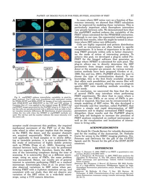
<strong>Pix<strong>FRET</strong></strong>: AN IMAGEJ PLUG-IN FOR PIXEL-BY-PIXEL ANALYSIS OF <strong>FRET</strong>Fig. 6. expN<strong>FRET</strong> reduces <strong>in</strong>tercellular variability <strong>in</strong> pixel-bypixel<strong>an</strong>alyses. Cos-7 cells were tr<strong>an</strong>sfected with expression vectors<strong>for</strong> PPARa-ECFP <strong>an</strong>d RXRa-EYFP. A: Images of two cells express<strong>in</strong>gboth PPAR-ECFP <strong>an</strong>d RXR-EYFP <strong>in</strong> the CFP <strong>an</strong>d YFP sett<strong>in</strong>g. AGaussi<strong>an</strong> blur of 1 was applied to the orig<strong>in</strong>al image. B: N<strong>FRET</strong> <strong>an</strong>dexpN<strong>FRET</strong> images generated by the <strong>Pix<strong>FRET</strong></strong> <strong>plug</strong>-<strong>in</strong>. C: Same as (B)but the cells are pseudocolorized with a different look-up table to bettervisualize <strong>in</strong>tensity differences. D: Distribution of pixel <strong>in</strong>tensitieswith<strong>in</strong> the two cells when the N<strong>FRET</strong> or expN<strong>FRET</strong> methods areused. The red bar <strong>an</strong>d the number <strong>in</strong>dicate the me<strong>an</strong> <strong>in</strong>tensity overthe entire cell.acceptor could circumvent this problem, the requiredmech<strong>an</strong>ical movement of the PMT (or of the filtercube wheel <strong>in</strong> other set-ups) implies that the images<strong>in</strong> the <strong>FRET</strong>, the donor, <strong>an</strong>d the acceptor ch<strong>an</strong>nelsare acquired sequentially. While this approach iswell applicable to fixed samples or to slowly diffus<strong>in</strong>gor immobile prote<strong>in</strong>s, it is more problematic <strong>for</strong>the study of <strong>in</strong>teractions of highly mobile prote<strong>in</strong>ssuch as PPARs (Feige et al., 2005). Sc<strong>an</strong>n<strong>in</strong>g eachch<strong>an</strong>nel between l<strong>in</strong>es, which c<strong>an</strong> only be per<strong>for</strong>medwith two separate PMTs, there<strong>for</strong>e, limits the diffusionof the <strong>in</strong>teract<strong>in</strong>g complexes dur<strong>in</strong>g the time ofsc<strong>an</strong>n<strong>in</strong>g. It is noteworthy that on a different microscope,us<strong>in</strong>g <strong>an</strong> array of PMTs, the same problemwas encountered. Hence, our results are of general<strong>in</strong>terest to scientists per<strong>for</strong>m<strong>in</strong>g <strong>FRET</strong> on a confocalmicroscope. Interest<strong>in</strong>gly, Chen et al. (<strong>in</strong> press) havealso observed variations <strong>in</strong> SBT ratios when per<strong>for</strong>m<strong>in</strong>g<strong>FRET</strong> with a 2-photon excitation set-up, <strong>an</strong>dconsistent with our study, they did not observe <strong>an</strong>yvariation of the SBT ratios on a wide-field microscope,us<strong>in</strong>g a CCD camera detection.In cases where SBT ratios vary as a function of fluorescence<strong>in</strong>tensity, we showed that <strong>FRET</strong> <strong>calculation</strong>c<strong>an</strong> be improved by model<strong>in</strong>g these variations. This isparticularly import<strong>an</strong>t when fluorophore <strong>in</strong>tensitiesvary greatly between cells. We demonstrated here thatthe expN<strong>FRET</strong> method reduces the variability of the<strong>FRET</strong> values calculated <strong>for</strong> the PPAR/RXR <strong>in</strong>teraction.Although <strong>in</strong> our case, the exponential model<strong>in</strong>g generatedthe best results, other fits might be tested <strong>in</strong> situationswhere SBT ratios vary differently.Cells are highly org<strong>an</strong>ized <strong>an</strong>d prote<strong>in</strong> distributionas well as <strong>in</strong>teractions are often limited to specificcompartments. It is hence of import<strong>an</strong>ce to be able tomap <strong>FRET</strong> precisely with<strong>in</strong> cells to better characterizethe mode of action of <strong>in</strong>teract<strong>in</strong>g prote<strong>in</strong>s. Toachieve this goal, we developed a <strong>plug</strong>-<strong>in</strong> called Pix-<strong>FRET</strong> <strong>for</strong> the <strong>ImageJ</strong> software that generates <strong>an</strong>image where N<strong>FRET</strong> is calculated <strong>for</strong> each pixel. The<strong>plug</strong>-<strong>in</strong> allows one to directly determ<strong>in</strong>e the SBTparameters from images acquired when only thedonor or the acceptor are present. As various normalizationmethods have been proposed (Gordon et al.,1998; Xia <strong>an</strong>d Liu, 2001), <strong>Pix<strong>FRET</strong></strong> allows the user tochoose the type of normalization desired. To ourknowledge, this is the first freely available programthat offers such possibilities <strong>an</strong>d the source files willbe available upon request, allow<strong>in</strong>g users to programspecific SBT ratio model<strong>in</strong>g methods accord<strong>in</strong>g totheir needs.In conclusion, we uncovered the bias that the useof several PMTs may <strong>in</strong>troduce when per<strong>for</strong>m<strong>in</strong>g<strong>FRET</strong> experiments. We show that <strong>in</strong> cases where aset-up with two PMTs or <strong>an</strong> array of PMTs is preferredor required, this bias c<strong>an</strong> be circumvented by asimple model<strong>in</strong>g of SBT ratios. We also developed auser-friendly <strong>an</strong>d free <strong>in</strong>terface, called <strong>Pix<strong>FRET</strong></strong>, thatallows a simple <strong>an</strong>d rapid determ<strong>in</strong>ation of SBTparameters <strong>an</strong>d to display normalized <strong>FRET</strong> images.Altogether, these results <strong>an</strong>d the <strong>Pix<strong>FRET</strong></strong> <strong>plug</strong>-<strong>in</strong>will help cell biologists to <strong>in</strong>crease the precision of<strong>FRET</strong> <strong>an</strong>alyses conducted on confocal microscopes aswell as the visualization <strong>an</strong>d localization of <strong>in</strong>teractions<strong>in</strong> liv<strong>in</strong>g cells.ACKNOWLEDGMENTSWe th<strong>an</strong>k Dr. Claude Berney <strong>for</strong> valuable discussions<strong>an</strong>d <strong>for</strong> the read<strong>in</strong>g of the m<strong>an</strong>uscript, Dr. NathalieGar<strong>in</strong> <strong>for</strong> access to the ISREC imag<strong>in</strong>g plat<strong>for</strong>m, Drs.Peterson <strong>an</strong>d DeGrazia <strong>for</strong> the gift of the GI-FITClig<strong>an</strong>d, <strong>an</strong>d Dr. Tavaré <strong>for</strong> the gift of the EYFP–ECFPconstruct.REFERENCESBerney C, D<strong>an</strong>user G. 2003. <strong>FRET</strong> or no <strong>FRET</strong>: a qu<strong>an</strong>titative comparison.Biophys J 84:3992–4010.Chen Y, El<strong>an</strong>gov<strong>an</strong> M, Periasamy A. 2005. <strong>FRET</strong> data <strong>an</strong>alysis: thealgorithm. In: Periasamy A, Day RN, editors. Molecular imag<strong>in</strong>g:<strong>FRET</strong> microscopy <strong>an</strong>d spectroscopy. Ox<strong>for</strong>d: Ox<strong>for</strong>d UniversityPress (<strong>in</strong> press).DeGrazia MJ, Thompson J, Heuvel JP, Peterson BR. 2003. Synthesisof a high-aff<strong>in</strong>ity fluorescent PPARgamma lig<strong>an</strong>d <strong>for</strong> highthroughputfluorescence polarization assays. Bioorg Med Chem 11:4325–4332.El<strong>an</strong>gov<strong>an</strong> M, Wallrabe H, Chen Y, Day RN, Barroso M,Periasamy A. 2003. Characterization of one- <strong>an</strong>d two-photon excitationfluorescence reson<strong>an</strong>ce energy tr<strong>an</strong>sfer microscopy. Methods29: 58–73.57