PDF(6.5mb) - Malaysian Dental Association
PDF(6.5mb) - Malaysian Dental Association
PDF(6.5mb) - Malaysian Dental Association
- No tags were found...
Create successful ePaper yourself
Turn your PDF publications into a flip-book with our unique Google optimized e-Paper software.
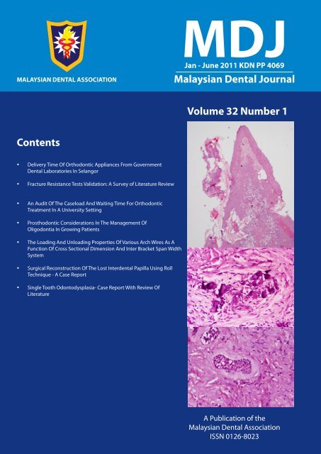
<strong>Malaysian</strong> <strong>Dental</strong> Journal Editorial Team 2010/2011Editor:Assistant Editor:Secretary:Treasurer:Ex-Officio:Associate Professor Dr. Dalia AbdullahDr Shalini KanagasingamDr Nurul Asyikin YahyaDr Mumtaj Nisah Abd RahimDr Ng Woan tyngDr Mohamad Muzafar HamirudinThe Editor of the <strong>Malaysian</strong> <strong>Dental</strong> <strong>Association</strong> wishes to acknowledge the tireless efforts of the following referees toensure that the manuscripts submitted are of high standard.Prof. Dr. Toh Chooi Gait Prof. Dr. Ong Siew Tin Dato’ Prof. Dr. Hashim b. YaacobProf. Dr. Lui Joo Loon Prof. Zubaidah Abdul Rahim Prof. Dr. Phrabhakaran NambiarProf Dr Francesco Mannocci Dr Philip Mitchell Dr Omar IkramDr. Zamros Yuzadi Dr. Siti Mazlipah Ismail Prof. Dr. Tara Bai Taiyeb AliDr. Elise Monorasinghe Prof. Dr. Siar Chong Huat Prof. Dr. Rahimah Abdul KadirDr. Lau Shin Hin Assoc. Prof. Dr. Shanmuhasuntharam Assoc. Prof. Dr. Datin Rashidah EsaDr. Loke Shuet Toh Dr. Fathilah Abdul Razak Assoc. Prof. Dr. Tuti Ningseh Mohd DomDr. Shahida Said Dr. Lam Jac Meng Assoc. Prof. Dr. Roszalina RamliDr. Zamri Radzi Dr. Nor Himazian Mohamed Assoc. Prof. Dr. Roslan Abdul RahmanDr. Siti Adibah Othman Dr. Norliza Ibrahim Dr. Dalia AbdullahDr. Zeti Adura Che Abd. Aziz Dr. Wey Mang Chek Dr. Wong Mei LingThe PublisherThe <strong>Malaysian</strong> <strong>Dental</strong> Journal is an official publication of the <strong>Malaysian</strong> <strong>Dental</strong> <strong>Association</strong> and is publishedhalf yearly (KDN PP4069/12/98).<strong>Malaysian</strong> <strong>Dental</strong> <strong>Association</strong>54-2, (2nd Floor), Medan Setia 2, Plaza Damansara,Bukit Damansara, 50490 Kuala LumpurTel: 603-20951532, 20947606, Fax: 603-20944670Website address: http://mda.org.myE-mail: mdentalassoc@gmail.com or mdaassoca@unifi.myCover page: Pictures courtesy of Dr Manpreet Kalra and co-authors1<strong>Malaysian</strong> <strong>Dental</strong> Journal Jan-Jun 2011 Vol 32 No 1© 2011 The <strong>Malaysian</strong> <strong>Dental</strong> <strong>Association</strong>
MALAYSIAN DENTAL JOURNALAim And ScopeThe <strong>Malaysian</strong> <strong>Dental</strong> Journal covers all aspects relevant to the science and practice of Dentistry, interdisciplinary fieldsand supporting aspects of Medicine. Within the scope of the journal, the following articles will be published:• Editorials - commentary by editors• Clinical articles - in depth discussions on clinical procedures or treatment techniques• Case reports - illustrating various aspects of clinical practice and management• Materials, instruments and technical innovations - reports and research on new innovations and dental products• Ask the Experts - questions posed to seek expert opinion and views on specific topics• Book reviews - critical commentary on the latest printed publications• Product reviews - critical opinion and feedback on newly introduced dental or dental-related products• Letter to editor - comments and feedback from readers pertaining to journal articles• Conference abstracts - proceedings of workshops, conferences and symposiums organized by the MDA• Cover photographs – Interesting photographs for the front cover of the journalThe mission is to promote and elevate the quality of patient care and to encourage the advancement of practice,continuing education and scientific research in Malaysia.PublicationThe <strong>Malaysian</strong> <strong>Dental</strong> Journal is an official publication of the <strong>Malaysian</strong> <strong>Dental</strong> <strong>Association</strong> and is published half yearly(KDN PP4069/12/98)Instructions for submissionOriginal articles, editorial, correspondence and suggestion for review articles should be sent to:Editor of <strong>Malaysian</strong> <strong>Dental</strong> Journal : mdjsubmission@gmail.comPlease ensure that you cc a copy to the Chief Editor: hzl@hotmail.comAuthors are requested to submit their typescript and illustrations via the email address provided. A paper is acceptedfor publication on the understanding that it has not been submitted simultaneously to another journal in the EnglishLanguage. The editor reserves the right to make editorial and literary corrections in the interest of conciseness, clarityand consistency. Any opinion expressed or policies advocated do not necessarily reflect the opinion or policies of theeditors.CopyrightAuthors submitting an article do so on the understanding that the work has not been published before. The submissionof the manuscript by the authors means that the authors automatically agree to sign exclusive copyright to the Editorand the publication committee if and when the publication is accepted for publication. The copyright transfer agreementcan be downloaded at the MDA webpage (www.mda.org.my). A copy of the agreement must be signed by the principalauthor before any paper can be published. There will be no limitation on your freedom to use material contained in thearticle (without having to request permission) provided that an acknowledgement is made to the journal as the originalsource of publication.Presentation of manuscriptPlease follow these instructions carefully as manuscripts which have not been prepared in the approved format will berejected.Manuscripts should be submitted in journal style, in UK English with font should be Arial (size 12). Articles should betyped with double spacing. Papers should be set out as follows with each beginning in a separate page: title page, keywords, abstract, text, acknowledgements, references, tables, caption to illustrations.• Title page. The title page should give the following information: 1) title of the article; 2) full names and professional/academic qualifications/positions of each author; 3) name, address, telephone, fax and e-mail address for the correspondingauthor, assumed to be the first listed author unless otherwise advised. If the paper was presented at an<strong>Malaysian</strong> <strong>Dental</strong> Journal Jan-Jun 2011 Vol 32 No 1© 2011 The <strong>Malaysian</strong> <strong>Dental</strong> <strong>Association</strong>2
Delivery Time Of Orthodontic Appliances From Government<strong>Dental</strong> Laboratories In SelangorAuthors:S T LokeHead of Orthodontic Unit, Orthodontic Specialist Clinic, Jalan Tengku Kelana, 41000 Klang, SelangorS Y Tan<strong>Dental</strong> Clinic, Jalan Tengku Kelana, 41000 Klang, SelangorABSTRACTCompliance to delivery of orthodontic appliances within specified times and factors which influenced compliance bydental technicians in Selangor was evaluated. This is a prospective 8-month study of 18 trainee/ trained techniciansfrom 4 main government dental laboratories in Kajang, Klang, Tanjong Karang and Shah Alam. Delivery timesspecified by orthodontists were 1 day for plastic retainer, 3 days acrylic retainer, 5 days active plates (URA), 10 daysfunctional appliances (FA), 10 days transpalatal arch (TPA) and 10 days for quadhelix. Punctual delivery was recordedas ‘compliant’. Compliance was compared between appliances, clinics, technicians, orthodontists and seniority oftechnicians. The sample comprised appliances from 365 patients; 38 (10.4%) Tanjong.Karang, 114 (31.2%) Kajang,191 (52.3%) Klang and 22 (6.0%) Shah Alam. The majority of appliances were retainers (66.3%), followed by URA(13.4%), functionals (9.3%), TPA (9.0%) and quadhelix (1.9%). Mean compliance for all appliances in Selangor was55%. Plastic retainers had highest compliance (77.8%), followed by acrylic retainers (59.9%), quadhelix (57.1%), FA(47.1%), TPA (45.5%) and URA (24.5%). Senior technicians (>3 years in service) were more compliant than juniors(
Loke / Tanfollowed by Kajang (35%), Tanjong.Karang (9.1%) and Shah Alam (7.1%). The highest number of plastic retainers(66.7%) and FA (88.2%) were from Klang. Kajang issued the highest number of TPA (42.4%) and Shah Alam thelowest (6.1%).Table 1: Appliance output from diIfferent clinicsClinicAppliance typen(%) Retainer Retainer FunctionalURA acrylic plastic appliance TPA Quadhelix TotalKajang 20(17.5%) 69(60.5% 10(8.8%) 0 14(12.3%) 1(.9%) 114(31.2%)Tanjong Karang 4(10.5%) 18(47.4%) 2(5.3%) 3(7.9%) 10(26.3%) 1(2.6%) 38(10.4%)Klang 24(12.6%) 96(50.3%) 30(15.7%) 30(15.7%) 7(3.7%) 4(2.1%) 191(52.3%)Shah Alam 1(4.5%) 14(63.6%) 3(13.6%) 1(4.5%) 2(9.1%) 1(4.5%) 22(6.0%)Total 49(13.4%) 197(54.0%) 45(12.3%) 34(9.3%) 33(9.0%) 7(1.9%) 365(100%)Figure 1: Mean Output of appliancesCompliance in different clinicsMean compliance was 54.8% and all clinics achieved more than 70% compliance for punctuality exceptfor Kajang. Although Shah Alam had the highest compliance (86.4%) it also produced the lowest output ofappliances. There was statistically significant difference between the clinics (p
Delivery Time Of Orthodontic Appliances From Government <strong>Dental</strong> Laboratories In SelangorCompliance in appliance typePlastic retainers have the highest compliance (77.8%), followed by acrylic retainers (59.9%), quadhelix(57.1%), FA (47.1%), TPA (45.5%) and URA (24.5%). There was statistically significant difference betweenappliance type (p
Delivery Time Of Orthodontic Appliances From Government <strong>Dental</strong> Laboratories In SelangorEstimated mean annual output of techniciansExcluding trainees and technicians who fabricated only one appliance, the estimated mean annual outputin Selangor was 43 appliances with about 61% compliance. The estimated mean annual output for seniortechnicians and junior technicians was 31.2 and 38.9 respectively; excluding two technicians who fabricated onlyone appliance each.Table 7: Mean output and compliance of technicians in different clinicsClinic *Mean output per Mean Estimated annual outputtechnician (8 mth) compliance per technicianKajang 56.5 14.9% 84.7Tg Karang 18.5 71.7% 27.7Klang 32.8 71.7% 49.2Shah Alam 7.0 86.4% 10.5Mean 28.7 61.2% 43.0*Trainees and technicians who fabricated only 1 appliance are excludedTable 8: Mean output and compliance of trained techniciansSeniority *Mean output per Mean Estimated annual outputtechnician (8 mth) compliance per technicianJunior technician 20.8 26.0% 31.2Senior technician 25.9 66.5% 38.9*2 Technicians who fabricated only 1 appliance were excludedDISCUSSIONAlthough the mean compliance in Selangor wasonly 55%, individual clinic achievement was reasonablygood from Klang, Tanjong.Karang and Shah Alam withmore than 70% compliance except for Kajang. Perhapsthe high workload for junior technician KJ1 in Kajangmay explain high non-compliance but it does notexplain the high non-compliance of KJ2 and KJ3 whohad much lighter workload. Al-Awadhi et al.(2006) 2reported that 20% of the contracted laboratory workserviced to the Orthodontic department in St.JamesHospital during their 3-month study period arrived lateand most of the delayed work was delayed for morethan 24 hours.There was a very wide variation of workloadand compliance of individual technicians. Orthodonticworkload per se did not appear to explain compliancesince senior technicians who had more than twice theworkload of juniors surprisingly had more than doubletheir compliance. Although compliance percentagein the junior technician group may be somewhatskewed due to the large workload by a single juniortechnician KJ1 compared with much lighter workloadof other junior technicians. One of the limitationsof the current study was the total scope of work inindividual technicians was not evaluated. Constructionof dentures, repair of dentures/appliances, setting ofstudy models and other non-clinical responsibilities maybe equally time-consuming and may affect fabricationof orthodontic appliances. Multiple linear regressionshowed that seniority and individual technicians werefactors influencing compliance. Hence, this indirectlyshowed that workload is probably not associated withcompliance.Despite the short delivery period specified forplastic retainers the high compliance rate is probablydue to the simple design and simpler procedureof thermoforming. It was surprising that there wasmore compliance with functional appliances than URAsince URA is technically simpler to fabricate6. Perhapssome orthodontists are more insistent on compliancefor functional appliances as it was more critical toissue them within a specified period for better fitand performance. Al-Awadhi et al.(2006) 2 similarlyobserved that most laboratory delays occurred withfunctionals, retainers and study models. The lowercompliance of TPA and quadhelix may be due totheir more complicated design and perhaps skill withsoldering 7 . However, appliance type and individualorthodontist were not shown to be associated withcompliance in the current study.Since there was a large disparity in orthodonticoutput, an attempt was made to calculatemathematically the possible mean annual output oftechnicians for better equity in workload distributionand to improve skills. The current study showed itwas possible for trained technicians to produce about<strong>Malaysian</strong> <strong>Dental</strong> Journal Jan-Jun 2011 Vol 32 No 110
Loke / Tan30-40 appliances a year within a compliance of about61%. However, the number of appliances fabricatedfrom each clinic is dependent on the order fromthe orthodontist and this is influenced by individualtreatment modality preference.Thus, it is not realistic to set a standard targetoutput for all technicians as a KPI. It is probably morepragmatic to have equitable overall workload fortechnicians in the same clinic and set a minimum annualtarget output for better compliance and improvementin skill. Perhaps better communication betweenorthodontists and technicians and continuous trainingand updating in knowledge will lead to improvementin skills 8,9 . Good dental care is delivered by a dedicatedteam; thus working holistically together for the patientand continuous quality improvement interventionsmay be effective quality assurance measures 10 .CONCLUSIONMean compliance in Selangor was about 55%with plastic retainers having the highest compliancefollowed by acrylic retainers, quadhelix, functionalappliance, TPA and URA. There was statisticallysignificant difference in compliance to deliveryin different clinics, orthodontists, seniority oftechnicians, individual technicians and appliance typewhen analyzed individually. However, multiple linearregression showed that only the seniority of technicianand individual technician were variables whichinfluenced compliance, although they contributed toonly 24% of factors.RECOMMENDATIONManagers should set minimum annual outputtargets for individual technicians based on their scopeof work. Those with more non-clinical responsibilitiesor with heavier denture workload may be allotted lessorthodontic work and vice versa. Technicians withless exposure to orthodontic work need to do morecases to improve and may be given more training andsupervision. Recognition for excellent work output bydepartment Heads is suggested as an incentive andmotivation for continuous improvement in technicians.ACKNOWLEDGEMENTThe author would like to acknowledge thecooperation of all orthodontists and staff in Selangorparticipating in this study and the assistance ofDr.Cheong Wai Sern and Dr.Praveen Gil Kaur. Wewould like to thank the Deputy Director of Oral HealthServices in Selangor for her support and the Ministry ofHealth Malaysia for permission to publish this studyREFERENCES1. Atta AE. Total quality management in orthodonticpractice. Am J Orthod Dentofacial Orthop 1999Dec:116(6):659-602. Al-Awadhi EA, Wp;stemcrpft SJ, Blake M. An auditof the laboratory service provided to the HealthService Executive Orthodontic Department, St.James Hospital, Dublin. J Ir Dent Assoc. 2006Winter;52(3):149-523. Laporan Tahunan Jabatan Kesihatan NegeriSelangor 2008. Jabatan Kesihatan Negeri Selangor.Published by Vermillion Network.4. Sistem Maklumat Pengurusan Kesihatan (Healthmanagement Information system) KementerianKesihatan Malaysia. Laporan Bulanan Individu/Klinik/Negeri bagi Perkhidmatan Pakar OrtodontikSelangor. PG208 Pind.2/2007 Tahun 2008.5. Mesyuarat Kepakaran Pergigian KKM Bil 1/2009(12-14 Julai 2009). Key Performance Indicators2009. Bahagian Kesihatan Pergigian, KementerianKesihatan Malaysia.6. Griffiths E. Functional forum: a laboratory surveyof 1,250 Twin Blocks. Funct Orthod 1995 May-Jul;12(3):34-87. Heidermann J, Witt E, Feeg M, Werz R, PiegerK.Orthodontic soldering techniques: aspects ofquality assurance in the dental laboratory. JOrofac Orthop 2002 Jul;63(4):325-388. Jusczyk AS, Clark Rk, Radford DR. UK dentallaboratory technicians’ views on the efficacy andteaching of clinical-laboratory communication. BrDent J 2009 May 23;206(10):E21: 532-39. Murphy MT. Collaboration interdisciplinaryagreements: A new paradigm in laboratory andspecialist communication and patient care. JADA2006 Aug; vol 137:1164-116710. Bluth EI, Lambert DJ, Lohmann TP, Franklin DN,Bourgeois M, Kardinal CG, Dalovisio JR, WilliamsMM, Becker AS. Improvement in ‘stat’ laboratoryturnaround time. A model continuous qualityimprovement project. Arch Intern Med. 1992Apr;152(4):837-40Address for correspondence:Dr S T LokeHead of Orthodontic UnitOrthodontic Specialist Clinic, Jalan Tengku Kelana,41000 Klang, SelangorTel:03-33713811Fax: 03-33737940e-mail: shuetl@yahoo.com11<strong>Malaysian</strong> <strong>Dental</strong> Journal Jan-Jun 2011 Vol 32 No 1
Ahmad / Zaripah / Adam / Aws H. Alidata should be used to determine the load that mustapplied during dynamic tests. Further more, whendynamic investigations are performed, specimens canbe first subjected to thermocycling to imitate aging ofmaterials, mocking the clinical functions over time. 12Static Mechanical Test (Fracture Resistance Test)The choice of appropriate restorations should beinfluenced by both physical properties and aesthetics.The aims of restoration are: to compensate the missingtooth structure, to reinforce the tooth and to provide aneffective isolation between the root canal system andthe oral environment 13, 14 . Coronal leakage may lead tobacterial contamination; it is well documented that, theprognosis of an endodontic treatment is significantlyincreased by accurate coronal restoration or seal 15,16 . Clinicians often experience the clinical fractureof endodontically treated teeth 17 . Most of thesefractures are the result of the loss of tooth structuredue to carious lesions and/or cavity preparation 18 .Furthermore the interfaces of restorative materialswith different module of elasticity represent the weakpoint of a restorative system which may be lead tothe failure in restoration 19, 20 . Many studies havebeen conducted to measure the mechanical fractureresistance of pulpless teeth 13, 21-29 . These studieshave been conducted to evaluate the mechanicalresistance to fracture in many teeth especially onmaxillary premolars 25, 26, 29 , as a high incidence offracture for this group of teeth has been reported 30 .Some of these studies focused on the materials andtechniques used to reinforce the tooth-restorationcomplex 31 . While other studies have been tried todetermine the best technique and combination ofmaterials to be used to reinforce the tooth restorationcomplex 13, 16, 32 . In all mechanical resistance to fracturetests of endodontically treated teeth share commonfeatures such as load form, load application area, loadfrequency, load jig characteristics, load speed, loadintensity, angle of load application, and simulation ofsupporting tissues 25-29, 33-37 .Load form (Static or Dynamic)The selection of load form depend the aim of thestudy and other convenient factors, like time and costs.The use of static load permits to simplify the studyunderstanding and requires universal testing machineswhich they are easier to use and less expensivethan fatigue and thermo-mechanical (dynamic) cyclingtest workstations. Static load tests allow investigatingthe mechanical properties of a material to evaluatestiffness, toughness, or static strength to differentkind of loads 38, 39 . While dynamic load tests allow toanalyze the mechanical performances of materials orrestorative systems during function over time 37, 40 .Load application areaIt is one of the principal factors to achievereliable laboratory results and this area changes inrelation to tooth anatomy and type of tooth to betested 9, 23, 41 . When anterior teeth are tested 41-43 ,particularly maxillary incisors, and the load has beenalways applied on the palatal aspect in an area 2 to 3mm below the incisal edge. This was due to the roleof incisors, designed to bear non axial loads ratherthan axial forces. While When the posterior teethis investigated 5, 44 , especially maxillary premolars,the load has been applied in areas ranging from thecenter of the occlusal surface to the supporting cusps.Furthermore the position of the loading site mayinfluence the failure mode, particularly when the postspresent 5 .Load frequencyThis has been considered only in dynamic testssuch as fatigue tests and thermomechanical cyclinganalyses 28, 37, 45, 51 . 1.3 Hz was selected as thefrequency that the specimens have been loaded, basedon the masticatory function investigating speed ofmandibular movements during functions.Load jig kindsDifferent types of jigs have been used; thewidth of these jigs had been selected accordingto tooth anatomy, occlusal morphology and/orrestoration type. Usually rounded tips have beenchosen to homogeneously apply loads 5, 9, 32, 36 ; on thecontrary, sharp tips are well known to develop stressconcentration areas. On the other hand, differentmaterials have been used to perform load application,just like ceramics 42 and steel 32, 33 .Load speedSpeed of load application should simulate oralfunctions just like chewing. High speed would causenot homogenoeus stress arising in both tooth tissuesand restorative materials, whereas low speed wouldnot represent the real oral functions. On such basis,specimens have been loaded at a speed ranging from0.5 to 2 mm/min 26, 34, 46,47, 49 . As in static investigations,specimens have always been loaded from 0 Newtontill fracture occurred, in order to record data aboutthe maximum fracture resistance 9, 52 . In most fatigueanalyses an arbitrary load of 30 Newton has beenapplied to assess the mechanical resistance to fractureof endodontically treated teeth 28, 37, 51 .Angle of load applicationIn clinical oral function teeth are subjectedto different loading condition due to their functionand location within dental arch. Such as, posteriorteeth are responsible for heavy grinding forces whileanterior teeth for tearing forces. It has been welldocumented that fracture resistance of teeth depends13<strong>Malaysian</strong> <strong>Dental</strong> Journal Jan-Jun 2011 Vol 32 No 1
Fracture Resistance Tests Validation: A Survey of Literature Reviewon the angle of applied load and axial forces are lessdetrimental than oblique forces 48 . Experimental loadangulation remains a controversial topic 32, 44, 45 .When anterior teeth are investigated 42, 45 , specificallymaxillary incisors, loads at 130° to the longitudinal axisof teeth have been widely described due to both theaverage value of interincisal angle and incisor guidancesuch teeth are devoted to. Conversely, in case of invitro assessments on posterior teeth 33, 34, 44 , forceswith an angulation ranging between 30° and 45° to thelongitudinal axis of teeth have been proposed.Simulation of supporting tissuesUsing rigid materials, such as acrylic resin, toembed extracted teeth during mechanical tests as theacrylic resin blocks will produce a ferrule effect and thatmay lead to misleading results of load values and modefailure of teeth 34, 49, 50 . Further more, the fracturestrength values of restored teeth without artificialligament were higher than those with simulatedligaments, Most of in vitro studies, specimens havebeen embedded in acrylic resin blocks and a self curingsilicone index has been used to simulate alveolar boneand periodontal ligament respectively 9, 45 .CONCLUSIONMechanical tests should be performed beforeclinical trials to be conducted on the restorativematerials. These tests must be designed and preformedin precise rolls to gain validity results for properunderstanding the mechanical properties of dentalrestoration complex and to prove the efficacy andsafety for the clinical implementation.REFERENCES1. Zarone F, Apicella D, Sorrentino R, Ferro V, AversaR, Apicella A. Influence of tooth preparationdesign on the stress distribution in maxillarycentral incisors restored by means of aluminaporcelain veneers: A 3d-finite element analysis.Dent Mater 2005;21: 1178-88.2. Zarone F, Epifania E, Leone G, Sorrentino R, FerrariM. Dynamometric assessment of the mechanicalresistance of porcelain veneers related totooth preparation: A comparison between twotechniques. The Journal of prosthetic dentistry2006;95: 354-63.3. Sorrentino R, Aversa R, Ferro V, Auriemma T,Zarone F, Ferrari M, et al. Three-dimensionalfinite element analysis of strain and stressdistributions in endodontically treated maxillarycentral incisors restored with different post, coreand crown materials. Dent Mater 2007;23: 983-93.4. Jokstad A, Bayne S, Blunck U, Tyas M, Wilson N.Quality of dental restorations. Fdi commissionproject 2-95. Int Dent J 2001;51: 117-58.5. Fokkinga WA, Le Bell AM, Kreulen CM, LassilaLV, Vallittu PK, Creugers NH. Ex vivo fractureresistance of direct resin composite completecrowns with and without posts on maxillarypremolars. International endodontic journal2005;38: 230-7.6. Hayashi M, Takahashi Y, Imazato S, Ebisu S.Fracture resistance of pulpless teeth restoredwith post-cores and crowns. Dent Mater 2006;22:477-85.7. Salameh Z, Sorrentino R, Papacchini F, Ounsi HF,Tashkandi E, Goracci C, et al. Fracture resistanceand failure patterns of endodontically treatedmandibular molars restored using resin compositewith or without translucent glass fiber posts.Journal of endodontics 2006;32: 752-5.8. Sabbagh J, Vreven J, Leloup G. Dynamic and staticmoduli of elasticity of resin-based materials. DentMater 2002;18: 64-71.9. Hayashi M, Sugeta A, Takahashi Y, Imazato S,Ebisu S. Static and fatigue fracture resistancesof pulpless teeth restored with post-cores. DentMater 2008;24: 1178-86.10. Goracci C, Tavares AU, Fabianelli A, MonticelliF, Raffaelli O, Cardoso PC, et al. The adhesionbetween fiber posts and root canal walls:Comparison between microtensile and push-outbond strength measurements. European journalof oral sciences 2004;112: 353-61.11. Kremeier K, Fasen L, Klaiber B, Hofmann N.Influence of endodontic post type (glass fiber,quartz fiber or gold) and luting material on pushoutbond strength to dentin in vitro. Dent Mater2008;24: 660-6.12. Dilmener FT, Sipahi C, Dalkiz M. Resistance ofthree new esthetic post-and-core systems tocompressive loading. The Journal of prostheticdentistry 2006;95: 130-6.Sedgley CM, Messer HH. Are endodonticallytreated teeth more brittle? Journal of endodontics1992;18: 332-5.13. Ferrari M, Vichi A, Garcia-Godoy F. Clinicalevaluation of fiber-reinforced epoxy resin postsand cast post and cores. Am J Dent 2000;13: 15B-18B.<strong>Malaysian</strong> <strong>Dental</strong> Journal Jan-Jun 2011 Vol 32 No 114
Ahmad / Zaripah / Adam / Aws H. Ali14. Creugers NH, Kreulen CM, Fokkinga WA, MentinkAG. A 5-year prospective clinical study on corerestorations without covering crowns. TheInternational journal of prosthodontics 2005;18:40-1.15. Tronstad L, Asbjornsen K, Doving L, Pedersen I,Eriksen HM. Influence of coronal restorations onthe periapical health of endodontically treatedteeth. Endodontics & dental traumatology2000;16: 218-21.16. Monticelli F, Sword J, Martin RL, Schuster GS,Weller RN, Ferrari M, et al. Sealing propertiesof two contemporary single-cone obturationsystems. International endodontic journal2007;40: 374-85.17. Trope M, Rosenberg ES. Multidisciplinaryapproach to the repair of vertically fracturedteeth. Journal of endodontics 1992;18: 460-3.18. Fuss Z, Lustig J, Katz A, Tamse A. An evaluationof endodontically treated vertical root fracturedteeth: Impact of operative procedures. Journal ofendodontics 2001;27: 46-8.19. Assif D, Gorfil C. Biomechanical considerationsin restoring endodontically treated teeth. TheJournal of prosthetic dentistry 1994;71: 565-7.20. Ausiello P, De Gee AJ, Rengo S, Davidson CL.Fracture resistance of endodontically-treatedpremolars adhesively restored. Am J Dent1997;10: 237-41.21. Mannocci F, Sherriff M, Ferrari M, WatsonTF. Microtensile bond strength and confocalmicroscopy of dental adhesives bonded to rootcanal dentin. Am J Dent 2001;14: 200-4.22. Grandini S, Balleri P, Ferrari M. Scanning electronmicroscopic investigation of the surface of fiberposts after cutting. Journal of endodontics2002;28: 610-2.23. Akkayan B. An in vitro study evaluating theeffect of ferrule length on fracture resistanceof endodontically treated teeth restored withfiber-reinforced and zirconia dowel systems. TheJournal of prosthetic dentistry 2004;92: 155-62.24. Grandini S, Goracci C, Tay FR, Grandini R, FerrariM. Clinical evaluation of the use of fiber postsand direct resin restorations for endodonticallytreated teeth. The International journal ofprosthodontics 2005;18: 399-404.25. Mezzomo E, Massa F, Suzuki RM. Fractureresistance of teeth restored with 2 different postand-coredesigns fixed with 2 different lutingcements: An in vitro study. Part ii. QuintessenceInt 2006;37: 477-84.26. Nissan J, Parson A, Barnea E, Shifman A, Assif D.Resistance to fracture of crowned endodonticallytreated premolars restored with ceramic andmetal post systems. Quintessence Int 2007;38:e120-3.27. Sengun A, Cobankara FK, Orucoglu H. Effect of anew restoration technique on fracture resistanceof endodontically treated teeth. Dent Traumatol2008;24: 214-9.28. Forberger N, Gohring TN. Influence of the typeof post and core on in vitro marginal continuity,fracture resistance, and fracture mode of lithiadisilicate-based all-ceramic crowns. The Journalof prosthetic dentistry 2008;100: 264-73.29. Nissan J, Barnea E, Bar Hen D, Assif D. Effect ofremaining coronal structure on the resistanceto fracture of crowned endodontically treatedmaxillary first premolars. Quintessence Int2008;39: e183-7.30. Soares PV, Santos-Filho PC, Queiroz EC, Araujo TC,Campos RE, Araujo CA, et al. Fracture resistanceand stress distribution in endodontically treatedmaxillary premolars restored with compositeresin. J Prosthodont 2008;17: 114-9.31. Ortega VL, Pegoraro LF, Conti PC, do Valle AL,Bonfante G. Evaluation of fracture resistanceof endodontically treated maxillary premolars,restored with ceromer or heat-pressed ceramicinlays and fixed with dual-resin cements. J OralRehabil 2004;31: 393-7.32. Hannig C, Westphal C, Becker K, Attin T. Fractureresistance of endodontically treated maxillarypremolars restored with cad/cam ceramic inlays.The Journal of prosthetic dentistry 2005;94: 342-9.33. Akkayan B, Gulmez T. Resistance to fractureof endodontically treated teeth restored withdifferent post systems. The Journal of prostheticdentistry 2002;87: 431-7.34. Newman MP, Yaman P, Dennison J, Rafter M, BillyE. Fracture resistance of endodontically treatedteeth restored with composite posts. The Journalof prosthetic dentistry 2003;89: 360-7.15<strong>Malaysian</strong> <strong>Dental</strong> Journal Jan-Jun 2011 Vol 32 No 1
Fracture Resistance Tests Validation: A Survey of Literature Review35. Yamada Y, Tsubota Y, Fukushima S. Effect ofrestoration method on fracture resistance ofendodontically treated maxillary premolars. TheInternational journal of prosthodontics 2004;17:94-8.36. Schwartz RS, Fransman R. Adhesive dentistry andendodontics: Materials, clinical strategies andprocedures for restoration of access cavities: Areview. Journal of endodontics 2005;31: 151-65.37. Heydecke G, Butz F, Binder JR, Strub JR. Materialcharacteristics of a novel shrinkage-free zrsio(4)ceramic for the fabrication of posterior crowns.Dent Mater 2007;23: 785-91.38. Salameh Z, Sorrentino R, Papacchini F, Ounsi HF,Tashkandi E, Goracci C, et al. Fracture resistanceand failure patterns of endodontically treatedmandibular molars restored using resin compositewith or without translucent glass fiber posts.Journal of endodontics 2006;32: 752-5.39. Roberto Sorrenrtino ZS, Fernando Zarone, FranklinR. Tay, Marco Ferrari. Effect of post-retainedcomposite restoration of mod prepartions onthe fracture resistance of endodontically treatedteeth. The journal of adhesive dentistry 2007;9:8.40. Papadogiannis Y, Helvatjoglu-Antoniades M, LakesRS. Dynamic mechanical analysis of viscoelasticfunctions in packable composite resins measuredby torsional resonance. J Biomed Mater Res BAppl Biomater 2004;71: 327-35.41. Davies S, Al-Ani Z, Jeremiah H, Winston D, SmithP. Reliability of recording static and dynamicocclusal contact marks using transparent acetatesheet. J Prosthet Dent 2005;94: 458-61.42. Heydecke G, Butz F, Hussein A, Strub JR. Fracturestrength after dynamic loading of endodonticallytreated teeth restored with different post-andcoresystems. The Journal of prosthetic dentistry2002;87: 438-45.43. Huang HM, Lee SY, Yeh CY, Wang MS, Chang WJ,Lin CT. Natural frequency analysis of periodontalconditions in human anterior teeth. Ann BiomedEng 2001;29: 915-20.44. Nagasiri R, Chitmongkolsuk S. Long-term survivalof endodontically treated molars without crowncoverage: A retrospective cohort study. TheJournal of prosthetic dentistry 2005;93: 164-70.45. Heydecke G, Butz F, Strub JR. Fracture strengthand survival rate of endodontically treatedmaxillary incisors with approximal cavities afterrestoration with different post and core systems:An in-vitro study. Journal of dentistry 2001;29:427-33.46. Goncalves LA, Vansan LP, Paulino SM, SousaNeto MD. Fracture resistance of weakenedroots restored with a transilluminating post andadhesive restorative materials. The Journal ofprosthetic dentistry 2006;96: 339-44.47. Moosavi H, Maleknejad F, Kimyai S. Fractureresistance of endodontically-treated teethrestored using three root-reinforcement methods.J Contemp Dent Pract 2008;9: 30-7.48. Loney RW, Moulding MB, Ritsco RG. The effect ofload angulation on fracture resistance of teethrestored with cast post and cores and crowns. TheInternational journal of prosthodontics 1995;8:247-51.49. Mendoza DB, Eakle WS, Kahl EA, Ho R. Rootreinforcement with a resin-bonded preformedpost. The Journal of prosthetic dentistry 1997;78:10-4.50. Sirimai S, Riis DN, Morgano SM. An in vitro studyof the fracture resistance and the incidenceofvertical root fracture of pulpless teeth restoredwith six post-and-coresystems. The Journal ofprosthetic dentistry 1999;81: 262-9.51. Cobankara FK, Unlu N, Cetin AR, Ozkan HB.The effect of different restoration techniques onthe fracture resistance of endodontically-treatedmolars. Operative dentistry 2008;33: 526-33.52. Akkayan B, Caniklioglu B. Resistance to fractureof crowned teeth restored with different postsystems. Eur J Prosthodont Restor Dent 1998;6:13-8.Address for correspondence:Dr. Ahmad mahmoodFaculty of Dentistry USIM,Level 15, Menara B, Persiaran MPAJJalan Pandan Utama, Pandan Indah, 55100 KualaLumpur.Phone: 603-42892563 Fax: 603-42892522Mobile: 0172159612Email: iden81@gmail.com<strong>Malaysian</strong> <strong>Dental</strong> Journal Jan-Jun 2011 Vol 32 No 116
An Audit Of The Caseload And Waiting Time For OrthodonticTreatment In A University SettingAuthors:Asma Alhusna Abang AbdullahBDS(Mal), MOrth (UKMalaysia)Lecturer, Department of Orthodontic, Faculty of Dentistry, Universiti Kebangsaan MalaysiaRuzawani RuslanDDS (UKMalaysia)Kapten/<strong>Dental</strong> Officer , Pusat Pergigian Angkatan Tentera,Kolej Tentera Udara,Kepala Batas, Alor Setar,Kedah.Siti Hajar Mohd. YashinDDS (UKMalaysia)<strong>Dental</strong> Officer, Klinik Pergigian Kota Tinggi, Kota Tinggi, Johor.ABSTRACTObjective: To audit the amount and complexity of case load and the waiting time for orthodontic treatment inOrthodontic Department of Universiti Kebangsaan Malaysia (UKM).Materials and Methods: This study involved three waiting list records in Orthodontic department, UKM and 484patients’ record were selected using Random Sampling technique. Demographic data of the patients were noted.Data on date of patient’s visit to ‘Klinik Rawatan Utama’ (KRU), Screening clinic and first orthodontic treatment(removable/fixed) clinics were also recorded. The severity of referred cases were graded using complexity scale(Russle et al, 1999).Results: Patients were mostly female (76%) with age ranging from 10 to 52 years old. 75% of the referred caseswere complex cases. From the year 2002-2007, 35% were referred for removable and 65% were referred for fixedclinic. In average, orthodontic screening waiting time was 6.9 ± 2.5 month. Patient would received removable andfixed appliance treatment after 4.4 ± 1.0 months and 14.5 ± 9.8 months respectively.Conclusions: Most patients were referred to fixed waiting list. The waiting time from 2002 until 2007 for orthodontictreatment in UKM was longest for fixed followed by screening. The shortest waiting time was for the removabletreatment.Key Words: orthodontic, waiting list, referralINTRODUCTIONOrthodontic treatment is one of the dentaltreatment that may change patients’ dental and facialaesthetic. Nowadays, patients are becoming moreeducated in term of their oral health and has becomemore concern regarding their personal appearance. 1Children as well as adult were asking for orthodontictreatment for two main reasons i.e. aesthetic and alsofunctional. 2However, the limited number of orthodonticsin Malaysia could not meet the high demand fororthodontics treatment. Currently there are only about200 registered orthodontics practising in Malaysia,thus this shortage of orthodontics rarely meets thethousands of orthodontic demand of <strong>Malaysian</strong>. 3 Eventhough there is an increasing number of orthodonticsentering the dental workforce each year, the numbercould still not meet the high demand. 4Universiti Kebangsaan Malaysia (UKM)orthodontic clinic is one of public dental clinicsthat provide orthodontic treatments. The types oforthodontic treatments offered are removableappliances (RA) and fixed appliances (FA). Normally,the removable appliance treatment is proviede by4th and 5th year dental students under orthodonticspecialists supervision. Meanwhile, orthodontic postgraduatestudents and orthodontic specialist will carryout the treatments for fixed appliance.Long waiting list time has always been associatedwith government orthodontic services. Study to17<strong>Malaysian</strong> <strong>Dental</strong> Journal Jan-Jun 2011 Vol 32 No 1© 2011 The <strong>Malaysian</strong> <strong>Dental</strong> <strong>Association</strong>
An Audit Of The Caseload And Waiting Time For Orthodontic Treatment In A University Settinginvestigate the amount of time patient has to wait before getting any orthodontic treatment has not been lookedinto especially in a university’s dental set-up.Thus, the aim of this study is to audit the amount and complexity of case load and the waiting time fororthodontic treatment in Orthodontic Department of UKM.MATERIAL AND METHODThis research was a retrospective study, conducted at Orthodontic Department of Universiti KebangsaanMalaysia (UKM). Referral pattern from year 2002 until 2007 was observed by recording the total number ofpatients in removable appliance (RA) and fixed appliance (FA) waiting list.In order to investigate the average waiting time in each waiting list, random sampling technique was usedin which every fifth patient in the waiting list were included into this study. Patient were excluded from this studyif :• Patient who did not respond at first call of appointment after screening.• Patient who postponed, denied or delayed the treatment voluntarily.• Patient who did not follow every stage of new patient flow for orthodontic treatment.Therefore, a total of 484 patients’ records which fulfilled the selection’s criteria were selected.From patient’s record, the demographic data of patient were recorded i.e. age and gender. The date of entryinto the waiting list were determined based on 3 main visits which are the ‘Klinik Rawatan Utama’(KRU) visit,screening visit and first visit for orthodontic treatment. Screening waiting time was calculated based on the timetaken from KRU to screening visit while time taken from screening to first visit for orthodontic treatment woulddetermined removable/fixed waiting time.The complexity of the referred cases were also graded according toComplexity Scale 5 as shown in Table 1.All the data collected were analysed descriptively using SPSS (Statistic Package for Social Science) version18.0. The decriptive data were presented in tables and line charts.Table 1: Complexity scale 5Complexity scaleCriteriaVery simpleSimpleModerateComplexVery complexExtractions only or a single removable appliance.More than one removable appliance required.Upper removable appliance with extra-oral anchorage/traction or single arch fixed appliance orupper and lower fixed appliances for alignment of Class I cases.Full fixed appliance in both arches for cases other than Class I but excluding cases defined as verycomplex. Treatment with functional appliance only.Full fixed appliance in both arches for all IOTN 5 cases or the additional difficulty that the caserequires interdisciplinary treatment or the additional involvement of a functional appliance.RESULTDemographic DataA total of 500 patients’ record involved in this study with 16 of them were excluded based on theexclusions criteria. 368 of the patients were female while the remaining 116 were male. The age of the patientsranges from 10 to 52 years old with mean of 21 ± 6 years old.Orthodontic case loadThere are two orthodontic treatment waiting lists i.e. the removable appliance (RA) and fixed appliance(FA) waiting lists.<strong>Malaysian</strong> <strong>Dental</strong> Journal Jan-Jun 2011 Vol 32 No 118
Asma / Ruzawani / Siti HajarTable 2 represents the total number of patients in a year from 2002 to 2007. The total number of patientsreferred to both orthodontic waiting lists from the year 2002 to 2007 was 3003. 1064 patients (35%) werereferred for RA treatment while 1939 patients (65%) were referred for FA treatment. RA waiting list had thehighest number of patients in 2006 with 274 patients while FA’s highest number of patients was in 2007 with 831patients. The total number of case load in year 2006 was the highest compared to other years.The heaviest RA case load was detected in the year 2006 whereby 22.8 ± 13.0 patients were referred permonth (Table 3). From the year 2004 to 2007, the number of FA referral were shown to be increasing with thehighest number of patient was 69.3 ± 35.2 per month as observed in the year 2007 (Table 3).Complexity of cases referredMajority of cases referred to the Orthodontic department of UKM were complex with percentage of74.6%. Simpler cases which graded as very simple and simple consist of only 16.7% at the total cases referred.7.2% of cases were very complex while only 1.4% of cases were in moderate category.Waiting list timingIn average, patients had to wait for 6.9 ± 2.5 months to be screened as calculated from the year 2002 to2007. Patients would receive RA treatment after an average of 4.4 ± 1.0 months. The shortest waiting time wasin 2005 with 3.1 ± 5.0 months while the longest waiting time was in 2004 with 5.7 ± 4.9 months of waiting.From the year 2002 to 2007, FA waiting time is 14.5 ± 9.8 months in average. In year 2002, patient had towait the longest time of 32.5 ± 21.9 months while in 2005, the waiting time was the shortest of 4.0 ± 5.3 months.Table 2: Number of patients in each waiting list2002 2003 2004 2005 2006 2007Removable appliance 131 163 138 228 274 130Fixed appliance 0 0 131 278 699 831Total 131 163 269 506 973 961Table 3 : Mean of cases seen per month for Removable appliance (RA) and Fixed appliance (FA)2000 2001 2002 2003 2004 2005 2006 2007RA 5.5 ± 5.9 4.1 ± 3.2 10.9 ± 7.1 13.6 ± 6.3 11.5 ± 7.2 19.0 ± 12.2 22.8 ± 13.0 10.8 ± 9.6FA 10.9 ± 9.1 24.9 ± 16.5 58.2 ± 28.4 69.3 ± 35.2DISCUSSIONFrom this study, majority of patient who seek fororthodontic treatment are female (76%) as proven byShaw, 1981 where dental dissatisfaction were found tobe more common in female than male. 6 Mean age ofpatients who seek orthodontic treatment in this studywas 21.3±6.06. A study has shown that patients in theirearly adulthood tend to be more concern about theiresthetic compared to children. 1From 2002 until 2008, the number of patients inwaiting list for removable appliance is increased exceptin 2004 and 2007. The number of patient in waitinglist for removable appliance is correlated with numberof fifth year dental students which is increased exceptin 2004 (41 students). In 2007, compare to a yearbefore, the total number of fourth and fifth year dentalstudents was decreased.Treatment for fixed appliance mainly started in2004 concurrent with the orthodontic postgraduateteaching clinic. By 2007, there are 8 postgraduatestudents and five orthodontists in the <strong>Dental</strong> Facultyof UKM. The caseload for fixed appliance were alsoincreased from the year 2002 unitl 2007 whichreflects the need to have fixed appliance cases by thepostgraduate students. Furthermore, the increment ofnumber of patient refered to fixed appliance list canbe because of the esthetic demands from the patients2 . Hamdan et al 2004 also found that most patientsare wiling to have fixed appliance as modalities oftheir orthodontic treatment compared to removableappliance. 2Most orthodontic cases that being referredto orthodontic department of UKM are complexcases. Studies have shown that patient with severemalocclusion are motivated by themselves to seekorthodontic treatment. 7,8,9 Marked improvement inthe facial and dental appearance from orthodontictreatment in patients with severe malocclusion mayproduce psychological benefit e.g. self-confidencewhich can accompanies those changes. 919 <strong>Malaysian</strong> <strong>Dental</strong> Journal Jan-Jun 2011 Vol 32 No 1
An Audit Of The Caseload And Waiting Time For Orthodontic Treatment In A University SettingThe average waiting time to be screened from2002 until 2007 is 6.9±2.5 months. The half a year ofscreening waiting time is because there were limitedscreening teaching clinic as it only involved the fifthyear student. Meanwhile, the waiting time to receiveremovable appliance was shorter (4.4±1.0 months).This is because the removable appliance cases weretaken care by both the fourth and fifth year dentalstudents. Due to the larger number of operators, thusmaking the removable appliance waiting time wasslightly shorter than in screening.For fixed appliance, the average waiting timewas the longest among all waiting list (14.5±9.8months). Fixed appliance can only be done byorthodontic postgraduate students and orthodonticspecialist. The limited number of orthodontic fixedappliance practitioners in UKM contributed to thislong waiting time. Furthermore, the treatment time forfixed appliance is longer thus operators may only takelesser number of patients in a month when comparedto removable treatment.Regarding the sample for this study, some data inbetween the year 2000-2003 were difficult to retrieveddue to some of the patients’ records were missingand the waiting list records were not systematicallyarranged. Further improvement in the records arecurrently being done in order to facilitate furtherauditing.The orthodontic dental services in UKM canbe improved by providing better orthodontic clinicfacilities and increasing number of orthodonticpractitioners. Therefore, UKM orthodontic clinic canaccommodate the expected increasing number ofpatient and accelerates the waiting time prior toreceiving orthodontic treatment.CONCLUSIONFrom the year 2002-2007, most patients (65%)were referred to fixed appliance waiting list while only35% patients were referred for removable appliancewaiting list. The longest waiting time was the fixedappliance of 14.5 ± 9.8 months. The average waitingtime to be screened was 6.9 ± 2.5 months. Meanwhile,the shortest waiting time was the removable appliancelist of 4.4 ± 1.0 months. Most of cases referred werecomplex (75%).2. Hamdan A.M. The relationship between patient,parent and clinician perceived need and normativeorthodontic treatment need. Eur J of Orthod2004;26: 265-2713. <strong>Malaysian</strong> <strong>Association</strong> of Orthodontics(http://www.mao.org.my)4. Oral Health Division, Ministry of HealthMalaysia(http://www.moh.gov.my/ohd)5. Russell J.I, Pearson A.I., Bowden D.E.J., WrightJ., O’Brien K.D. The consultant orthodonticservice-1996 service. Br Dent J 1999;187(3): 149-1536. Shaw WC. Factors influencing the desire fororthodontic treatment. Eur J Orthod, 1981; 3:151–162.7. Liepa A., Urtane I., Richmond S., Dunstan F.Orthodontic treatment need in Latvia. Eur JOrthod. 2003; 25: 279-2848. Li X, Tang Y, Huang X, Wan H, Chen Y. FactorsInfluencing Subjective Orthodontic TreatmentNeed and Culture-related Differences amongChinese Natives and Foreign Inhabitants Int J OralSci, 2010;2(3): 149–1579. De Oliveira C.M., Sheiham A.. Orthodontictreatment and its impact on oral health-relatedquality of life in Brazilian adolescent. J Orthod.2004;31:20-27Address for correspondence:Dr Asma Alhusna Abang AbdullahJabatan Ortodontik, Fakulti Pergigian, UniversitiKebangsaan Malaysia,Jalan Raja Muda Abdul Aziz, 50300 Kuala Lumpur.Office no: 03-92897727HP no.: 0193329852Fax no.: 03-92897856email: asmaabdullah@yahoo.comREFERENCES1. Gherunpong S., Tsakos G., Sheiham A. Asocio-dental approach to assessing children’sorthodontic need. Eur J of Orthod 2006;28: 393-399<strong>Malaysian</strong> <strong>Dental</strong> Journal Jan-Jun 2011 Vol 32 No 120
Prosthodontic Considerations In The Management Of OligodontiaIn Growing PatientsAuthors:Dr Yew Hsu ZennDepartment of Operative Dentistry, Faculty of Dentistry, Universiti Kebangsaan MalaysiaABSTRACTCongenitally missing teeth creates significant challenges to the clinicians in both diagnosis and management. Theneed for interdisciplinary involvement is essential for optimum dental care. The purpose of this clinical report is todescribe an interdisciplinary management of a 15-year-old adolescent presented with non-syndromic oligodontia.The principle of management is presented with special emphasis on prosthodontic aspects. Various restorativetreatment modalities specified for oligodontia patients are outlined.Key Words: Oligodontia, resin bonded bridge, composite resinsINTRODUCTION<strong>Dental</strong> agenesis is one of the most common dentalanomalies 1 . Conventionally, the term ‘hypodontia’ hasbeen used to describe the condition in which one ormore teeth excluding third molars are congenitallymissing. In a more severe case, ‘oligodontia’, ischaracterized by developmental absence of morethan 6 teeth 2 . <strong>Dental</strong> literature reported significantcontinental variations in the prevalence rates ofhypodontia. In Malaysia, Nik Hussein (1989) 3 foundthat 2.8% of children were affected with a femalepredilection. The general incidence of oligodontia,being a relatively rare condition, is estimated tobe only about 0.1-0.2%. Nevertheless, patientswith oligodontia almost always have accompanyingectodermal abnormalities and syndromes that affectthe maxillofacial growth and ultimately the facialappearance. Therefore, management of patient witholigodontia is often complex and challenging 4 .The general consensus in the literaturesupports the need for interdisciplinary approach inthe management of these patients 5 . The primary aimin the management of hypodontia is to restore bothfunction and esthetic of the patient 6 . Frequently,a joint collaboration of dental disciplines such asprosthodontics, orthodontics, oral surgery andpaediatric dentist is essential to ensure successfultreatment outcome 5,7 . From the standpoint ofprosthodontics, the restorative treatment optionsavailable for management of oligondontia are similar tothe replacement of any missing teeth and the choicesinclude tooth and implant-supported prostheses 8 . Thecomplexity in the prosthetic management of this group ofpatient lies in the presentation of supplemental clinicalfeatures associated with oligodontia. Microdontia 4 ,alveolar ridge atrophy, disruption of occlusal planefrom infraoccluded retained deciduous teeth, reducedvertical dimension and anterior-posterior skeletalrelationship discrepancies 9 are common accompanyingdento-facial features that may complicate restorativetreatment planning and management.In growing individuals, further consideration ofage and timing of intervention is necessary. Jepsonet al (2003) 10 recommended early developmentand planning of the possible long term definitiverestoration. Once a final restorative treatment plan isin place, the necessary adjunctive treatment to achievethe aesthetic and functional space requirements canbe carried out. In a case report of 20 years follow upof a prosthetic rehabilitation of hypodontia, Bergendalet al (2001) 8 adviced the importance of considerationof the changing needs and wishes of the patient duringgrowth while refraining from giving promises on thetreatment outcome.Broadly speaking, prosthodontic managementof oligodontia can be divided into three phases, takinginto account the age and developmental stages ofthe patient. The initial restorative phase is vital notonly to maintain the remaining dentition but also topromote emotional and psychosocial well being of thepatient. During this phase of treatment, it is importantto inculcate good dental attitude through behaviouralmanagement as the dental care of these patients isusually over an extensive period of time. The nextphase is referred to as the interim restorative phase.21<strong>Malaysian</strong> <strong>Dental</strong> Journal Jan-Jun 2011 Vol 32 No 1© 2011 The <strong>Malaysian</strong> <strong>Dental</strong> <strong>Association</strong>
Prosthodontic Considerations In The Management Of Oligodontia In Growing PatientsTreatment in this phase is designed to maintain therestorative space created with the provision of semipermanent prosthesis. Upon completion of skeletalmaturity, definitive restorative phase can be instituted.The purpose of this paper is to illustrate aninterdisciplinary management of a 15-year-oldadolescent presented with non-sydromic oligodontia,focusing upon treatment planning and interimrestorative phase from prosthodontic perspective. Thelong term treatment outcome will be presented in asubsequent publication.CLINICAL REPORTA 15-year-old male teenager visited the dentalClinic with a chief complaint of difficulty in chewing andtearing chewy food such as steak. His accompanyingmother expressed cosmetic concern with missingteeth. She stated that patient had been teased due tohis pointy teeth. His lower teeth were also noted to betoo “forward”.There was no remarkable medical history. Heis the only child of his normal healthy parents. Hismother indicated that there were no other similarcases in the family. He was born after full termpregnancy and his childhood was relatively uneventfulexcept for occasional ear and nose infections. Thepatient reported normal sweating capacity and wasactive in sport and outdoor activities.The patient was noted to have retained primaryincisors at the age of 9-10 years. At the age of 8, hehad a lower denture constructed. Nevertheless, theplan was abandoned due to poor tolerance. He wasprescribed a space maintainer again at the age of 9, butpatient had not been able to tolerate the appliance.Clinical examination showed a symmetrical facewith mild concave profile and diminished lower facialheight (Figure 1). The free way space recorded wasapproximately 8 mm. There was no evidence of mentaland development disability, in particular hair, skin,nails or sweat glands anomalies. He presented withlow smile line with only incisal third of lower incisorsdisplayed on broad smile.Intraorally, there were missing upper lateralincisors, lower incisors, lower second premolars, firstand second molars for both quadrants. Presence ofa submerged tooth 75 and slight tipping of tooth 36was noted. (Figure 2) His upper incisors includinga retained tooth 53 appeared to have conical andatypical morphologies. The patient exhibited significantresidual alveolar ridge deficiency with a knife-edgeconfiguration in the lower anterior region. Both upperand lower arches had constricted U-shaped morphologywith high palatal vault.Figure 1: Frontal and lateral facial profile.Figure 2: Intraoral photos; (A) palatal view, (B) frontalview and (C) lower occlusal view.<strong>Malaysian</strong> <strong>Dental</strong> Journal Jan-Jun 2011 Vol 32 No 122
HZ YEWPatient displayed Angles Class III incisorrelationship with bilateral posterior cross bites. Reverseoverjet (-3mm) and complete overbite were recorded.Occlusal analysis revealed no mandibular displacementor significant shift from retruded cuspal position (RCP)to intercuspal positon (ICP).The panoramic tomography (Figure 3) confirmedthe absence of missing permanent dentition as seenclinically. The remaining dentition including theretained deciduous teeth (53 and 75) showed normalroot length, although slight distal root resorption wasevident. The pretreatment cephalometric evaluation(Table 1) showed: mild retrusive maxilla (SNA 81.2°) andprognathic mandible (SNB 88.7°) relative to the cranialbase, with low FMA and low facial height. The ANB(−7.5°) indicated a Class III skeletal relationship. Thehand-wrist radiograph (Figure 4) indicated calcificationof left sesamoid bone, capping of middle phalanges ofthird finger and a non-fusion of epiphysis-diaphysis,which correlates with skeletal maturity of 96.8%.Figure 3: Orthopantomogram at initial presentation.Figure 4: Hand-wrist radiographTable 1: Cephalometric analysiValueNormSNA ( 0 ) 81.2 82.0SNB ( 0 ) 88.7 80.9ANB ( 0 ) -7.5 1.6Wits Appraisal (mm) -13.1 -1.0FMA (MP-FH) 14.7 23.5Interincisal angle (U1-L1) ( 0 ) 161.9 130Lower face height (ANS-Xi-Pm) ( 0 ) 38.4 45.0PFH/AFH (%) 78.5 64.5Lower lip to E-Plan -2.0 -2.0Primary impression was taken and study modelswere constructed. On the articulated models, additionsof wax to the upper teeth were made to establishproper tooth form and function based on the ‘Golden–Proportion’ (Figure 5). Using the duplicated models ofthe diagnostic wax-up, Kesling’s set up (Figure 6) wasperformed by sectioning all the upper anterior teethfrom the cast and rearranging them to the desiredposition according to the arch alignment and prostheticspace requirement for replacement of missing 22 . Alongwith the orthodontist, the future tooth movement tothe prescribed position was determined. Orthognathicsurgery was indicated to correct the anterior-posteriorand vertical skeletal discrepancies.From the diagnostic wax-up, it was apparentthat the restorative space for the lower anteriorregion was only adequate for three lower incisors.To recreate space for an additional incisor can beorthodontically challenging due to issues concerninganchorage control. After thorough discussion with thepatient and his parents, the treatment plan as shownin Table 2 was finalized.23 <strong>Malaysian</strong> <strong>Dental</strong> Journal Jan-Jun 2011 Vol 32 No 1
Prosthodontic Considerations In The Management Of Oligodontia In Growing PatientsTable 2: Restorative Treatment PlanInitial Restorative Phase (Phase I)UpperOrthodontics:Retention of 53 for as long as possibleRedistribution of spacesRestorative space creation for missing 22Increase lower facial height, increase OVDRestorative:Composite build up: 53,11,21,13LowerRetention of 75 for as long as possibleComposite build up to occlusal planeComposite build up 43,33Figure 5: Diagnostic wax-upRemovable partial denture with overlaying tooth 75ORResin bonded bridge 43-33 (replacement with 3 incisors),46-45Interim Restorative Phase (Phase II)Upper:22 space maintenance:Removable orthodontic retainer ORResin bonded bridge ORRemovable partial dentureLower:Maintain phase IDefinitive Restorative Phase (Phase III)Orthognathic surgeryUpper:Options for replacement of 22Resin bonded bridge ORSingle implant supported crown ORRemovable partial dentureLower:Options for replacement of 32-42, 85 / exfoliation of 75Resin bonded bridge ORRemovable partial denture ORImplant supported prosthesis with bone graftsFigure 6: Kesling set-upTreatment objective (Initial restorative phase):1. To change the conical teeth morphology and toprovide reference for tooth 22 restorative spacecreation2. To redistribute spaces and to create restorativespace for missing tooth 223. To provide space maintenance for the lower arch.4. To increase OVD5. Pre-surgical orthodonticsAfter establishing the posterior centric stop,composite build-ups were performed on teeth13,11,21,23 with aid from a silicon template derivedfrom the diagnostic wax-up. The mesio-distal widthof retained 53 was built up to simulate a lateralincisor. Following placement of the composite resins,aesthetic, comfort and function including speech wereevaluated (Figure 7).<strong>Malaysian</strong> <strong>Dental</strong> Journal Jan-Jun 2011 Vol 32 No 124
HZ YEWtherapy and was scheduled on recall restorative visitsat each 3 months. Patient was satisfied with thepresent aesthetic and functional outcomes.DISCUSSIONFigure 7: Composite build-ups 13,53,11,21,23Figure 8: Postoperative photos.The patient was keen of a fixed prostheticoption for replacement of his missing lower teeth.However, due to financial constraints, the patientdeferred replacement of the missing 85 and acceptedthe risk of possible tipping or migration of tooth 46.The missing lower incisors were replaced with 3-unitsresin bonded bridge (RBB). No tooth preparation wasdone on the abutment teeth 33 and 43 (Figure 8)The patient is currently undergoing orthodonticIn the literature, much has been written aboutdental agenesis. Both environmental and geneticfactors have been implicated as the aetiology ofthis condition 11 . Congenitally missing teeth can occureither in isolation, which has been linked to mutationsof MSX1 and PAX9 or associated with syndromes suchas ectodermal dysplasia 12 . In the present case, geneticassessment was not performed. However, familyhistory and clinical evaluation excluded the presence ofsyndromes and revealed no definite familial aetiology.It is invariably necessary to obtain routine records ofpatient including detailed case history, radiographsincluding orthopantomograph and lateral cephalographas well as study models. Additional hand wristradiograph was taken in the present case to determinethe skeletal growth status. The level of skeletal maturitycan be appraised by comparing the wrist radiographto the standardized atlas by Greulich & Pyle, whichwas based on a longitudinal growth study 13 . Thedetermination of the skeletal age is crucial so thatdefinitive restorative modalities can be instituted uponcessation of growth. This patient demonstrated only96.8% of skeletal maturity 14 . Further facial growthcan be expected, hence, at this stage, the plannedrestorative therapy is of interim in nature. It should beborne in mind that this procedure imposes additionalradiation to the patient 13 .When it comes to achieving a desirableaesthetic and functional result, a diagnostic wax-up isan important tool in any restorative treatment plan.It allows prediction of treatment outcome as well asa visual aid to the communication between patient,parents and clinician 15 . It is especially pertinent in thiscase where the upper anterior teeth were dysmorphic.Several guidelines have been suggested to determinethe appropriate restorative space including the GoldenProportion. The ‘Golden Proportion’ proposed that thefrontal widths of anterior teeth are at 1:0.618 ratiowith tooth adjacent to it 16 . The upper anterior teethwere wax-upped according to this classic wisdom.Kesling set-up was made based on the diagnostic waxup.This process allows the orthodontist to visualizethe space needed after the ‘modification’ of the teethmorphology 17 .Management of the upper arch in thispatient essentially involves replacement of missinglateral incisors. A review by Kinzer & Kokich 18-20listed three treatment options: canine substitution,tooth-supported restoration and implant supportedrestoration. Tooth supported restorations includeresin bonded bridge (RBB), conventional cantileveror fixed-fixed bridge 18 . Canine substitution may not25 <strong>Malaysian</strong> <strong>Dental</strong> Journal Jan-Jun 2011 Vol 32 No 1
Prosthodontic Considerations In The Management Of Oligodontia In Growing Patientsbe a suitable option in Class III occlusal relationshipwith bulky canines. Significant tooth reduction maybe required to simulate the appearance of a lateralincisor. Therefore, definitive replacement with eithera tooth or implant supported restoration is morefavourable. Both options require adequate restorativespace, which necessitate orthodontic intervention.If implant is contemplated, consideration of bothocclusal and inter-radicular restorative space isimportant 19 . To modify the morphology of the anteriorteeth, composite build –ups were preferred to invasiveindirect restorations at this stage. This method offersa simple, conservative and reversible approach ofrehabilitation of forms and function in a growingpatient. Following orthodontic treatment, the spacefor tooth 22 can be maintained with either resinbonded bridge or removable orthodontic retainer 18 .The replacement options of the lowerincisors may include the use of removable partialdentures (RPD), conventional and adhesive bridgeas well as implant supported prostheses 6, 10 . In thiscase, removable partial overdenture replacing bothanterior and posterior teeth overlaying the submergedretained lower left E’s may be a reasonable andeconomic option. In growing patient, RPD allows theflexibility for adjustment in accordance to the needof the developing oro-facial structures 21 . For longterm provisional reasons, a metal cast denture isusually indicated. However, the patient has reportedpoor tolerance of removable appliance. Studies haveshown that the acceptance of removable prosthesisis highly dependent on the patient’s perception andattitude towards the treatment 21 . Moreover, themarked atrophy of the lower alveolar bone and thediscrepancies between both upper and lower archescomplicate the provision of RPD.Similar to the upper jaw, the rehabilitationof lower dentition can also include implants. Thepredictability of implant supported prosthesis asprosthetic rehabilitation of partially edentulous patienthas been well established by many studies. The generalsurvival rates of implants reported are mostly morethan 90% after 5-10 years 22 . Another long term studyby Lekholm et al 2006, a cumulative survival rate of 91%has been reported for both implants and prosthesisafter 20 years 23 . Early clinical reports showed that thefeasibility of using dental implants in growing adultsespecially in cases with multiple teeth agenesis 24 .Despite excellent result reported in the literature, theprimary concern for the use of implant in this group ofpatient was the possibility of infraocclusion due to theankylotic nature of osseointegrated implant that lacksbehind the continous eruption and growth of adjacentteeth or alveolar ridge 24 . Sharma & Vargerik (2006) 25outlined several practical guidelines with respectto implant placement in children. According to theauthors, implants can be placed in partially edentulouschildren keeping in mind the need for the prosthesisto be remade or future surgery to correct the jaw sizediscrepancy. In patient with missing single tooth withintact adjacent teeth such as in the upper jaw of thepresent case, the authors recommend abstaining fromimplant placement until cessation of growth.Nevertheless, data on dental long term successof implant remains scarce. Furthermore, severe atrophyof the alveolar ridge in the lower anterior edentuloussites resulted in the need for hard and soft tissuegrafting procedure prior to implant placement 24 . At thepresent stage, due to financial limitation, the patientkept away from this relatively expensive treatmentmodality.Another possible restorative option for thelower incisors is a fixed prosthesis spanning from 33to 43. Generally, the growth in the anterior region ofmandible is completed following fusion of mandibularsymphisis at around 1 year of age. After the age of 5,minimal changes in this region are expected. Therefore,placement of rigid prosthesis bridging the symphisismay not interfere with growth 26 . Resin bonded bridgehas become a viable long term treatment option.A meta analysis study by Creuger et al 27 reported apromising 4-year survival rate of 74%. However, therehas been a concern over the poor survival rate inpatients below the age of 20 especially in long spanprosthesis. The poor performance of RBBs was thoughtto be due to inadequate bonding area of young teeth 28 .Another disadvantage of using RBB in this patient isthat any future orthodontic tooth movement in theanterior region would be limited.On the other hand, a conventional bridgemay involve preparation of sound and healthy lowercanines. In addition, large pulpal chamber and shortclinical crown associated with the newly eruptedcanines add further risk to the teeth preparation 10 .In view of the limitations posed by the conventionalbridge, a less invasive resin bonded bridge seems to bea sensible fixed restorative choice.With regard to the retained tooth 75, extractionof deciduous molar before becoming submerged hasbeen advocated 29 . However in the present case, thedecision to maintain the submerged lower molar wasmade based on the need to preserve the alveolar ridgeuntil the provision of final restoration upon completionof his growth. Composite resin build-up of the affecteddeciduous molar was necessary to prevent overuptionof the antagozing teeth.CONCLUSIONThere is no doubt that optimal managementof patient with oligodontia requires multidisciplinarycollaboration .From the prosthodontist point of view,early determination of the type of definitive therapyforms the ‘road map’ for the joint managementduring growing phase. While treatment planning forfuture restorations is crucial, provi¬sion of an interim<strong>Malaysian</strong> <strong>Dental</strong> Journal Jan-Jun 2011 Vol 32 No 126
HZ YEWprosthetic solution that satisfies the current estheticand func¬tional needs of the patient during theindividual’s growth is equally important.REFERENCES1. De Coster PJ, Marks LA, Martens LC, Huysseune A.<strong>Dental</strong> agenesis: genetic and clinical perspectives.J Oral Pathol Med. 2009;38:1-17.2. Singer SL, Henry PJ, Lander ID. A treatmentplanning classification for oligodontia. Int JProsthodont. 2010;23:99-106.3. Nik-Hussein NN. Hypodontia in the permanentdentition: a study of its prevalence in <strong>Malaysian</strong>children. Aust Orthod J. 1989;11:93-5.4. Dhanrajani PJ. Hypodontia: etiology, clinicalfeatures, and management. Quintessence Int.2002;33:294-302.5. Hobkirk JA, Nohl F, Bergendal B, et al. Themanagement of ectodermal dysplasia and severehypodontia. International conference statements.J Oral Rehabil. 2006;33:634-7.6. Pigno MA, Blackman RB, Cronin RJ, Jr., CavazosE. Prosthodontic management of ectodermaldysplasia: a review of the literature. J ProsthetDent. 1996;76:541-5.7. Nunn JH, Carter NE, Gillgrass TJ, et al. Theinterdisciplinary management of hypodontia:background and role of paediatric dentistry. BrDent J. 2003;194:245-51.8. Bergendal B. Prosthetic habilitation of a youngpatient with hypohidrotic ectodermal dysplasiaand oligodontia: a case report of 20 years oftreatment. Int J Prosthodont. 2001;14:471-9.9. Chung LK, Hobson RS, Nunn JH, et al. An analysisof the skeletal relationships in a group of youngpeople with hypodontia. J Orthod. 2000;27:315-8.10. Jepson NJ, Nohl FS, Carter NE, et al. Theinterdisciplinary management of hypodontia:restorative dentistry. Br Dent J. 2003;194:299-304.11. Larmour CJ, Mossey PA, Thind BS, et al. Hypodontia--a retrospective review of prevalence and etiology.Part I. Quintessence Int. 2005;36:263-70.12. Matalova E, Fleischmannova J, Sharpe PT, TuckerAS. Tooth agenesis: from molecular genetics tomolecular dentistry. J Dent Res. 2008;87:617-23.13. Verma D, Peltomaki T, Jager A. Reliability ofgrowth prediction with hand-wrist radiographs.Eur J Orthod. 2009;31:438-42.14. Greulich WW PS. Radiographic atlas of skeletaldevelopment of the hand and wrist. StanfordUniversity Press; 1959.15. Malik K, Tabiat-Pour S. The use of a diagnosticwax set-up in aesthetic cases involving crownlengthening--a case report. Dent Update;37:303-4,6-7.16. Nikgoo A, Alavi K, Mirfazaelian A. Assessmentof the golden ratio in pleasing smiles. World JOrthod. 2009;10:224-8.17. Sandler J, Sira S, Murray A. Photographic 'Keslingset-up'. J Orthod. 2005;32:85-8.18. Kinzer GA, Kokich VO, Jr. Managing congenitallymissing lateral incisors. Part II: tooth-supportedrestorations. J Esthet Restor Dent. 2005;17:76-84.19. Kinzer GA, Kokich VO, Jr. Managing congenitallymissing lateral incisors. Part III: single-toothimplants. J Esthet Restor Dent. 2005;17:202-10.20. Kokich VO, Jr., Kinzer GA. Managing congenitallymissing lateral incisors. Part I: Canine substitution.J Esthet Restor Dent. 2005;17:5-10.21. Bidra AS, Martin JW, Feldman E. Complete dentureprosthodontics in children with ectodermaldysplasia: review of principles and techniques.Compend Contin Educ Dent. 2011;31:426-33; quiz34, 44.22. Bragger U, Karoussis I, Persson R, et al. Technicaland biological complications/failures with singlecrowns and fixed partial dentures on implants:a 10-year prospective cohort study. Clin OralImplants Res. 2005;16:326-34.23. Lekholm U, Grondahl K, Jemt T. Outcome of oralimplant treatment in partially edentulous jawsfollowed 20 years in clinical function. Clin ImplantDent Relat Res. 2006;8:178-86.24. Kramer FJ, Baethge C, Tschernitschek H. Implantsin children with ectodermal dysplasia: a casereport and literature review. Clin Oral ImplantsRes. 2007;18:140-6.25. Sharma AB, Vargervik K. Using implants for thegrowing child. J Calif Dent Assoc. 2006;34:719-24.26. Yap AK, Klineberg I. <strong>Dental</strong> implants in patientswith ectodermal dysplasia and tooth agenesis: acritical review of the literature. Int J Prosthodont.2009;22:268-76.27. Creugers NH, Van 't Hof MA. An analysis of clinicalstudies on resin-bonded bridges. J Dent Res.1991;70:146-9.28. Dunne SM, Millar BJ. A longitudinal study of theclinical performance of resin bonded bridges andsplints. Br Dent J. 1993;174:405-11.29. Sabri R. Management of over-retainedmandibular deciduous second molars with andwithout permanent successors. World J Orthod.2008;9:209-20.Address for correspondence:HZ YEWDepartment of Operative Dentistry, Faculty ofDentistry, National University of Malaysia, JalanRaja Muda Abdul Aziz, 50300 Kuala Lumpur. Emailaddress: hsuzenn81@yahoo.com27 <strong>Malaysian</strong> <strong>Dental</strong> Journal Jan-Jun 2011 Vol 32 No 1
The Loading And Unloading Properties Of Various Arch Wires AsA Function Of Cross Sectional Dimension And Inter Bracket SpanWidth SystemAuthors:Thomas MathewMDS Orthodontics, Lecturer, Faculty of Dentistry, International Medical University.ABSTRACTIt is the aim of all clinicians to accomplish biological tooth movement, which implies the use of low, continuousforce. Constant unrelented search for a better wire, which can deliver optimal orthodontic force, has led to theinvention of a lot of orthodontic wires such as Stainless steel, Beta Titanium, Nickel Titanium and multi strandedwires. In this study, the loading and unloading properties of 0.016 inch, 0.016x0.022 inch and 0.017x0.025 inchdimensions of stainless steel, conventional NiTi, Super elastic NiTi, and TMA arch wires were determined by meansof a modified three point bending test for two inter bracket widths of 5 mm and 6.5 mm for deflection of 1 to 3mm. The applied forces dependence on cross-sectional size differs from the linear-elastic prediction in super elasticNiTi wires. The stainless steel wires had the highest force values on all the three dimensions and cross section. Onloading and unloading, TMA wires had force values in-between stainless steel, conventional NiTi and super elasticNiTi. The conventional NiTi had much lower force values compared to stainless steel and TMA and were linearlyprogressing compared to Super elastic NiTi. On loading and unloading the super elastic NiTi had force values in therange of conventional NiTi and had constant forces on higher deflection. The studies showed that the force valuewas comparatively higher in 5 mm inter bracket width than the 6.5 mm inter bracket width for all the cross sectionand dimension of wires.Key Words: Optimal Orthodontic force, Inter bracket span, Cross sectional dimension.INTRODUCTIONOrthodontic tooth movement is greatlyinfluenced by the characteristics of applied force byorthodontic wires. Kusy has enumerated many factorswhich is required for an ideal arch wire namely strength,stiffness, range, frictional properties, formability andbiocompatibility. 1 Ideally, biomechanical considerationsrequire forces that are low in magnitude and continuousin nature.Different phases of tooth movement in edgewiseor pre-adjusted edgewise system requires arch wires ofdifferent stiffness with varying degree of flexibilityand modulus of elasticity. Stiffness as defined byBrian “is the amount of force required per unitdeflection”. 2 The factors that affect wire stiffnessinclude wire alloy composition, strength, state of heattreatment and cross section. Stiffness is also affectedby bracket width, inter bracket distance, length of wireand incorporation of loops.In mechanotherapy, the periodic change of wirefrom the initial phase of treatment to the finishingphase was mandatory. In the period prior to seventies,when gold and stainless steel were the only availablematerials, the stiffness of wire was affected by alteringthe cross section of the wire from round to rectangularand its dimension from small to large. This strategyof wire selection and usage is called variable crosssection orthodontics. 3 In mid seventies, a host of newarch wires such as Nitinol and Beta Titanium becameavailable with widely varying moduli. It became possiblefor clinicians to select wires with lower moduli for theearly stages of treatment. This approach has beentermed by Burstone as variable modulus orthodontics. 4In nineties, Nickel Titanium arch wire thathave super elastic and thermodynamic propertieswere introduced. By taking advantage of the bodytemperature and setting the alloys transformationtemperature to the martensitic phase, transformationcontrol of the memory phenomenon can be affected.This is called varying transformation temperatureorthodontics. 5Studies on Stainless steel and TMA wires havedemonstrated linear loading characteristics andNickel-Titanium alloy wires have demonstrated a linearloading and unloading characteristic. 6,7,8 Howevernewer Nickel-Titanium alloys that exhibit the effectof super elasticity have shown to demonstrate nonlinearloading and unloading behaviours with relativelyconstant force levels throughout their deactivations. 9Hence this present study was undertaken to evaluatethe loading and unloading properties of stainless steel,<strong>Malaysian</strong> <strong>Dental</strong> Journal Jan-Jun 2011 Vol 32 No 1© 2011 The <strong>Malaysian</strong> <strong>Dental</strong> <strong>Association</strong>28
ThomasConventional NiTi, Super Elastc NiTi and Beta Titanium arch wires of cross sectional dimension, 0.016 inch,0.016x0.022 inch and 0.017x0.025 inch on two different inter bracket widths of 5 mm and 6.5 mm.MATERIALS & METHODThe sample for the study consists of archwires of different cross section, dimension and composition asgiven in table 1.Table:1 Sample of arch wires for the study.Group No of Sample Composition Dimension Cross Section1a 10 Stainless steel 0.016” round1a 10 Stainless steel 0.016”x0.022” rectangular1c 10 Stainless steel 0.017”x0.025” rectangular2a 10 NiTi 0.016” round2b 10 NiTi 0.016”x0.022” rectangular2c 10 NiTi 0.017”x0.025” rectangular3a 10 Super elastic NiTi 0.016” round3b 10 Super elastic NiTi 0.016”x0.022” rectangular3c 10 Super elastic NiTi 0.017”x0.025” rectangular4a 10 TMA 0.016” round4b 10 TMA 0.016”x0.022” rectangular4c 10 TMA 0.017”x0.025” rectangularAll the wires were from the same manufacturer(Ormco Sybron dental specialities) to minimize thevariation of the mechanical properties due to differentmanufacturing process, chemical composition and heattreatment of the alloy. Samples were divided into12 groups based on composition, cross section anddimension. 30 mm of wire from the distal end ofpreformed arches were utilized for the study. Testswere carried on 5 wires from each group with twodifferent interbracket span of 5 mm and 6.5 mm andthus a total of 120 wires.Acrylic BlockTwo acrylic blocks were fabricated using selfcure acrylic. A cut was made in the 1st block to a depthof 10x10 mm and 13x10 mm in the centre of the 2ndblock to allow deflection. A row of four pre-adjustededgewise stainless steel brackets of Roth prescriptionof manufacturer GAC were embedded on to the acrylicblocks. In first partial acrylic block (Fig:1), 4 bracketswere embedded in position representing both thelower central incisors, canines and 1st premolar, withthe load cell acting as the displaced lateral incisor.The inter bracket width between central incisor andcanine is 10 mm and load cell (acting as lateral incisor)in-between at 5 mm. In second Partial acrylic block(Fig:2), 4 brackets were embedded in the positionsrepresenting both upper central incisors, canines and1st premolar, with the load cell acting as the displacedlateral incisor. The inter bracket width between centralincisor and canine is 13 mm and load cell (acting aslateral incisor) in-between at 6.5 mm.Figure 1: 1st partial acrylic block.Figure 2: 2nd Partial acrylic block.29 <strong>Malaysian</strong> <strong>Dental</strong> Journal Jan-Jun 2011 Vol 32 No 1
The Loading And Unloading Properties Of Various Arch Wires As A Function Of Cross Sectional Dimension And Inter Bracket Span WidthModified Three - Point BendingA modified three point bending test enumeratedby Wilkinson was carried out on various wires withan Instron 4500 universal testing machine (Fig:3)fitted with a 100N load cell calibrated on 2 kg rangeand a deflecting rod (Fig:4) of 2mm diameter steelwith a groove. 10 The testing machine was operatedat a crosshead speed of 2 mm/min and the analogueoutput was passed through computer and digitaldisplay.Figure 3: Instron 4500 universal testing machine.The wire was loaded in buccolingual directionin the brackets on flat acrylic block model by elasticmodules. The wires were deflected upto 3 mm in theloading deflection test. The loading and unloadingforce value measured in Newton for all the 120samples were recorded in the computer and subjectedto statistical analysis.Statistical AnalysisThe values tabulated were subjected to analysisof variance. One way ANOVA was used to comparethe mean values between different study groups.Mean, standard deviation and test of significance werecalculated between different types of wires withineach group and enumerated in tables. P value ≤ 0.05denoted the statistical significance.RESULTSFigure 4: Deflecting rod.Loading and unloading of 0.016” arch wires at 5mm and 6.5 mm inter bracket Span.On loading of 0.016 inch wires on deflectionof 1 mm to 3 mm with 5 mm inter bracket width(Graph:1,Table:2). Stainless steel required the highestforce for deflection from 14.54N for 1 mm to 21.9Nfor 3 mm deflection and stainless steel had a lesserforce value (8.6N-15.2N) for 6.5 mm interbracket width(Graph:1,Table:3). Conventional NiTi had force valuesthat were linearly increasing for 5 mm inter bracketspan (2.8N-6.7N) and (2.1N-5.2N) for 6.5 mm interbracket span. Super elastic NiTi had a force value of3.16N on 1 mm and 5.9 N on 3 mm deflection for 5mm inter bracket span. For 6.5 mm inter bracket span,Super elastic NiTi did not linearly progress from 2 mmto 3mm (2.6N-3.6N). For TMA wires the force valuewere (5.0N-12.8N) on deflection of 1 mm to 3 mm.<strong>Malaysian</strong> <strong>Dental</strong> Journal Jan-Jun 2011 Vol 32 No 130
ThomasTable:2 Loading and unloading of 0.016” arch wires at 5 mm inter bracket Span in 1mm,2mm and 3mm.N – No of sample.Loading in1mmSuper elasticNiTiN5Mean± SD3.168±.193PvalueSignificanceUnLoading in1mmSuper elasticNiTiN5Mean± SD3.34±.152PvalueSignificanceConventionalNiTi52.820±.130.000 SignificantConventionalNiTi52.214±4.8.002 SignificantStainless Steel514.54±2.12Stainless Steel5000TMALoading in2mmSuper elasticNiTi5N55.040±.207Mean± SD4.720±.205PvalueSignificanceTMAUnLoading in2mmSuper elasticNiTi5N4.208±7.6Mean± SD3.40±3.46PvalueSignificanceConventionalNiTi54.320±.179.002 SignificantConventionalNiTi2.64±8.99.001 SignificantStainless Steel519.78±.672Stainless Steel000TMALoading in3mmSuper elasticNiTi5N510.82±.928Mean± SD5.942±.115PvalueSignificanceTMAUnLoading in3mmSuper elasticNiTiN55.33±.268Mean± SD4.33±.187PvalueSignificanceConventionalNiTi56.760±.313.002 SignificantConventionalNiTi53.06±.106.001 SignificantStainless Steel521.98±1.02Stainless Steel58.54±.658TMA512.86±1.00TMA57.50±.20031 <strong>Malaysian</strong> <strong>Dental</strong> Journal Jan-Jun 2011 Vol 32 No 1
ThomasX axis -- forces in Newton. ------------------ 6.5 mm interbracket spanY axis -- deflection in mm . ___________ 5 mm interbracket spanL0, L1,L2,L3 – Loading on 0 mm, 1 mm, 2 mm and 3 mm deflection.UL3, UL2, UL1- Unloading on 3 mm, 2 mm and 1 mm deflection.SE NiTi – Superelastic NiTi, NiTi – Conventional NiTiSS – Stainless Steel, TMA – Titanium Molybednum alloyGraph:2 - UnLoading of 0.016” arch wire at 5 mm and 6.5 mm inter bracket span.Loading & Unloading of 0.016”x0.022” arch wires in 5 mm and 6.5 mm inter bracket span.On loading of 0.016 x0.022 inch arch wires (Graph:3,Table:4) on inter bracket width of 5 mm, StainlessSteel had the highest force value on 1 mm deflection (45.4N) and linearly progressed to (58.5N) on 2mmdeflection and (61.1N) on 3 mm deflection. For 6.5 mm interbracket span(Graph:3,Table:5), stainless steel had(36.4N-41.4N) force values. Conventional NiTi linearly progressed (9.1N-19.7N) for both 5 mm interbracket spanand (6.7N-13.2N) 6.5 mm interbracket span. Super elastic NiTi had much lesser force value (6.7N-13.8N) andremained constant on higher deflection. For 6.5 mm inter bracket span, Super elastic NiTi had the least forcevalue (2.9N-8.3N) and was remaining constant from 2 mm to 3 mm deflection (6.8N-8.2N). TMA had force values(2O.3N-49.0N) between Stainless Steel and NiTi.Table:4 Loading and unloading of 0.016”x0.022” arch wires at 5 mm inter bracket Span at 1mm, 2 mm and 3mm. N – No of sample.Loading in1mmSuper elasticNiTiN5Mean± SD6.7±.464PvalueSignificanceUnLoading in1mmSuper elasticNiTiN5Mean± SD4.28±.239PvalueSignificanceConventionalNiTiStainless Steel559.18±.31145.4±.7.001 SignificantConventionalNiTiStainless Steel554.4±.182000.002 SignificantTMALoading in2mmSuper elasticNiTi5N520.3±.565Mean± SD10.8±.482PvalueSignificanceTMAUnLoading in2mmSuper elasticNiTi5N4.34±.244Mean± SD4.28±4.6PvalueSignificanceConventionalNiTi516.3±.7.002 SignificantConventionalNiTi5.50±.462.001 SignificantStainless Steel558.5±.604Stainless Steel000TMA527.1±1.9TMA3.79±.37233 <strong>Malaysian</strong> <strong>Dental</strong> Journal Jan-Jun 2011 Vol 32 No 1
The Loading And Unloading Properties Of Various Arch Wires As A Function Of Cross Sectional Dimension And Inter Bracket Span WidthLoading in3mmSuper elasticNiTiN5Mean± SD13.8±.23PvalueSignificanceUnLoading in3mmSuper elasticNiTiN5Mean± SD5.4±.245PvalueSignificanceConventionalNiTi519.7±.381.001 SignificantConventionalNiTi57.51±.104.001 SignificantStainless Steel561.1±.524Stainless Steel519.5±3.0TMA532.0±1.0TMA510.2±4.2On Unloading of 0.016x0.022 inch arch wires on 5 mm inter bracket width (Graph:4,Table:4), StainlessSteel had the highest force on 3mm deflection (19.5N) and no value on 2 mm unloading. For 6.5mm inter bracketspan (Graph:4, Table 5) Stainless steel had the highest force value (17.9N) on 3 mm deflection and did not givevalues for 2 mm and 1 mm deflection. Super elastic NiTi had constant force value on loading (5.4N-4.3N) andunloading (2.5N-2.2N) on 2 mm and 3 mm deflection. Conventional NiTi had force values linearly decreasing andhad recovered to original position for both 5 mm and 6.5 mm inter bracket span. TMA had force value in-betweenStainless Steel and NiTi (10.2N) for 3 mm deflection.Table:5 Loading and unloading of 0.016”x0.022” arch wires at 6.5 mm inter bracket Span at 1mm, 2mm and3mm. N – No of sample.Loading in1mmSuper elasticNiTiN5Mean± SD2.94±.305PvalueSignificanceUnLoading in1mmSuper elasticNiTiN5Mean± SD2.14±.230PvalueSignificanceConventionalNiTi56.73±.301.000 SignificantConventionalNiTi52.484±4.7.004 SignificantStainless Steel536.4±.646Stainless Steel5000TMALoading in2mmSuper elasticNiTi5N515.7±1.85Mean± SD6.7±.251PvalueSignificanceTMAUnLoading in2mmSuper elasticNiTi5N2.3±2.12Mean± SD2.59±.124PvalueSignificanceConventionalNiTi511.9±.447.002 SignificantConventionalNiTi3.72±.296.001 SignificantStainless Steel538.8±.920Stainless Steel000TMALoading in3mmSuper elasticNiTi5N525.2±.798Mean± SD8.38±8.3PvalueSignificanceTMAUnLoading in3mmSuper elasticNiTiN53.16±.241Mean± SD4.54±.195PvalueSignificanceConventionalNiTi513.1±.466.002 SignificantConventionalNiTi55.34±.288.001 SignificantStainless Steel541.4±1.66Stainless Steel517.9±.713TMA528.3±1.43TMA57.58±.517<strong>Malaysian</strong> <strong>Dental</strong> Journal Jan-Jun 2011 Vol 32 No 134
The Loading And Unloading Properties Of Various Arch Wires As A Function Of Cross Sectional Dimension And Inter Bracket Span WidthTable:6 - Loading and unloading of 0.017”x0.025” arch wires at 5 mm inter bracket Span at 1mm,2 mm and 3mm. N – No of sample.Loading in1mmSuper elasticNiTiN5Mean± SD9.42±.363PvalueSignificanceUnLoading in1mmSuper elasticNiTiN5Mean± SD5.34±.195PvalueSignificanceConventionalNiTi516.8±.522.001 SignificantConventionalNiTi54.86±.209.000 SignificantStainless Steel542.4±1.4Stainless Steel5000TMALoading in2mmSuper elasticNiTi5N524.8±.733Mean± SD13.5±.377PvalueSignificanceTMAUnLoading in2mmSuper elasticNiTi5N4.32±.277Mean± SD6.16±.195PvalueSignificanceConventionalNiTi522.7±.638.002 SignificantConventionalNiTi6.96±.647.004 SignificantStainless Steel551.23±.836Stainless Steel000TMALoading in3mmSuper elasticNiTi5N545.18±.683Mean± SD19.76±.377PvalueSignificanceTMAUnLoading in3mmSuper elasticNiTiN56.0±.235Mean± SD7.64±.422PvalueSignificanceConventionalNiTi531.3±1.4.002 SignificantConventionalNiTi510.6±.402.001 SignificantStainless Steel557.4±2.84Stainless Steel533.4±.515TMA550.94±26.9TMA531.6±.539<strong>Malaysian</strong> <strong>Dental</strong> Journal Jan-Jun 2011 Vol 32 No 136
ThomasTable:7 - Loading and unloading of 0.017”x0.025” arch wires at 6.5 mm inter bracket Span at 1mm,2 mm and 3mm. N – No of sample.Loading in1mmSuper elasticNiTiN5Mean± SD5.96±.699PvalueSignificanceUnLoading in1mmSuper elasticNiTiN5Mean± SD4.32±.192PvalueSignificanceConventionalNiTi513.07±.697.000 SignificantConventionalNiTi57.36±.416.000 SignificantStainless Steel534.2±.857Stainless Steel5000TMALoading in2mmSuper elasticNiTi5N520.42±.795Mean± SD9.72±.422PvalueSignificanceTMAUnLoading in2mmSuper elasticNiTi5N4.9±.495Mean± SD4.54±.297PvalueSignificanceConventionalNiTi515.1±.451.002 SignificantConventionalNiTi8.7±.311.001 SignificantStainless Steel548.24±.680Stainless Steel000TMALoading in3mmSuper elasticNiTi5N536.86±2.13Mean± SD12.34±.472PvalueSignificanceTMAUnLoading in3mmSuper elasticNiTiN511.7±3.54Mean± SD5.98±.164PvalueSignificanceConventionalNiTi517.22±.725.002 SignificantConventionalNiTi59.3±.567.000 SignificantStainless Steel551.88±.960Stainless Steel537.2±1.89TMA553.8±.937TMA514.3±.195Stainless Steel did not return to the original position because of the permanent deformation. TMA wireshad force values in-between Stainless steel and NiTi wires (31.6N- 6.8N). Super elastic NiTi had constant forcevalues on higher deflection (5.9N for 1 mm, 10.9N for 2 mm, 12.3N for 3 mm). Conventional NiTi had force valueslinearly progressing (13.0N-17.2N). For Stainless Steel, the force values linearly progressed from (34.2N- 51.8N)and for TMA wires, the force values were in-between Stainless Steel and NiTi (20.4N- 53.8N). On unloading of0.017”x0.025” arch wires on 6.5 mm inter bracket width (Graph:6, Table:7), Super elastic NiTi had constant forcevalue (5.9-4.3N). Conventional NiTi had force values linearly decreasing from (14.1N-7.3N). Stainless Steel andTMA did not recover to the original position because of the permanent deformation.37 <strong>Malaysian</strong> <strong>Dental</strong> Journal Jan-Jun 2011 Vol 32 No 1
The Loading And Unloading Properties Of Various Arch Wires As A Function Of Cross Sectional Dimension And Inter Bracket Span WidthGraph:5 - Loading of 0.017”x0.025” arch wire at 5 mmand 6.5 mm inter bracket span.X axis -- forces in Newton. ------------------ 6.5 mminterbracket spanY axis -- deflection in mm . ___________ 5mm interbracket spanL0, L1,L2,L3 – Loading on 0 mm, 1 mm, 2 mm and 3mm deflection.UL3,UL2,UL1- Unloading on 3 mm, 2 mm and 1 mmdeflection.SE NiTi – Superelastic NiTi, NiTi – Conventional NiTiSS – Stainless Steel, TMA – Titanium MolybednumalloyGraph:6 - Unloading of 0.017”x0.025” arch wire at 5mm and 6.5 mm inter bracket span.DISCUSSIONThe ideal requisite for Orthodontic toothmovement is the application of light and continuousforce. 11 Orthodontic wires which generate thebiomechanical forces are the most important factorin the application of light and continuous force. Inselection of wires for particular stage of treatment,the orthodontist should consider a variety of factorsincluding the elastic range or spring back, formabilityor ease of manipulation and load deflection rate. 12Bending tests have been popular for evaluationof the mechanical properties of archwires, especiallythe load deflection rate because of the relevanceof bending deformation to the activation received. 13Three types of bending tests are usually performed:a cantilever bending, a three point bending andfour point bending test. 12 The standard method forevaluating orthodontic wires not containing preciousmetals is the three-point elastic bending test accordingto ADA specification no.32. 11A modified version of the three point bendingby Wilkinson etal was performed in this study toevaluate the load deflection rate, the most importantparameter determining the biologic nature of toothmovement. 10 This test was chosen mainly because ofits close simulation to clinical application, reproducibleresults and the ability to differentiate wires withsuperelastic property. Cross section speed 10 for theload cell was chosen as 2 mm per minute to simulatethe clinical situation of biologic tooth movement. Theinterbracket span was chosen as 5 mm simulating amalaligned lower lateral incisor and 6.5 mm simulatinga malaligned upper lateral incisor. 8Large interbracket distance, smaller dimensionwires and as much as intra bracket space as possibleis needed to get the greatest range or flexibility oforthodontic wires. 14 The use of 0.018 inch slot bracketincreases control, but sacrifices some of the flexibilityassociated with intrabracket space. So in this study,0.022” slot pre-adjusted edgewise brackets of Rothprescription were embedded in the partial acrylicblocks 1 and 2 to simulate the clinical condition. 10,15The modulus of elasticity in bending can becalculated from the force deflection plot using equationfrom solid mechanics. For three point bending, themaximum deflection (y) of the beam is given byy=FL3/48EI, where F is the applied force, L is the testspan length, E represents the modulus of elasticity andI is the moment. 9The vertical axis of the force deflection plotrepresents force or bending moment and the horizontalaxis represents the deflection or range. The clinicalrelevance of the load/deflection plot are the stiffnesswhich is the slope of the initial linear region, the forceor moment at which yielding takes place and the valueof deflection at the elastic limit termed the range. 16Since Stainless steel is the most commonlyused arch wire material, it was compared to superelastic NiTi, beta titanium (TMA) and conventional NiTiwires to differentiate their mechanical properties. Theresults revealed a linear progression during loadingof stainless steel arch wire for different wire crosssection and additionally there was an increase inforce value with increasing wire cross section. Onunloading, Stainless steel did not show any force valuebecause of the permanent deformation. This findingwas similar for the varying inter bracket span in spite ofthe reduced force values with increased inter bracketspan. 17 The stainless steel wires delivered twice the<strong>Malaysian</strong> <strong>Dental</strong> Journal Jan-Jun 2011 Vol 32 No 138
Thomasforce than TMA wires and four times the force ofnickel-titanium wires which was statistically significantin this study for a comparable degree of wire deflection.According to Drake etal , the modulus data indicatedthat stainless steel alloy deliver twice the force thanTMA and four times the force of NiTi. 18 In this study,the Stainless steel arch wires had the highest forcevalues on both interbracket distance for 0.016”,0.016”x0.022” and 0.017”x0.025” archwires and onUnloading, the Stainless steel wire did not recover tooriginal position due to permanent deformation similarto studies of Kusy. 19According to Andersan Etal , the properties ofNiTi arch wires are the good spring back and flexibilitycompared to stainless steel and TMA. 8 In this study,the conventional NiTi wires exhibited linear progress offorce values on loading on all the three cross sectionaldimension (0.016”, 0.016”x0.022”, 0.017”x0.025”). Theforce values are lesser than the stainless steel and TMAwhich was statistically significant and similar to studyof Rucker etal. On unloading, the NiTi wires returned totheir original position due to shape memory effect. 20The force values were lesser compared to the loading 8 .Hurst evaluated the recovery of commercial availableNiTi wires which was around 90% and the recoverypattern was similar to this study. 21The super elastic property according to Brantleyis the ability of wires to withstand much higherelastic strain before permanent deformation and toprovide constant forces at large deformation duringunloading. 21 In this study the superelastic NiTi wiresexhibited constant forces on higher deflection of 2 mmand 3 mm on loading at both inter bracket width. Onunloading of these super elastic NiTi wires, the forcevalues were smaller than that on loading and there wasconstant level of force compared to conventional NiTi,Stainless steel and TMA. 24 These findings demonstratesuper elasticity and the results were similar to Khieretal and Garrec eta studies. 22Beta titanium wire was popularized in the 1980s.It is commercially available as TMA. In this study, TMAarch wires had load deflection values in-betweenStainless steel and NiTi arch wires for both interbracket width. Beta titanium wires delivered abouthalf the amount of force compared to stainless steel.The TMA archwires deflected almost twice as much asstainless steel without permanent deformation similarto the studies by Burstone etal and Johnson. 23CONCLUSIONThe study showed that the force values werecomparatively higher in 5 mm inter bracket width thanthe 6.5 mm inter bracket width for all cross sectionand dimension of wires.The force values were loweron unloading compared to the loading for all the wirestested. The arch wires exhibited linear progress offorce values on loading on all the three cross sectionaldimension expect the Super elastic NiTi wires whichshowed constant force.REFERENCES1. Kusy P. Orthodontic biomaterials: From past topresent. Angle orthodontics 2002; 72 : 509-510.2. Brian K and Kusy P. Elastic flexural properties ofmulti stranded stainless steel versus conventionalNickel Titanium arch wires. Angle Orthodontics2002; 72: 302-309.3. Burstone J C and Goldberg J. Maximum forcesand deflections from orthodontic appliances.American J orthod Dentofacial Orthop 1983; 84:95-103.4. Burstone J C. Variable modulus of orthodontics.American J orthod Dentofacial Orthop 1981; 80:1-16.5. Burstone J C, Qin B and Morton J. Chinese NiTiwire: A new orthodontic alloy. American J orthodDentofacial Orthop 1985; 87:445-452.6. Otljen J M, Manville G and Nanda R S. Stiffnessdeflection behavior of selected orthodontic wires.Angle Orthodontics 1997; 67: 209-218.7. Johnson E. Relative stiffness of Beta Titaniumarch wires. Angle Orthodontics 2003; 73: 259-269.8. Andersan F G and Morrow E R. Laboratory andclinical analysis of Nitinol wire. American J orthodDentofacial Orthop 1978; 73: 142-151.9. Garrec P and Jordan L. Stiffness in bending ofsuper elastic Ni-Ti orthodontic wire as a functionof cross sectional dimension. Angle Orthod 2004;74:691-696.10. Wilkinson D P, Dsart S and James. A Load deflectioncharacteristics of super elastic nickel titaniumorthodontic wires. American J orthod DentofacialOrthop 2002; 121: 483-495.11. Theodosia N. Bartzela, Christiane Senn, AndreaWichelhaus Load-Deflection Characteristics ofSuperelastic Nickel-Titanium Wires. Angle Orthod:November 2007, Vol. 77, No. 6, pp. 991-998.12. Oltjen JM, Manville GDJ, Ghosh J. Stiffnessdeflectionbehavior of selected orthodontic wire.Angle Orthod.1997; 67:209–218.39 <strong>Malaysian</strong> <strong>Dental</strong> Journal Jan-Jun 2011 Vol 32 No 1
The Loading And Unloading Properties Of Various Arch Wires As A Function Of Cross Sectional Dimension And Inter Bracket Span Width13. Brantely A, Augat S and Winders V. Bendingdeformation studies of orthodontic wires. J DentRes 1978; 557: 609-615.14. Schudy G and Schudy F. Inter bracket space andinter bracket distance: Clinical factors in clinicalorthodontics. American J orthod DentofacialOrthop 1989; 281-294.15. Hemingway R, Williams R and Hunt. The influenceof bracket type on the force delivery of NiTi archwires. Eur J of Orthod 2001; 23:233-241.Address for correspondence:Dr Thomas Mathew. MDS Orthodontics.Lecturer, Faculty of Dentistry.International Medical University.No 126,Jalan Jalil Perkasa 19,Bukit Jalil,57000Kuala Lumpur,Malaysia.drthomas_m@yahoo.co.inH/P No – 0164844537.16. Parvizi F and Pock W .The load deflectioncharacteristics of thermally activated orthodonticarch wires. Eur J of Orthod 2001; 25: 417-421.17. Jones M L, Stainford and Chan C. Comparisonof superelastic NiTi and Multistranded Stainlesssteel wires in initial alignment. JCO 1990; 10:611-613.18. Drake R S, Wayne M D and Powers M J.Mechanical properties of orthodontic wires intension, bending and torsion. . American J orthodDentofacial Orthop 1982; 82: 206-210.19. Kusy P R. A review of contemporary arch wirestheir properties and charechtreistics. AngleOrthod 1997 ; 67 : 197-208.20. Rucker K and Kusy P R .Elastic flexural propertiesof stainless steel versus conventional NiTi archwires Angle orthod 2002;72:302-309.21. Brantley A W and Augat S. Bending deformationstudies of orthodontic wires. J Dent Res 1978;557:609-615.22. Khier E S and Brantely A. Bending properties ofsuper elastic and non super elastic NiTi wires. .American J orthod Dentofacial Orthop 1991;310-318.23. Goldberg J and Burstone J C. An evaluation of BetaTitanium alloys for use in orthodontic appliances.J Dent Res 1979; 593-600.24. Miura F, Mogi M and Ohura Y. The super-elasticproperty of the Japanese NiTi alloy wire for usein orthodontics. American J Orthod DentofacialOrthop. 1986; 90:1–10.<strong>Malaysian</strong> <strong>Dental</strong> Journal Jan-Jun 2011 Vol 32 No 140
Surgical Reconstruction Of The Lost Interdental Papilla Using RollTechnique - A Case ReportAuthors:Rizwan M SanadiMDS, Reader, Dept of Periodontics, Yerala Medical Trust & Research Centre’s <strong>Dental</strong> College & Hospital, Kharghar,Navi Mumbai- 410210 Maharashtra, India.Asha A PrabhuMDS, Reader, Dept of Periodontics, Yerala Medical Trust & Research Centre’s <strong>Dental</strong> College & Hospital, Kharghar,Navi Mumbai- 410210 Maharashtra, India.Kavita G PolMDS, Lecturer, Dept of Periodontics, Yerala Medical Trust & Research Centre’s <strong>Dental</strong> College & Hospital, Kharghar,Navi Mumbai- 410210 Maharashtra, India.ABSTRACTThe open gingival embrasure, also called the “black triangle,” is a visible triangular space caused by the lack ofinterdental gingival papilla filling this area resulting in esthetic impairments, phonetic problems and food impaction.The central and lateral incisors are the most dominant anterior teeth in the maxillary arch because they can be fullyvisible in patients with a broad smile. In such patients the open gingival embrasure between the maxillary centraland lateral incisors interferes with the esthetics of their smile. Several surgical and non-surgical techniques havebeen proposed to treat the loss of interdental papilla and manage the interproximal space. The present case reportdescribes surgical reconstruction of the interdental papilla using roll technique.Key Words: Interdental papilla loss, esthetic impairment, phonetics, black triangleINTRODUCTIONThe absence or loss of interdental papilla is ofmajor concern in smile designing. The loss of interdentalpapilla may create esthetic impairments, phoneticproblems and food impaction. 1 The interdental papillais formed by a dense connective tissue covered byoral epithelium. The contact relationships betweenthe teeth, width of the approximal tooth surfacesand course of the cemento-enamel junction (CEJ)determine the shape of the interdental papilla. Inthe anterior region, it is pyramidal in shape. In thepremolar/ molar regions, the buccal and lingual papillaare separated by a concavity known as the “col.” 2The presence or absence of the interdentalpapilla is correlated with the vertical distance betweenthe contact point and the crest of the bone. Whenthe tissue fills the embrasure completely, the papillais considered to be present. When the space is visibleapical to the contact point, the papilla is deemedmissing. When the vertical distance from the contactpoint to the crest of bone is 5mm or less, the papilla ispresent almost 100% of the time. When the distance is6mm or more, the papilla is usually missing. 2The interdental papilla can be lost as a resultof several distinct clinical situations like, naturallyoccurring diastemas, divergent roots, a triangularshaped clinical crown, periodontal disease or as aresult of periodontal surgical procedures. 3 The opengingival embrasure, also called the “black triangle,”is a visible triangular space caused by the lack ofinterdental gingival papilla filling this area. The centraland lateral incisors are the most dominant anteriorteeth in the maxillary arch because they can be fullyvisible 1 in patients with a broad smile. In such patientsthe open gingival embrasure between the maxillarycentral and lateral incisors interferes with the estheticsof their smile. 4Several surgical and non-surgical techniqueshave been proposed to treat the soft tissue deformitiesand manage the interproximal space. The nonsurgicalapproaches (orthodontic, prosthetic and restorativeprocedures) modify the interproximal space, therebyinducing modifications to the soft tissues. The surgicaltechniques aim to recontour, preserve or reconstructthe interdental papilla. 1Surgical techniques aimed at correcting thelost interdental papilla include: free epithelializedgingival grafts, repeated interproximal curettage, 4 or41<strong>Malaysian</strong> <strong>Dental</strong> Journal Jan-Jun 2011 Vol 32 No 1© 2011 The <strong>Malaysian</strong> <strong>Dental</strong> <strong>Association</strong>
Surgical Reconstruction Of The Lost Interdental Papilla Using Roll Technique - A Case Reportdisplacement of the interproximal palatal tissue in thebuccal direction. 5The present case report describes a case ofreconstruction of the interdental papilla using rolltechnique (involving displacement of the interproximalpalatal tissue in the buccal direction).CASE REPORTA 24 years old male patient reported to theDepartment of Periodontics with the chief complaint ofan unpleasant smile. On examination there was partialloss of the interdental papilla in the midline betweenthe maxillary central incisors (Fig. 3). The interdentalpapilla occupied one-third of the interproximalembrasure space. The soft tissues presented a healthyclinical aspect with minimal sulcus depth and theinitial therapy consisted of oral prophylaxis. Patientwas instructed on the significance and maintenanceof good oral hygiene. The surgical procedure wascarried out under an adequate antibiotic coverage[Doxycycline 100mg (twice on the first day and oncedaily for the next 5days)] and anti-inflammatorycoverage [Ibuprofen 400mg thrice daily for 4days}starting one day prior to the surgery. 2% Lignocainehydrochloride with 1:80,000 adrenalin was used as thelocal anaesthetic agent.Surgical ProcedureAfter adequate local anaesthesia was obtained,a William’s graduated periodontal probe was used tomeasure the distance between the alveolar bony crest(abc) to the desired height of the papilla reconstruction(pr). A partial thickness incision was made using aNo.15 blade mounted on a Bard Parker handle. Thisincision was made such that it started from the mesiofacialline angle of right central incisor, extended alongits mesial surface upto its mesio-palatal line angle.From here the incision was continued onto the palateextending upto a distance of twice abc-pr. The secondincision was started on the mesio-facial line angle ofthe adjacent left central incisor, extended in the samemanner along its mesial surface upto its mesio-palatalline angle. Then extending onto the palatal upto adistance of twice abc-pr. Then the two incisions wereconnected by a horizontal incision (Fig. 1). A partialthickness flap was then dissected using a periostealelevator and the flap was pushed with gentle pressurethrough the embrasure space onto the labial side (Fig.2, 4, 5). The unhealthy granulation tissue, if present,was curetted and the root surfaces of the adjacentteeth thoroughly planed. The elongated papilla wasthen folded upon itself to approximate the connectivetissue sides in a manner similar to the roll techniquefor ridge augmentation. The lateral aspects of thepapilla were contoured using a scissor to create thedesired pyramidal form. Then Ethicon suture material(3-0 black silk swaged onto the needle) was used tobind the laminated papilla together and suspend itbetween the two central incisors with a sling suturearound adjacent tooth (Fig. 6). The same procedurewas repeated on the left lateral incisior.Periodontal dressing was applied to the palatalaspect to act as a scaffold for the reconstructed papillaand on the labial aspect to position the papilla properlyand immobilise it (Fig. 7).Figure 1: Partial thickness incision made on palatalgingivaFigure 2: Cross-sectional view of roll techniqueprocedureFigure 3: Pre-operative view of the unesthetic openembrasure between central incisors<strong>Malaysian</strong> <strong>Dental</strong> Journal Jan-Jun 2011 Vol 32 No 142
Rizwan / Asha / KavitaFigure 4: Labial view of transposed palatal graftspedicled on the labial gingivaThe postoperative care consisted of 0.2%chlorhexidine rinses twice daily for 4 weeks, withno mechanical cleaning of the interproximal area.Sutures were removed after 1week (Fig. 8).Flossing wasinitiated at the beginning of the ninth postoperativeweek. Healing following the surgical procedure wasuneventful.The patient was recalled after 3weeks (Fig. 9),6weeks (Fig. 10), 9weeks (Fig. 11) and 6months (Fig.12) post-operatively and photographs were taken toevaluate the reconstructed papilla. The interdentalpapilla occupied two-thirds of the embrasure spacepost surgically (in comparison to the one-third spaceoccupied by the interdental papilla pre-operatively)Figure 8: Labial view -1week post-operative of thereconstructed papillaeFigure 5: Palatal view of the reconstructed papillafolded upon themselves prior to suturingFigure 9: Labial view -3week post-operative of thereconstructed papillaeFigure 6: Labial view of the reconstructed papillaestabilised into position by interrupted sling sutureFigure 10: Labial view -6week post-operative of thereconstructed papillaeFigure 7: Periodontal dressing placed43 <strong>Malaysian</strong> <strong>Dental</strong> Journal Jan-Jun 2011 Vol 32 No 1
Surgical Reconstruction Of The Lost Interdental Papilla Using Roll Technique - A Case Reportoutcome is essential for a successful clinical outcomeof the therapy and patient satisfaction.REFERENCESFigure 11: Labial view -9week post-operative of thereconstructed papillaeFigure 12: Labial view -6months post-operative of thereconstructed papillaeDISCUSSIONThe present case demonstrates the successfulreconstruction of the papilla. The reconstructedpapilla remained stable and without any signs ofclinical inflammation. The advantages of this techniqueinclude: reconstruction of interdental papilla witha contiguous (pedicle) palatal graft, restoration ofgingival architecture close to normal and thus a betteresthetic result, filling of the open embrasure spaceswith gingival tissue, so that the patient’s complaintof unpleasant smile is resolved, reduction of speechdifficulties caused by open embrasures, one surgicalsite, less pain and discomfort to the patient. Howevercertain disadvantages do exist like: severe haemorrhagein the incisive papilla region, tearing or perforation theflap, difficulty in stabilising the graft during / aftersuturing as it is a loose mass of tissue and possibilityof shrinkage of the reconstructed papilla due to lack ofbone support. 6The ever-increasing demand for a pleasingpersonality and good esthetics in every walk of life hasmade people conscious of their appearance. Hence,open embrasures are no longer acceptable to thepatient. A thorough evaluation of the clinical conditionand selection of appropriate technique is of primeconcern is enhancement of the smile of an individual.Hence knowledge of different techniques and their1. Pini Prato GP, Rotundo R, Cortellini P, Tinti C, AzziR. Interdental papilla management: a review andclassification of the therapeutic approaches. Int JPeriodontics Restorative Dent 2004; 24: 246-255.2. Tarnow Dp, Magner AW, Fletcher P. The effectof the distance from the contact point to thecrest of bone on the presence or absence of theinterproximal dental papilla. J Periodontol 1992;63: 995-9963. Carnio J. Surgical Reconstruction of InterdentalPapilla using an interposed subepithelialconnective tissue graft: A Case Report. Int JPeriodontics Restorative Dent 2004; 24: 31-37.4. Shapiro A. Regeneration of interdental papillausing periodic curettage. Int J PeriodonticsRestorative Dent 1985; 5(5): 27-33.5. Beagle JR. Surgical reconstruction of theinterdental papilla: Case report. Int J PeriodonticsRestorative Dent 1992; 12: 145-151.6. Abrams L. Augmentation of the deformed residualedentulous ridge for fixed prosthesis. Comp ContEdu Dent 1980; 1(3): 205-214.7. Miller PD Jr, Allen EP. The development ofperiodontal plastic surgery. Periodontol 20001996; 11: 7-17.Address for correspondence:Dr Rizwan M SanadiReader, Dept of Periodontics, Yerala Medical Trust& Research Centre’s<strong>Dental</strong> College & Hospital, Kharghar, NaviMumbai- 410210 Maharashtra, India .Ph no: 09730858235Fax: 022-27744427Email: drriz28@yahoo.com<strong>Malaysian</strong> <strong>Dental</strong> Journal Jan-Jun 2011 Vol 32 No 144
Single Tooth Odontodysplasia- Case Report With Review OfLiteratureAuthors:Kalra Manpreet 1 , Gurunathan. Deepa 2 , Radhakrishnan Raghu 3 , Dave Aparna 4 , Kanwardeep Singh Nanda 4 ,Madan Parul 41,4 Department of Oral and Maxillofacial Pathology, SGT <strong>Dental</strong> College Hospital and Research institute, Gurgaon,India2 Department of Pedodontic, Saveetha Institute of Medical and Technical Sciences, Chennai3 Department of Oral and Maxillofacial Pathology, Manipal college of <strong>Dental</strong> sciences Manipal, IndiaABSTRACTRegional odontodysplasia (RO) is a rare, non-hereditary anomaly of the dental hard tissues that results incharacteristic ghost like appearance of affected teeth. This paper reports a unique case in a 11yr year old boy with“single tooth” in maxillary arch affected instead of a segment or a quadrant/arch. Final diagnosis of RO was givenon the basis of typical radiological and histopathological investigations. Clinical management of cases RO has givenrise to controversy. The purpose of the article is to provide valuable information and review pathogenesis, specialclinical, radiographical, histological feature and various treatment alternatives for ROD.Key Words: Single tooth, odontodysplasia, ghost teeth, pathogenesis,treatment alternatives.INTRODUCTIONHitchin was first to recognize this conditionin 1934 1 , however it was McCall and Wald 2 whowere credited for publishing the first report ofodontodysplasia in 1947, in which they termed thecondition ‘arrested tooth development’. In 1954 theterm ‘shell teeth’ was introduced by Rushton 3 , whichthe author used to describe the radiographic findingsof the anomaly. In 1963, Zegarelli et al. 4 were thefirst to suggest the term ‘odontodysplasia’. The term‘regional’ was added because the condition affects agroup of several adjacent teeth in a particular segmentof the jaw.The etiology of RO is uncertain; numerousfactors have been suggested and considered as localtrauma, irradiation, hypophosphatasia, hypocalcemia,hyperpyrexia. Generally, the disturbance is localizedto one arch and the maxilla is involved twice as oftenas the mandible 5 . The left side of the maxilla is themost frequently affected site followed, in decreasingfrequency, by the maxillary right, mandibular rightand mandibular left regions 5 . The condition is usuallyunilateral, without tendency to cross the midline 6, 7 .However in some cases, RO has been seen to crossthe midline, particularly in lower jaw 6,8,9 . The numberof affected teeth is variable and the affected teethare usually in a continuous series. Sometimes thecondition has been seen to ‘skip’ a tooth or a group ofteeth 10 .45CASE REPORTAn eleven year old boy came to dentalpractitioner with the complaint of an uneruptedpermanent maxillary right central incisor. According tothe mother there was a history of trauma to the upperanterior region of the child when he was two year oldand the deciduous right maxillary central incisor hadturned mobile which was extracted as advised by thedentist. Predecessors were reported to be normal withno atypical appearance. No relevant medical historywas found and there were no similar cases in thefamily.On examination the patient had no facialasymmetry or other extra oral abnormalities. Onintraoral examination all teeth were present accordingto age and were normal with a good oral hygieneexcept for the missing upper right central incisor. Thegingival tissue covering the missing tooth was bluish incolour and softer than the adjacent area (Fig 1).Radiographic examination showed impactedpermanent right upper central incisor. The crown of theaffected tooth was surrounded by a large radiolucencysuggestive of an enlarged dental follicle. The teethhad thin radio-opaque contours with no distinctionbetween enamel and dentin, and wide pulp chambersgiving a ghost- like appearance. (Fig 2)On basis of clinical and the radiographic findings,a provisional diagnosis of RO was made.<strong>Malaysian</strong> <strong>Dental</strong> Journal Jan-Jun 2011 Vol 32 No 1© 2011 The <strong>Malaysian</strong> <strong>Dental</strong> <strong>Association</strong>
Single Tooth Odontodysplasia- Case Report With Review Of LiteratureUnder local anaesthesia and intravenous sedation,unerupted maxillary incisor was enucleated. Theenucleated tooth was of altered morphology, yellowishin colour with pitted enamel and short root that had adistal curvature in the apical third ( Fig 3).Following decalcification, the extracted teethwere prepared for microscopic examination in the usualmanner and stained with haematoxylin and eosin. Lightmicroscopic examination reveals poorly formed hardtissue. The enamel was irregularly mineralized andhypoplastic while dentin has large areas of interglobularsubstance. The dental follicle was hyperplastic andwas composed of dense fibrous connective tissuewith scattered collection of basophilic enamel likecalcification suggestive of enameloid conglomeratesand islands of odontogenic epithelium (Fig 4,5,6).Based on histologic features, the provisional diagnosisof RO was confirmed.Figure 3: Gross viewFigure 1: Frontal view of the patientFigure 4 :(4X magnification). Section shows poorlyformed hard tissue. The enamel is irregularlymineralized and hypoplastic while dentin has largeareas of interglobular substanceFigure 2: Occlusal view showing “ghost teeth”Figure 5: (40X magnification). Basophilic enamel-likecalcification called enamel conglomerates<strong>Malaysian</strong> <strong>Dental</strong> Journal Jan-Jun 2011 Vol 32 No 146
Kalra / Gurunathan / Radhakrishnan / Dave / Kanwardeep / Madanhydrocephalus 21 , dolichocephaly and clinodactyly 22 ,epidermal nevus syndrome 23 , hypophosphatasia 24 , andothers abnormalities 25 .Pathogenesis and evolution of RO have raisednew questions. Initially it was thought that prefunctionaland postfunctional active odontogenic tissue ofepithelium and mesenchymal origin could be primarilyinvolved, because morphogenetic disturbances areseen in hard and soft odontogenic tissue. In this since,structural disorganization of the affected teeth hasbeen associated with imbalance in amounts of matrixmetalloproteinase (MMP1,2 and 9) and their naturalinhibitors (TIMP 1 and 2) in the gingival tissue aroundthe follicle. Decrease in the collagen network appearsto favour deleterious morphogenetic effects on dentalFigure 6: (40x magnification). Scattered islands of tissue development in RO 25 . Moreover, multipleodontogenic epitheliumcalcified nodules intermingling collagenous stroma, andcementum like deposits, as seen in histopathologicalDISCUSSIONspecimen and reported by other authors, seem to beassociated with follicle dysfunction 5, 26-28 .The apparent compartmentalization in theTo the best of our knowledge, the present caseexpressivity of the disease could reflect specific geneticof single tooth odontodysplasia is rare to be reported.regulation of coronal and radicular morphogenesis.The unique feature of this case is that a single toothIn this sense, deficient mice for the sonic hedgehogwas affected instead of a segment or a quadrant/arch.protein (SHH) develop abnormal teeth, presenting withA pubmed search from 1965 till date using key wordssevere retardation in tooth growth and small crowns“Regional Odontodysplasia AND single tooth” andwith anomalous morphology 29 . These alterations are“single ghost teeth” revealed no such reports.very similar to those seen in RO. Sonic hedgehogThe denomination odontodysplasia wasproposed by Zegarelli et al 4 protein regulates the growth and determines the shapein 1963, while the termof teeth, acting directly in dental follicle 29 . In additionregional was added after the observation that RO tendsNakatomi et al 30 showed that in SHH deficient mice,to involve several adjacent teeth of the same quadrant,usually without crossing the midline 11, 12 the tooth eruption were disturbed and all of the roots. When itwere shorter, albeit morphologically normal. Theseaffects two or three segments or multiquadrants thenfindings suggest that deregulation of the SHH pathwayit is called generalized odontodysplasia. Althoughis a potential mechanism to explain the pathogenesisconditions such as dentinal dysplasia, amelogenesisof RO.and dentinogenesis imperfecta show some similaritiesThe age of the patient at presentation isto RO, these conditions affect the entire dentitionvariable, although the condition typically manifestswithout segmental involvement.RO is a condition of unknown aetiology 13 during the time of primary tooth eruption or during. Thethe mixed dentition 31 . RO is slightly more common incondition is non hereditary and it appears to be thefemales and there is no tendency for its occurrence in aresult of local factors affecting the tooth-formingtissues during development 14 specific ethnic group6. Clinically, the affected teeth are. Several factors haveatypically shaped with pits and grooves, hypoplastic,been suggested as causes in the literature, such as:hypocalcified, and show yellowish or brownishthe activation of a latent viral infection of the toothdiscoloration 12 . Some of the affected teeth are whitishgerm during development; local trauma or ischaemia;in colour at eruption, and later become yellowishirradiation; metabolic and nutritional disturbances;or brownish 14 . Because their structure is defectiverhesus incompatibility; local somatic mutation; geneticthey are usually small in size and more susceptibletransmission; medication taken during pregnancy;to dental caries 32 . The eruption of the affected teethfailure of migration of the neural crest cells; and localvascular defects and hemangiomas 15-18 is behind schedule or does not occur at al 33,34 . When. However, nocuretted, the unerupted teeth are extremely friable,one factor has been positively identified as the singleand the dentine is very soft and could be mistaken forcause of condition. In addition it remains hard toadvanced caries 33 .explain why, in the vast majority of cases, particularRadiographically, the affected teeth showcontiguous teeth are affected with no involvementabnormal morphology and hypoplastic crowns. Theof others. In our case there was history of traumaenamel and dentine are less radio-opaque thanwhen the child was 2 yr old. In some reports, affectedpatient also had vascular naevi 19 unaffected counterparts, and there is little demarcation, hypoplasia of theaffected side of the face and facial asymmetry 17,20, between enamel and dentine. This faint outline of the47 <strong>Malaysian</strong> <strong>Dental</strong> Journal Jan-Jun 2011 Vol 32 No 1
Single Tooth Odontodysplasia- Case Report With Review Of Literatureaffected teeth was the reason for the term ‘ghostteeth’ 16,35 . The pulp chambers and root canals arewide, and the roots are shorter with wide and openapices15. Calcifications are occasionally seen withinthe pulp chambers or rootcanals36. In some cases,the unerupted teeth are surrounded by a pericoronalradiolucency representing an enlarged dental follicle 15 .Histologically, all structures of the dental germare affected. In ground section, the enamel is ofvariable thickness, producing an irregular surface 5 .The enamel prisms are irregular and the enamelmay occasionally lack a prismatic structure 5 , andit is hypoplastic and contains degenerated globularcalcifications 37 . There is also hypocalcification of theenamel because residual enamel matrix is frequentlyseen in demineralised section 38 . The dentine is thin,and the tubules are reduced in number and tortuousin shape 32 . Interglobular dentin and globular massesinterrupting the dentinal tubules are frequentlyseen. Clefts within dentine some of which establishcommunication between the pulp and the oral cavity,are common findings 5 . Cellular dentine and amorphousares within the coronal dentine are usually evident 10 .Closer to the dentino-enamel junction, dentine is moreevenly calcified 32 . Although predentine is of variablethickness, it is usually wider than that seen in normalteeth 39 . Prominent interglobular dentine is also seenin the radicular dentine; however the radicular dentineand cementum are generally less abnormal comparedwith coronal dentine 40 . The pulp chamber is large withoccasional long pulp horns, and often contains largeirregular, calcified globules or stones 32 . Pulp necrosisis often noted as a result of the communication withthe oral cavity through the dentinal clefts and pulphorns 10 . There is strong trend for teeth which developrelatively late to have more normal structure thanthose which develop early in life<strong>Dental</strong> follicle is composed of dense fibrousconnective tissue 16,37,41 . odontogenic epitheliumrests, whorled fibrous tissue and foci of calcificationwhich occasionally coalesce into large globules arefrequently seen within dental follicle 5 . The enlargedgingival which accompanies the affected teeth insome cases usually shows a parakeratinized surfaceepithelium with acanthosis and very hyperplastic reteridges. The lamina propria is composed of fibrousconnective tissue and contains chronic inflammatorycell infiltrate 32 . Calcified globules and odontogenicepithelium rosettes similar to those found in thedental follicle are also sometimes observed insidethis tissue 32 . The calcifications are thought to be theresult of: degenerative changes of the reduced enamelorgan 38 ; degenerative change of the connectivetissue cells 27 ; earlier inflammation 42 ; or formation bymetaplastic epithelial cells 14 .Treatment of RO is controversial and no consensushas yet been reached, thus individualized managementis the best option. Most dentist elect to extract theteeth involved immediately and later rehabilitate thepatient with a temporary removable partial acrylicprosthesis because, even if they erupt, the teeth aredefective and of undesirable appearance 8,31,43,44 . Thelonger the teeth are retained, the higher the chances ofpathology developing. This will necessitate extractionof the teeth, and their removal may be more difficult,especially if they are unerupted 39 . The temporaryprosthesis can be maintained till the age of 17 or 18years, a time when the gingival margin is stable andrestoration with a fixed prosthesis can be considered.In our case also surgical removal of maxillary centralincisor was done, followed this aesthetic and functionalacrylic space was installed. The patient was kept underobservation and periodic recall was advised to monitorthe development of dentition and craniofacial growth.Others clinicians have argued that removal of teeth at ayoung age may lead to undesirable psychological effects,and a substantial reduction in alveolar ridge height. Thisloss of vertical dimensions on the affected side mightlead to defective jaw development and subsequentfacial asymmetry 14 . However, it is questionable ifnegative influences on the facial skeleton growth couldbe potentiated by this approach 12,32 .Placement of osteo-integrated implants ingrowing children with hypodontia is well documentedin literature 45-47 . Since the general quality of bone isnot affected in RO, there may be a role for implantsin such cases. Nevertheless, care must be taken whencarrying out such procedures since there have beenreports of a lower density of bone around affectedteeth 7 .Recents reports have shown dental structuredevelopment in RO patients, suggesting alternatives forthe radical approach 12,33 . One case of RO was followedradiographically for 9-year period, during which timethe ghost teeth exhibited progressive development ofdental tissues including complete root formation 48 .Some clinicians have even suggested movingthese teeth which have the most – developed rootsorthodontically with subsequent restorations or fixedprosthesis on the pillar elements remaining 49 .Finally the goal of treatment is to recover speechand masticatory function, improving aesthetics, andreduce psychological impact, while allowing normaljaw growth and development.In conclusion, a single tooth can manifest asodontodysplasia. It is quite possible the cliniciansmisdiagnose this rare entity; however a good historyand appropriate radiograph can help in the diagnosisof this typical “ghost tooth” appearance.<strong>Malaysian</strong> <strong>Dental</strong> Journal Jan-Jun 2011 Vol 32 No 148
Kalra / Gurunathan / Radhakrishnan / Dave / Kanwardeep / MadanREFERENCES1. Hitchin AD. Unerupted deciduous teeth in a youthaged 151/2. British <strong>Dental</strong> Journal 1934;56:631-633.2. McCall JO, Wald SS. Clinical <strong>Dental</strong>Roentgenography,3rd edn. Philadelphia, PA: W.B.Saunders, 1947.3. Rushton MA. A new form of dental dysplasia. OralSurgery 1954;7:543-549.4. Zegarelli E, Kutscher A, Applebaum E, Archard A.Odontodysplasia. Oral Surgery, Oral Medicine,Oral Pathology 1963;16:187-193.5. Crawford PJ, Aldred MJ, Regional odontodysplasia:a bibliography. Journal of Oral Pathology andMedicine 1989;18:251-263.6. Lustmann J, Klein H, Ulmansky M. Odontodysplasia.Report of two cases and review of literature.Oral Surgery, Oral Medicine, Oral Pathology1975;39:781-793.7. Ansari G, Reid JS, Fung DE, Creanor SL.Regional odontodysplasia: report of four cases.International Journal of Paediatric Dentistry1997;7:107-113.8. Lowry L, Welbury RR, Soames JV. An unusual caseof regional odontodysplasia. International Journalof Paediatric Dentistry 1992;2:171-176.9. Galeone RJ, Philips JF, Pincock DG. Odontodysplasia.Oral Surgery, Oral Medicine, Oral Pathology1970;29:879-880.10. Gibbard PD, Lee KW, Winter GB. Odontodysplasia.British <strong>Dental</strong> Journal 1973;135:525-532.11. Chinn C., Kohli K. Regional odontodysplasia. Acase report. New York <strong>Dental</strong> J 2003;69:27-9.12. Marques AC, Castro WH, Carmo MA. Regionalodontodysplasia: an unusual case with aconservatie approach. British <strong>Dental</strong> Journal1999;186:522-4.13. Vaikuntam J, Tatum NB, McGuff HS. Regionalodontodysplasia: review of the literature andreport of a case. Journal of Clinical PediatricDentistry 1996;21:35-40.14. Gerlach RF, Jorge J Jr, de Almmeida OP, ColletaRD, Zaia AA. Regional odontodysplasia. Reportof two cases. Oral Surgery, Oral Pathology, OralRadiology and Endodontics 1998;85:308-313.15. Rushton MA. Odontodysplasia: ghost teeth.British <strong>Dental</strong> Journal 1965;119:10-13.16. Gardner DG, Sapp JP. Regional odontodysplasia.Oral Surgery 1973;35:351-365.17. Schmid-Meier E. Unilateral odontodysplasia withipsilateral hypoplasia of mid-face. A case report.Journal of Maxillofacial Surgery. 1982;10:119-122.18. Sadeghi EM, Ashrafi MH. Regional odontodysplasia:clinical, pathologic and therapeutic considerations.Journal of the American <strong>Dental</strong> <strong>Association</strong>1981;102:336-339.19. Steinman HR, Cullen CL, Geist JR. Bilateralmandibular regional odontodysplasia withvascular nevus. Pediatric Dentistry 1991;13:303-306.20. Pandis N, Polido C, Bell WH. Regionalodontodysplasia. A case associated withasymmetric maxillary and mandibulardevelopment. Oral Surgery, Oral Medicine, OralPathology 1991;72:492-496.21. Dahllof G, Lindskog S, Theorell K, Ussisso R.Concomitant regional odontodysplasia andhydrocephalus. Oral Surgery, Oral Medicine, OralPathology 1987;63:354-357.22. Fanibunda KB, Soames JV. Odontodysplasia,gingival manifestations and accompanyingabnormalities. Oral Surgery, Oral Medicine,Oral Pathology, Oral Radiology and Endodontics1996;81:84-88.23. Slootweg PJ, Meuwissen PR. RegionalOdontodysplasia in epidermal nevus syndrome.Journal of Oral Pathology 1985;14:256-262.24. Russell K, Yacobi R. Generalized odontodysplasiaconcomitant with mild hypophosphatasia – acase report. Journal of the Canadian <strong>Dental</strong><strong>Association</strong> 1993;59:187-190.25. Wise GE, Frazier – Bowers S, D’Souza RN. Cellular,molecular and genetic determinants of tootheruption. Crit Rev Oral Biol Med 2002;13:323-34.26. Kerebel B, Kerebel LM, Heron D, Le Cabellec MT.Regional odontodysplasia: new histopathologicaldata. J Biol Buccale 1989;17:121-8.27. Kerebel LM, Kerebel B. Soft-tissue calcification ofthe dental follicle in regional odontodysplasia: asteuctural and ultrastructural study. Oral Surgery,Oral Medicine, Oral Pathology 1983;56:396-404.28. Courson F, Bdeoui F, Danan M, Degrande M, GoglyB. Regional odontodysplasia: expression of matrixmetalloproteinases and their natural inhibitors.Oral Surgery, Oral Medicine, Oral Pathology, OralRadiology and Endodontics 2003;95:60-6.29. Dassule HR, Lewis P, Bei M, Mass R, McMahonAP. Sonic hedgehog regulates growth andmorphogenesis of the tooth. Development2000;127:4775-85.30. Nakatomi M, Morita I, Eto K, Ota MS. Sonichedgehog signalling is important in toothdevelopment. J Dent Res 2006;85:427-31.31. Kahn MA, Hinson RL. Regional odontodysplasia.Case report with etiologic and treatmentconsiderations. Oral Surgery, Oral Medicine, OralPathology 1991;72:462-467.32. Cabral LA, Carvalho YR, Moraes E, NogueiraTde O, Cavalcante AS, de Moraes LC. Regionalodontodysplasia: a report of three cases.Quintessence International 1994;25:141-45.33. Sabah E, Eden E, Unal T. Odontodysplasia: reportof a case. Journal of Clinical Pediatric Dentistry1992;16:115-18.49 <strong>Malaysian</strong> <strong>Dental</strong> Journal Jan-Jun 2011 Vol 32 No 1
Single Tooth Odontodysplasia- Case Report With Review Of Literature34. Ferguson FS, Creath CJ, Buono B. Infraorbitalinfection related to odontodysplasia. Case report.Pediatric Dentistry 1990;12:397-400.35. Anneroth G, Ramstrom G. Unilateralodontodysplasia. Swedish <strong>Dental</strong> Journal1980;4:93-100.36. Fearne J, Williams DM, Brook AH. Regionalodontodysplasia: a clinical and histologicalevaluation. Journal of the International<strong>Association</strong> of Dentistry for Children. 1986;17:21-25.37. Hintz CS, Peter RA. Report of an unusual case andreview of literature. Oral Surgery, Oral Medicine,Oral Pathology 1972;34:744-50.38. Bergman G, Lysell L, Pindborg JJ. Unilateral dentalmalformation: report of two cases. Oral Surgery1963;16:48-60.39. Hanks PA, Williams B. Odontodysplasia: report oftwo cases. Pediatric Dentistry 1998;20:199-203.40. Gardner DG. The dentinal changes in regionalodontodysplasia. Oral Surgery, Oral Medicine,Oral Pathology 1974;38:887-897.41. Gould AR, Farman AG, Marks ID. Pericoronalfeatures of regional odontodysplasia. Journal ofOral Medicine1984;39:236-242.42. Kinirons MJ, O’Brien FV, Gregg TA. Regionalodontodysplasia: an evaluation of three casesbased on clinical, microradiographic andhistopathological findings. British <strong>Dental</strong> Jounal1988;165:136-139.43. Ferguson JW, Geary CP. Regional odontodysplasia.Australian <strong>Dental</strong> Journal 1980;25:148-151.44. O’Neil DW, Koch MG, Lowe JW. Regionalodontodysplasia: report of case. ASDC Journal ofDentistry for Children 1990;57:459-61.45. Guckes AD, McCarthy GR, Brahim J. Use ofendosseous implants in a 3-year old child withectodermal dysplasia: case report and 5-yearfollow up. Pediatric Dentistry 1997;19:282-285.46. Bergendal T, Eckerdal O, Hallonsten AL, KochG, Kurol J, Kvint S. Osseointegrated implants inthe oral habilitation of a boy with ectodermaldysplasia: a case report. International <strong>Dental</strong>Journal 1991;41:149-156.47. Becktor KB, Bector JP, Keller EE. Growth analysisof a patient with ectodermal dysplasia treatedwith endosseous implants: a case report.International Journal of Oral and MaxillofacialImplants. 2001;16:864-874.48. Spini TH, Sargenti-Neto S, Cardoso SV, Souza KC,de Souza SO, de Faria PR et al. Progressive dentaldevelopment in regional odontodysplasia. OralSurg Oral Med Oral Pathol Oral Radiol Endod.2007;104:e40-e45.49. Gomes MP, Modesto A, Cardoso AS, HespanholW. Regional odontodysplasia: report of a caseinvolving two separate affected areas. ASDCJournal of Dentistry for Children 1999;66:203-207.Address for correspondence:Dr. Manpreet KalraBDS, MDS (Oral and Maxillofacial Pathology)Associate ProfessorDepartment of Oral and Maxillofacial PathologySGT <strong>Dental</strong> College Hospital and ResearchInstituteGurgaon, INDIAPhone no: +919871173531E mail: drmanpreetkalra@gmail.com<strong>Malaysian</strong> <strong>Dental</strong> Journal Jan-Jun 2011 Vol 32 No 150
Synergizing Practice ExcellencewithModern Technological AdvancesDate : 7-12 May 2013Venue : Kuala Lumpur Convention Centre, Kuala Lumpur, MalaysiaBringing together international dental experts and leaders at a world-class venue...Discover <strong>Malaysian</strong> hospitality, endless opportunities to enjoy nature-based adventures, enriching culturalexperiences and fabulous shopping sprees...Come join us for an unforgettable experience!LOG ONwww.35thAPDC2013.comfor updatesMain / Platinum Sponsor:Supported by:Speaker sponsors:
Distinguish yourselfGraduate study by flexiblelearning in dentistryOne of the top five centresof excellence for dentaleducation in the world, andis the premier place to studydentistry in the UK.DENTAL INSTITUTE Distance LearningA unique education experience with access toworld-leading experts from your home, officeor practice.MClinDent Fixed & Removable ProsthodonticsMSc Advanced General <strong>Dental</strong> PracticeMSc Aesthetic DentistryMSc Endodontics*MSc Maxillofacial Prosthetic Rehabilitation*Subject to approvalwww.kcl.ac.uk/distancedentistry


