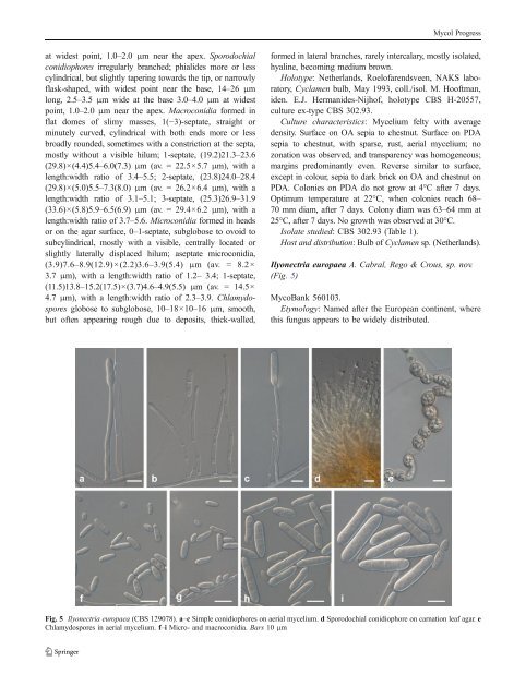
Mycol Progressat widest point, 1.0–2.0 μm near the apex. Sporodochialconidiophores irregularly branched; phialides more or lesscylindrical, but slightly tapering towards the tip, or narrowlyflask-shaped, with widest point near the base, 14–26 μmlong, 2.5–3.5 μm wide at the base 3.0–4.0 μm atwidestpoint, 1.0–2.0 μm near the apex. Macroconidia formed inflat domes of slimy masses, 1(−3)-septate, straight orminutely curved, cylindrical with both ends more or lessbroadly rounded, sometimes with a constriction at the septa,mostly without a visible hilum; 1-septate, (19.2)21.3–23.6(29.8)×(4.4)5.4–6.0(7.3) μm (av. = 22.5×5.7 μm), with alength:width ratio of 3.4–5.5; 2-septate, (23.8)24.0–28.4(29.8)×(5.0)5.5–7.3(8.0) μm (av. = 26.2×6.4 μm), with alength:width ratio of 3.1–5.1; 3-septate, (25.3)26.9–31.9(33.6)×(5.8)5.9–6.5(6.9) μm (av. = 29.4×6.2 μm), with alength:width ratio of 3.7–5.6. Microconidia formed in headsor on the agar surface, 0–1-septate, subglobose to ovoid tosubcylindrical, mostly with a visible, centrally located orslightly laterally displaced hilum; aseptate microconidia,(3.9)7.6–8.9(12.9)×(2.2)3.6–3.9(5.4) μm (av. = 8.2×3.7 μm), with a length:width ratio of 1.2– 3.4; 1-septate,(11.5)13.8–15.2(17.5)×(3.7)4.6–4.9(5.5) μm (av. = 14.5×4.7 μm), with a length:width ratio of 2.3–3.9. Chlamydosporesglobose to subglobose, 10–18×10–16 μm, smooth,but often appearing rough due to deposits, thick-walled,formed in lateral branches, rarely intercalary, mostly isolated,hyaline, becoming medium brown.Holotype: Netherlands, Roelofarendsveen, NAKS laboratory,Cyclamen bulb, May 1993, coll./isol. M. Hooftman,iden. E.J. Hermanides-Nijhof, holotype <strong>CBS</strong> H-20557,culture ex-type <strong>CBS</strong> 302.93.Culture characteristics: Mycelium felty with averagedensity. Surface on OA sepia to chestnut. Surface on PDAsepia to chestnut, with sparse, rust, aerial mycelium; nozonation was observed, and transparency was homo<strong>gene</strong>ous;margins predominantly even. Reverse similar to surface,except in colour, sepia to dark brick on OA and chestnut onPDA. Colonies on PDA do not grow at 4°C after 7 days.Optimum temperature at 22°C, when colonies reach 68–70 mm diam, after 7 days. Colony diam was 63–64 mm at25°C, after 7 days. No growth was observed at 30°C.Isolate studied: <strong>CBS</strong> 302.93 (Table 1).Host and distribution:BulbofCyclamen sp. (Netherlands).Ilyonectria europaea A. Cabral, Rego & Crous, sp. nov.(Fig. 5)MycoBank 560103.Etymology: Named after the European continent, wherethis fungus appears to be widely distributed.Fig. 5 Ilyonectria europaea (<strong>CBS</strong> 129078). a–c Simple conidiophores on aerial mycelium. d Sporodochial conidiophore on carnation leaf agar. eChlamydospores in aerial mycelium. f–i Micro- and macroconidia. Bars 10 μm
Mycol ProgressIlyonectriae robustae morphologice similis, sed longitudinemedia macroconidiorum breviore, 29.7–31.5 μm,distinguitur.Conidiophores simple or complex to sporodochial.Simple conidiophores arising laterally or terminally fromaerial mycelium, solitary to loosely aggregated, unbranchedor sparsely branched, bearing up to threephialides, 1–3-septate, 50–120 μm long; phialides monophialidic,cylindrical to subulate, 26–60 μm long, 2.5–3.5 μm wide at the base, 1.5–2.5 μm near the apex.Complex conidiophores aggregated in small sporodochia(on carnation leaf), repeatedly and irregularly branched.Macroconidia predominating, formed on both type ofconidiophores, on SNA formed in flat domes of slimymasses, 1(−3)-septate, straight or minutely curved, cylindricalwith both ends more or less broadly rounded, butmay narrow towards the tip, mostly without a visiblehilum; 1-septate, (16.4)21.9–23.4(34.0)×(4.0)5.2–5.6(7.8)μm (av.=22.7×5.4μm), with a length:width ratio of 3.2–5.4; 2-septate, (22.0)26.4–28.1(34.0)×(4.4)5.9–6.4(8.0)μm (av. = 27.2×6.1 μm), with a length:width ratio of3.4–6.4; 3-septate, (22.0)29.7–31.5(40.0)×(5.0)6.5–6.9(8.6) μm (av.=30.6×6.7μm), with a length:width ratioof 3.5–6.0. Microconidia 0–1-septate, ellipsoid to ovoid,more or less straight, without a visible hilum; aseptatemicroconidia sometimes curved towards one end, (3.0)8.5–9.8(17.0)×(1.7)3.3–3.5(5.0) μm (av.=9.1×3.4μm),with a length:width ratio of 1.5–3.4; 1-septate, (9.2)13.4–14.6(18.9)×(3.0)4.0–4.4(5.9) μm (av. = 14.0×4.2 μm),with a length:width ratio 2.6–4.0. Conidia formed in headsor on simple conidiophores as white (OA) or unpigmented(SNA) masses. Chlamydospores globose to subglobose, 9–14×7–14 μm, smooth, but often appearing rough due todeposits, thick-walled, terminal on short or long lateralbranches or intercalary, single, in chains or in clumps,golden-brown.Holotype: Portugal, Vidigueira, at basal end of a 2-yearoldVitis vinifera plant; scion Petit Verdot, <strong>root</strong>stock 110R,2008, coll./isol. C. Rego, holotype <strong>CBS</strong> H-20558, cultureex-type <strong>CBS</strong> 129078=Cy241=CPC 19165.Culture characteristics: Mycelium felty with averagedensity. Surface on OA chestnut, with saffron aerialmycelium. Sienna to saffron on PDA, with luteous aerialmycelium. Concentric zonation, with homo<strong>gene</strong>ous transparency,margins predominantly even. Reverse similar tosurface, except in the colour; sepia on OA, and chestnut toumber on PDA. Colonies on PDA grow poorly, 1–5 mmdiam at 4°C after 7 days. Optimum temperature for growthis 22°C, when colonies reach 43–57 mm diam, after 7 days.Colony diam was 37–47 mm at 25°C, after 7 days. Nogrowth was observed at 30°C.Isolates studied: Cy131; Cy155; <strong>CBS</strong> 537.92; <strong>CBS</strong>102892; <strong>CBS</strong> 129078 (Table 1).Hosts and distribution: Actinidia chinensis ‘Hayward’(internal lesion of stem) (France), Aesculus hippocastanum(wood) (Belgium), Phragmites australis (stem) (Germany),Vitis vinifera (Portugal).Ilyonectria gamsii A. Cabral & Crous, sp. nov. (Fig. 6)MycoBank 560112.Etymology: Named after Prof. dr. Walter Gams, who hasmade a major contribution to our knowledge of Hypocrealeansoil fungi.Ilyonectriae panacis morphologice similis, sed longitudinemedia macroconidiorum breviore, 34.3–38.5 μm,distinguitur.Conidiophores simple or complex to sporodochial.Simple conidiophores arising laterally or terminally fromaerial mycelium, solitary to loosely aggregated, unbranchedor sparsely branched, bearing up to two phialides, 1–3-septate, 50–150 μm long; phialides monophialidic, cylindricalto subulate, 30–60 μm long, 2.5–3.5 μm wide at thebase, 1.5–2.0 μm near the aperture. Sporodochial conidiophoresirregularly branched; phialides cylindrical, mostlywidest near the base. Macroconidia predominating, formedon simple conidiophores, on SNA formed in flat domes ofslimy masses, 1–3-septate, straight, cylindrical with bothends broadly rounded, with mostly visible, centrally locatedhilum; 1-septate, (22.0)25.7–27.9(33.0)×(4.0)5.1–5.5(6.0)μm (av. = 26.8×5.3 μm), with a length:width ratio of 4.3–6.2; 2-septate, (25.0)28.2–31.7(39.0)×(5.0)5.5–5.9(6.5) μm(av. = 29.9×5.7 μm), with a length:width ratio of 4.2–7.1; 3-septate, (24.0)34.3–38.5(44.0)×(5.0)5.9–6.3(7.0) μm (av.=36.4×6.1 μm), with a length:width ratio of 4.3–7.3. Microconidia0–1-septate, ellipsoid to subcylindrical, more or lessstraight, mostly with a visible hilum; aseptate microconidia(4.0)6.9–8.0(10.0)×(3.0)4.0–4.5(5.0) μm (av. = 7.4×4.3 μm), with a length:width ratio of 1.3–2.9; 1-septate,(8.0)12.9–15.7(18.0)×(4.0)4.2–4.7(5.5) μm (av. = 14.3×4.4 μm), with a length:width ratio 1.8–4.0. Chlamydosporesglobose to subglobose to ellipsoidal, 8–14×7–12 μm,smooth, but often appearing rough due to deposits, thickwalled,mostly intercalary, rarely terminal on short lateralbranches, single, in chains or in clumps, hyaline, becomingmedium brown.Holotype: Netherlands, Lelystad, soil, June 1997, coll./isol. J.T. Poll, iden. W. Gams, holotype <strong>CBS</strong> H-20559,culture ex-type <strong>CBS</strong> 940.97.Culture characteristics: Mycelium cottony, dense. Surfaceon OA cinnamon, with sparse, buff aerial mycelium,on PDA umber to chestnut, with buff to saffron aerialmycelium; zonation absent, transparency homo<strong>gene</strong>ous,margin even; reverse similar to surface, but chestnut onPDA. Colonies on PDA grow 6–7 mm diam at 4°C after7 days. Optimum temperature at 22°C when colonies reach