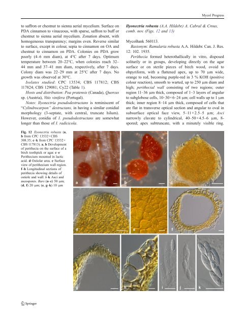
Mycol Progressto saffron or chestnut to sienna aerial mycelium. Surface onPDA cinnamon to vinaceous, with sparse, saffron to buff orchestnut to sienna aerial mycelium. Zonation absent, withhomo<strong>gene</strong>ous transparency; margins even. Reverse similarto surface, except in colour, sepia to cinnamon on OA andchestnut to cinnamon on PDA. Colonies on PDA growpoorly (4–6 mm diam), at 4°C after 7 days. Optimumtemperature between 20–22°C, when colonies reach 32–44 mm and 37–41 mm diam, respectively, after 7 days.Colony diam was 22–29 mm at 25°C after 7 days. Nogrowth was observed at 30°C.Isolates studied: CPC 13534; <strong>CBS</strong> 117812; <strong>CBS</strong>117824; <strong>CBS</strong> 129081; Cy22 (Table 1).Hosts and distribution: Poa pratensis (Canada), Quercussp. (Austria), Vitis vinifera (Portugal).Notes: Ilyonectria pseudodestructans is reminiscent of“<strong>Cylindrocarpon</strong>” destructans, in having a similar conidialmorphology (3-septate, with central, truncate hilum).However, conidia of I. pseudodestructans are somewhatlonger than those of I. radicicola.Ilyonectria robusta (A.A. Hildebr.) A. Cabral & Crous,comb. nov. (Figs. 12 and 13)MycoBank 560113.Basionym: Ramularia robusta A.A. Hildebr. Can. J. Res.12: 102. 1935.Perithecia formed hete<strong>rot</strong>hallically in vitro, disposedsolitarily or in groups, developing directly on the agarsurface or on sterile pieces of birch wood, ovoid toobpyriform, with a flattened apex, up to 70 μm wide,orange to red, becoming purple-red in 3 % KOH (positivecolour reaction), smooth to warted, up to 250 μm diam andhigh; perithecial wall consisting of two regions; outerregion 11–36 μm thick, composed of 1–3 layers of angularto subglobose cells, 10–30×6–24 μm; cell walls up to 1 μmthick; inner region 8–14 μm thick, composed of cells thatare flat in transverse optical section and angular to oval insubsurface optical face view, 5–11 × 2.5–5 μm; Ascinarrowly clavate to cylindrical, 40–50×4.5–6 μm, 8-spored; apex subtruncate, with a minutely visible ring.Fig. 12 Ilyonectria robusta (a,b from CPC 13532×<strong>CBS</strong>308.35; c–k from CPC 13532×<strong>CBS</strong> 117813). a, b Developmentof perithecia on the surface of abirch toothpick or agar. c–ePerithecium mounted in lacticacid. d Ostiolar area. e Surfaceview of perithecium wall region.f–h Longitudinal sections ofperithecia showing details ofostiole and wall. i–k Asci andascospores. Bars (a–c) 50μm;(d, f) 20μm; (e, g–k) 10μm
Mycol ProgressFig. 13 Ilyonectria robusta (All from <strong>CBS</strong> 129084, except f from <strong>CBS</strong> 605.92). a–c Simple conidiophores on aerial mycelium. d Sporodochialconidiophore on carnation leaf agar. e Chlamydospores on mycelium f–i Micro- and macroconidia. Bars 10 μmAscospores medianly 1-septate, ellipsoid to oblongellipsoid,somewhat tapering towards both ends, smoothto finely warted, frequently guttulate, hyaline, (8.2)9.4–9.7–10.0(11.5)×(2.5)2.9–3.0–3.1(3.7) μm. Conidiophores simpleor complex or sporodochial. Simple conidiophoresarising laterally or terminally from aerial mycelium, solitaryto loosely aggregated, unbranched or sparsely branched,bearing up to three phialides, 1–4-septate, 55–160 μm long;phialides monophialidic, cylindrical to subulate, 20–60 μmlong, 2.0–3.0 μm wide at the base, 1.5–2.0 μm near theapex. Complex conidiophores aggregated in small sporodochia(on carnation leaf agar; Crous et al. 2009b),repeatedly and irregularly branched; phialides more or lesscylindrical, but tapering slightly in the upper part towardsthe apex, or narrowly flask-shaped, mostly with widestpoint near the middle, 15–20 μm long, 2.5–3.5 μm wide atthe base, 3.0–4.0 μm at the widest point, and 1.0–2.0 μmwide near the apex. Macroconidia predominating, formedon simple conidiophores, on SNA formed in flat domes ofslimy masses, 1–3-septate, straight, minutely curved orsometimes distorted, cylindrical with both ends more or lessbroadly rounded, but may narrow towards the tip, mostlywithout a visible hilum; 1-septate, (15.0)22.8–24.6(35.0)×(4.5)6.3–6.7(8.0) μm (av. = 23.7×6.5 μm), with a length:width ratio of 2.7–5.2; 2-septate, (20.0)26.2–28.1(38.0)×(5.0)6.9–7.2(8.0) μm (av. = 27.2×7.0 μm), with a length:width ratio of 2.9–5.2; 3-septate, (24)32.3–34.7(58)×(6.0)7.2–7.5(9.0) μm (av. = 33.5×7.4 μm), with a length:widthratio of 3.1–7.3. Microconidia 0–1-septate, ellipsoid to ovoidto subcylindrical, more or less straight, without a visiblehilum; aseptate, (4.0)8.0–9.3(14.0)×(2.5)3.6–4.0(5.5) μm(av. = 8.7×3.8 μm), with a length:width ratio of 1.3–4.0;1-septate, (9.0)13.5–14.7(18.0)×(3.5)4.7–5.1(6.0) μm (av.=14.1×4.9 μm), with a length:width ratio 1.5–4.5. Conidiaformed in heads on simple conidiophores or as white (OA)or unpigmented (SNA) masses. Chlamydospores globose tosubglobose, 7–14×6–13 μm, smooth, but often appearingrough due to deposits, thick-walled, mostly occurringintercalary in chains, hyaline, becoming golden-brown.Lecto- and teleotype: Canada, Ontario, on living <strong>root</strong>s ofPanax quinquefolium, 1935, A.A. Hildebrand, lectotypedesignated here <strong>CBS</strong> H-20565, as dried culture of <strong>CBS</strong>308.35; teleotype designated here <strong>CBS</strong> H-20566, includingfertile perithecia of the teleomorph (CPC 13532×<strong>CBS</strong>308.35), culture ex-lectotype <strong>CBS</strong> 308.35.Fertile matings: Perithecia observed after 4 wk incrossings of strains: CPC 13532×<strong>CBS</strong> 308.35, CPC13532×<strong>CBS</strong> 773.83, CPC 13532×<strong>CBS</strong> 605.92, CPC13532×<strong>CBS</strong> 117813, <strong>CBS</strong> 129084×<strong>CBS</strong> 308.35, <strong>CBS</strong>129084×<strong>CBS</strong> 605.92, <strong>CBS</strong> 129084×<strong>CBS</strong> 117813.