Imaging Tissue Motion Using MRI DENSE - Displacement Encoding ...
Imaging Tissue Motion Using MRI DENSE - Displacement Encoding ...
Imaging Tissue Motion Using MRI DENSE - Displacement Encoding ...
- No tags were found...

You also want an ePaper? Increase the reach of your titles
YUMPU automatically turns print PDFs into web optimized ePapers that Google loves.
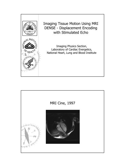
<strong>MRI</strong> Assessment of VentricularFunction I – Cine <strong>Imaging</strong><strong>MRI</strong> Assessment of VentricularFunction II – TaggingZerhouni, 1988, Axel, 1989.
<strong>MRI</strong> Assessment of VentricularFunction II – Tagging? Tagging measurement resolution is limiteddue the grid spacing.Victor Ferrari, private communicationA New Approach: <strong>Displacement</strong><strong>Encoding</strong> with Stimulated-Echo(<strong>DENSE</strong>)? Encode positional information into the phase ofeach image pixel (Aletras, Ding, Balaban, andWen, MRM Proc. 1998).? Higher spatial resolution.? Amenable to automatic processing.
<strong>DENSE</strong> Pulse Sequencefew ms ~ 1s90 ° 90 ° 90 °TmImage Acq.Initial phase mapsPhase shiftsY-encodedX-encodedStriping ArtifactsAletras AH, Ding SJ, Balaban RS, Wen H. Journal of Magnetic Resonance 1999; 137(1):247-252.
Removal of Artifacts? Inversion, through-slice encoding and phasecycling help suppress other echoes andseparate out the stimulated echoAletras AH, Wen H. Magnetic Resonance in Medicine 2001; 46(3):523- 534.Strain Vector Maps% Circumferential shorteningAletras, Ding, Balaban, and Wen, JMR 1999% wall thickening
3D displacement fieldECG triggered,respiratory gatedscan time = 4minutes/slice.Time resolved <strong>DENSE</strong>
Image Processing: from <strong>MRI</strong> Data toClinically Useful Parameters? Image segmentation: removing unwantedpixels.? Following material points over time tomeasure peak strain, torsion and timing.<strong>DENSE</strong> is Amenable to AutomaticImage Processing? Automatic masking of solid tissue pixels:§ <strong>Tissue</strong> movement should be continuous betweenneighboring pixels and adjacent time points.§ Flowing blood appears dark, which aids separation ofblood pool and solid tissue.? <strong>Tissue</strong> tracking-following material points over time§ <strong>Displacement</strong> vectors at all time points are available.
<strong>DENSE</strong>-ViewImage ReconstructionPhase unwrappingConverting to displacement mapsImagesegmentationShaped-smoothing<strong>Tissue</strong> trackingEulerian strain calculationDyssynchronyDyskinesisTorsionMagnitudePhase XPhase YRaw dataPhase singularityremovedSlip points removedIntensity thresholdedand phase unwrapped
Unsmoothed Circumferential StrainMovie? Strain calculation magnifies phase noise inthe data.How to smooth the noisy data?? Low-pass filtering averages over aneighborhood of fixed shapeLow-pass filtered
Shaped Smoothing? Kernel conforms to local anatomical co-oridinates, preserves transmural resolution.Shaped-Smoothing Smoothing is Superiorto Low-Pass FilteringLow-pass filteredShaped-smoothing
Strain Maps from Shaped-SmoothedSmoothed<strong>Displacement</strong> FieldsTracking Material Points over Time? Converting Eulerian displacement toLagrangian displacement
Dyskinesis and Dyssynchrony MapsDyskinesis and Dyssynchrony Maps
Patient Data – Epstein Kinase AlleleProtocol, NHLBI/NIH73, F, hypertension 55, M, hypertensionSummary of <strong>DENSE</strong> Cardiac FunctionAssessment? <strong>Displacement</strong> imaging enables automatedProcessing for regional ventricular function.? Future direction: test/retest reproducibility,correlation with echo TDI is being studied innormal volunteers and patients.
Arterial Wall Strain <strong>Imaging</strong> andMechanical Instability in VulnerablePlaques? A vulnerable plaque is a future culprit plaque -70% of culprit coronary lesions rupture.(Consensus paper by Naghavi et al., Circ.108:1664-1672, 1672, 1772-1778). 1778).Burke et al., NEJM 1997Mechanical Aspects Plaque Rupture? Rupture involves the fissuring of plaque capin high stress/strain spots.
Tensile Stress Distribution Predicts Rupture Sites inCoronary Balloon AngioplastyOhayon, Teppaz, Finet and Rioufol, Coronary Artery Disease 2001, 12:655Simulation of Strain Patterns forDifferent Plaque MorphologyLumen Cap Lipid coreBaldewsing, Schaar, Mastik, Oomens, Van der Stern, IEEETrans. Med. <strong>Imaging</strong>, 2005, 24(4):514
Simulation of Strain Patterns ofDifferent Plaque MorphologyBaldewsing, Schaar, Mastik, Oomens, Van der Steen, IEEETrans. Med. <strong>Imaging</strong>, 2005, 24(4):514Clinical strain imaging with IVUSIVUS strain mapping in a PCTA procedure patient, DerKorte and Van der Steen, Ultrasonics, 40:859-865,2002.
Frequency of Regional High Strain SpotsIs Correlated with Clinical IndicatorsSchaar et al., n = 55, IVUS elastography of culprit vesselbefore angioplasty (Circ. 109(22): 2716-2719, 2719, 2004).Positive correlation with presentation. hsCRP R 2 = 0.65.Circumferentially Averaged Distensibility IsCorrelated with Angioscopy PlaqueClassificationTakano et al., n = 38, post ACS. JACC 38(1):99-104, 104,2001.
<strong>DENSE</strong> <strong>Imaging</strong> of Registered Strain andMorphology Is Feasible in Human Carotid Artery? <strong>DENSE</strong> has inherent T 1 weighting and dark-blood contrast.<strong>Displacement</strong> Field around Carotid LumenCurrent 600µm in-planeresolution, 5 mm slicethickness.
Circumferential Strain around CarotidLumenWen, Vignaud, Rodriguez, submitted to AHA 2005Another example
Summary of vessel wall strainimaging? Simultaneous strain and morphology imaging ofthe carotid artery is feasible in humans.? Correlation of <strong>MRI</strong> findings with other markersneeds to be studied in patients with carotidlesions.? The spatial resolution at 1.5T is 500 – 600 µm,and is expected to improve with 3T scanners.
Acknowledgement? Engineering? Clinical applicationShujun Ding, PhD, NIHAnthony Aletras, PhD, NIHVinay Pai, PhD, NYUIgnacio Rodriguez, PhD, NIHAlex Vignaud, PhD, NIHEric Bennett, MS, NIHJon Plehn, MD, NIH/GWNeal Epstein, MD, NIHAndrew Arai, MD, NIH