FOCUS ON GLAUCOLIGHT - DORC
FOCUS ON GLAUCOLIGHT - DORC
FOCUS ON GLAUCOLIGHT - DORC
- No tags were found...

Create successful ePaper yourself
Turn your PDF publications into a flip-book with our unique Google optimized e-Paper software.
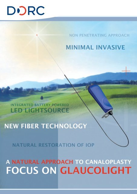
N<strong>ON</strong> PENETRATING APPROACHMINIMAL INVASIVEINTEGRATED BATTERY POWEREDLED LIGHTSOURCENEW FIBER TECHNOLOGYNATURAL RESTORATI<strong>ON</strong> OF IOPA NATURAL APPROACH TO CANALOPLASTY<strong>FOCUS</strong> <strong>ON</strong> <strong>GLAUCOLIGHT</strong>
D.O.r.C. GlauCOliGhtGlaucoliGhtcanaloplasty DeviceNEW FIBER TECHNOLOGY FOR SMOOTHAND MINIMAL INVASIVE SURGERY3269.SG Scharioth Glaucome Fiber(Box/3, sterile). Patent PendingThe Glaucolight is a surgical ophthalmiccanaloplasty device to facilitate the treatmentof open angle glaucoma. The Glaucolightfacilitates the new stretching-suture technologyto re-establish the natural aqueous outflowand reduce the intraocular pressure.The Glaucolight is a lightfiber based devicewith an integrated (battery powered) LEDsource and an atraumatic tip-design for asmooth transfer through the Schlemm’s canal.The bright LED illuminated fiber tip helpsvisualize the position of the fiber during the360 degree Schlemm’s canal passage.The Glaucolight has a 40G/0,15mm microdiameterfor minimally invasive surgeryand flexible 360 degree followability ofthe Schlemm’s canal.INTEGRATED LED LIGHTSOURCEN<strong>ON</strong> PENETRATING APPROACHEC<strong>ON</strong>OMICAL AND EFFECTIVEALTERNATIVE FOR CURRENTGLAUCOMA TREATMENTSA special suture fixation notch at thedistal end of the fiber assures a firm fixationof the stretching-suture in combinationwith minimizing injury effect of the sutureknot in the Schlemm’s canal.RESTORING THENATURAL FUNCTI<strong>ON</strong>OF THE EYEA NATURAL APPROACH TO CANALO<strong>FOCUS</strong> <strong>ON</strong> GLAUCOLI
STEP 1Begin by opening the conjunctiva and tenon markinga parabolic 5x5mm superficial flap with Scharioth’sscleral marker. Cut in to one third of the scleralthickness with a paracentesis knife to prepare asuperficial scleral flap up to the clear cornea.Use no or minimal diathermy.REDUCES THE NEEDFOR GLAUCOMAMEDICINESSTEP 2Prepare a deeper scleral flap approximately 3x3mmpassing scleral spur opening and un-roofing theSchlemm’s canal. Preparation of Descemetic windowcutting sharp at the edges with blunt preparationin the center. At this stage, a paracentesis incisionwhilst lowering the IOP reduces the risk ofperforation.STEP 3Introduce Glaucolight into the surgical ostia ofSchlemm’s canal using Scharioth’s Glaucolightforceps for easier manipulation. Use methyl cellulosefor lubrication of the Glaucolight fiber to facilitateeasier catheterization. Advance the Glaucolight 360ºthrough Schlemm’s canal. If blockage occurs, retryin opposite direction through Schlemm’s. Bendingthe tip may help slightly.STEP 4Tie a 10.0 suture at the distal end of the Glaucolight fiber. Then withdraw the Glaucolightimplanting the suture. Cut the deep scleral flap at its base (deep sclerotomy), tighten the10.0 suture and knot it so that the Descemet’s window is visibly pulled inwards.PLASTYGHTSTEP 5Check perfusion of aqueous humor at the surgicalsite. If this is inadequate, peel the juxtacanaliculartrabecular meshwork and inject high viscousHyaluronic Acid in to both Ostia of the Schlemm’scanal. Close the superficial scleral flap (watertight)using 5-7 10.0 monofil absorbable sutures. Injecthigh viscous Hyaluronic Acid under the superficialflap to form a scleral lake preventing collapse. Closethe conjunctiva with 10.0 monofil absorbable sutures.
![OCT Setting: CORNEA RADIAL 6( 6.0mm[1024] ) - innova](https://img.yumpu.com/48127738/1/190x146/oct-setting-cornea-radial-6-60mm1024-innova.jpg?quality=85)