Fig. 21 − Dr. V<strong>in</strong>icio Sosa dur<strong>in</strong>g a foray <strong>in</strong> “Aguita Fria” cloud fo<strong>re</strong>st, Veracruz, Mexico, tak<strong>in</strong>gsome pictu<strong>re</strong>s from the samples.............................R. biseptatum (Fig. 25)Conidia not navicular ..........................1414(13)Conidia ellipsoid to oval, (2−)3−4-septate, brown, apical cell pale brown,smooth, 23−32 × 11−13 µm .............................................. R. dennisii (Figs 30−33)Conidia obovate to subglobose, 3-septate,with dark brown middle <strong>and</strong> basal cells,<strong>and</strong> pale brown at the apex, smooth,16−21 × 8−13 µm ..........................................................R. quadrilocula<strong>re</strong> (Fig. 52)Conidia moniliform, 8−22-septate, brown<strong>and</strong> verrucose at the base <strong>and</strong> middle part,<strong>and</strong> pale brown <strong>and</strong> smooth towards theapex, 55−280 × 6−10 µm .................................................. R. moniliforme (Fig. 47)AcknowledgementsThe authors a<strong>re</strong> grateful to Dr Lori Carris(Wash<strong>in</strong>gton State University) for p<strong>re</strong>submission<strong>re</strong>view of this paper. Gabriela He<strong>re</strong>dia,R.M. Arias <strong>and</strong> R.F. Castañeda Ruiz a<strong>re</strong>thankful to CONABIO (Mexico) for f<strong>in</strong>ancialsupport through project IE004. T. Iturriaga isgrateful to FONACYT, Venezuela, for f<strong>in</strong>ancialsupport for project S1-2001000663. Theauthors thank Dr. Uwe Braun, Dr. Lori Carris,Dr. De-Wei Li, Dr. Felipe Wartchow, Dr.Antonio Hernández-Gutiér<strong>re</strong>z, Dr. Melissa282
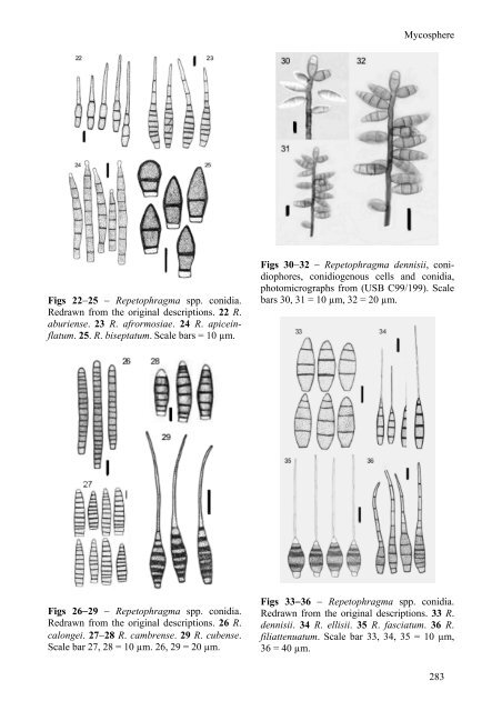
Mycosphe<strong>re</strong>Figs 22−25 − <strong>Repetophragma</strong> spp. conidia.Redrawn from the orig<strong>in</strong>al descriptions. 22 R.aburiense. 23 R. afrormosiae. 24 R. apice<strong>in</strong>flatum.25. R. biseptatum. Scale bars = 10 µm.Figs 30−32 − <strong>Repetophragma</strong> dennisii, conidiopho<strong>re</strong>s,conidiogenous cells <strong>and</strong> conidia,photomicrographs from (USB C99/199). Scalebars 30, 31 = 10 µm, 32 = 20 µm.Figs 26−29 − <strong>Repetophragma</strong> spp. conidia.Redrawn from the orig<strong>in</strong>al descriptions. 26 R.calongei. 27−28 R. camb<strong>re</strong>nse. 29 R. cubense.Scale bar 27, 28 = 10 µm. 26, 29 = 20 µm.Figs 33−36 − <strong>Repetophragma</strong> spp. conidia.Redrawn from the orig<strong>in</strong>al descriptions. 33 R.dennisii. 34 R. ellisii. 35 R. fasciatum. 36 R.filiattenuatum. Scale bar 33, 34, 35 = 10 µm,36 = 40 µm.283