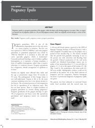
Review ArticleFIG. 6CO 2lased specimens showed melting of dentin andnarrowing of exposed dentinal orifices. (x 2500)It was concluded that compared to conventionalfluoridation or laser irradiation in the treatment ofdentin hypersensitivity, CO 2lasers in combinationwith Fluoride varnish appear to show better efficacythan either treatment modality alone [Fig.1-6].Conclusion: In the future, laser will be an importanttool in the armamentarium of a modern dental office. Itspotential for dental application in dentistry merits closerattention. Efforts should be focused on the search for a laserwavelength with optimal irradiation parameters that willenable the clinician to produce ideal modification of thedentin surface and other hard tissues of the tooth. Completefamiliarity with a safe and recommended protocol isessential at all times when treating vital teeth with lasers toalleviate the pain associated with hypersensitive dentin.References:1. Holland, G. R, Narhi, M. N. Addy, M.Gangarosa, L.&Orchardson, S. Guidelines for the design and conductof clinical trials on dentine hypersensitivity, Journal ofClinical Periodontology1997; 24:808–82. Adam Stabholz, SharonitSahar-Helft, Joshua Moshonov.Lasers in endodontics. Dent Clin N Am 2004; 48: 809–832.3. Rees J. The prevalence of dentine hypersensitivity ingeneral dental practice in the UK. Journal of ClinicalPeriodontology 2000; 27: 860–865.4. Branstrom M. The surface of sensitive dentin. OdontRev 1965;16:293-95. Kimura Y, Wilder-Smith P, Yonaga K, Matsumoto K.Treatment of dentine hypersensitivity by lasers: a review.J Clin Periodontal 2000;27: 715-216. Pashley D, Tay,FVaywood V, Collins, Drisco C. DentinHypersensitivity – Consensus based recommendations forthe diagnosis & management of dentin hypersensitivity,AEGIS Publications,20087. Strassler H, Serio ,F , Dentinal Hypersensitivity Etiology,Diagnosis and Management, ineedce.com8. Branstrom M, Johnson G, Nordenvall K. Transmissionand control of dentinal pain: resin impregnation for thedesensitization of dentin. J Am Dent Assoc 1979;99:612-89. Nordenvall KJ, Malmgren B, Brännström M.Desensitization of dentin by resin impregnation: aclinical and light-microscopic investigation. ASDC JDent Child1984 ; 51: 274-6.10. Plagmann HC, König J, Bernimoulin JP, Rudhart AC,Deschner J. A clinical study comparing two high-fluoridedentifrices for the treatment of dentinal hypersensitivityQuintessence Int 1997; 28: 403-811. Senda A, Gomi A, Tani T, Yoshino H, Hara G. A clinicalstudy on “soft laser 632”, a HeNelow energy medicallaser. Aichi-Gakuin J Dent sciences 1985;23:773-8012. Moritz A, Gutknecht N, Schoop U, Goharkhay K, EbrahimD, Wernisch J, Sperr. The advantage of CO2-treated dentalnecks, in comparison with a standard method: results of anin vivo study. J Clin Laser Med Surg 1996; 14: 27-3213. Renton-Harper P, Midda M.NdYAG laser treatment ofdentinal hypersensitivity. Br Dent J. 1992; 11:13-6..14. Wan-Hong Lan, Bor-Shiunn Lee, Hsin-Cheng Liu,Chun-Pin Lin. Morphologic study of Nd:YAG laserusage in treatment of dentinal hypersensitivity.Journal ofEndodontics 2004; 30: 131-13415. Ipci SD, Cakar G, Kuru B, Yilmaz S. Clinical evaluationof lasers and sodium fluoride gel in the treatment ofdentine hypersensitivity. Photomedicine and LaserSurgery 2009; 27:85-91.16. Zhang C, Lin Q, Zhao B. The combined occluding effectof fluor protector and Nd:YAG laser irradiation on humandentinal tubules Zhonghua Kou Qiang Yi XueZaZhi2001 Mar; 36:105-7.17. Matsumoto K, Funai H, Wakabayashi H, Oyama T. Studyon the treatment of hypersensitive dentine by GaAlAslaser diode. Japanese J Conserv Dent 1985;28:766-7118. Moritz A, Gutknecht N, Schoop U, Goharkhay K,Ebrahim D, Wernisch J, Sperr. The advantage of CO2-treated dental necks, in comparison with a standardmethod: results of an in vivo study. J Clin Laser MedSurg 1986; 14: 27-32.19. Senda A,GomiA,TaniT, A clinical studyon ~Soft Laser632~,a He Ne low energy medical laser Aichi-Gakuin JDent Sciences 1985;23:773-80.20. Matsumoto K,FunaiH, WakabayashiH, Oyama T. Studyon the treatment of hypersensitive dentine by GaAlAslaser diode. Japanese J Conserv Dent 1985;28:766-71.21. Renton-HarperP, MiddaM,.Nd YAGlaser treatment ofdentinal hypersensitivityBr.Dent J1992;172:13-6.22. StabholzA, NeevJ, LiawLL, StabholzAy, Torabinejad M.Sealing ability of dentinal tubulesbyXeCl308nm excimerlaser. Endod1993;19:267-71.23. Ahamed S, Gurucharan N, Meyappan R, Arun K, Deepa V,Babu S. A comparative analysis between Nd:YAG and CO2lasers for their sealing ability of dentinal tubules with andwithout fluoride varnish an in-vitro study. Journal of DentalLasers 2012;6:51-56.572Indian Journal of Multidisciplinary Dentistry, Vol. 2, <strong>Issue</strong> 4, <strong>Aug</strong>ust-<strong>Oct</strong>ober 2012
Alloplastic Bo n e Gr a f t i n g Materialsreview articleAnurag Ashok Shendre*, Deepti Gattani @ , Arshad Sayed % , Narpat Singh Rajput ~ABSTRACTBone grafting has become a valuable mainstream clinical procedure in today’s dentistry in a variety of reconstructiveapplications. Various materials and formulations have been developed for this purpose. The synthetic bone graftsubstitutes as yet offer only a part solution to the management of localized bone loss. Ideally a synthetic bone graft substituteshould mimic the native bone in both mechanical and osteogenic properties. They possess some of the desiredmechanical qualities of bone as well as osteointegrative/conductive properties. It has given the clinicians the abilityto offer patients alternative treatment modalities that can serve to restore the missing bone anatomy. Present reviewdeals with the various alloplastic bone graft materials. Various advantages and disadvantages with these materials arediscussed.Key words: Reconstruction, Bone grafts, Osteointegration , Osteoconduction, OsteogenesisIntroductionBone grafting has become a valuable mainstreamclinical procedure in today’s dentistry in avariety of reconstructive applications. Ridgepreservation after tooth extraction is very importantfor making any kind of prosthesis, be it dentures, fixedbridge or implants. 1 Various materials and formulationshave been developed for this purpose. It has given theclinicians the ability to offer patients alternative treatmentmodalities that can serve to restore the missing boneanatomy for ridge maintenance or preservation, ridgeaugmentation and for enhanced bone foundation forendosseous implant placement and to fill in any bonydefects or voids such as caused by removal of a cyst ortumor. 2There are four characteristics that an ideal bone graftmaterial should exhibit which include: 61. Osteointegration, the ability to chemically bond to thesurface of bone without an intervening layer of fibroustissue.2. Osteoconduction, the ability to support the growth ofbone over its surface.3. Osteoinduction, the ability to induce differentiation ofpluripotential stem cells from surrounding tissue to anosteoblastic phenotype.*PG Student,@Professor,%Dean and Head , Dept of Periodontics, Swargiya DadasahebKalmegh Smruti Dental College and Hospital, Nagpur.~ Post Graduate, Dept of Periodontics. Sree Balaji Dental Collegeand Hospital, Bharath University, Pallikaranai, Chennai.Corresponding Author:Dr.Narpat Singh RajputE mail: narpat_singh_rajput@yahoo.co.in4. Osteogenesis, the formation of new bone by osteoblasticcells present within the graft material. Synthetic bonegraft substitutes currently possess only osteointegrativeand osteoconductive properties.The synthetic bone graft substitutes as yet offer onlya part solution to the management of localized boneloss. They possess some of the desired mechanicalqualities of bone as well as osteointegrative /conductive properties but are largely reliant onviable periosteum/bone for their success. Ideallya synthetic bone graft substitute should mimic thenative bone in both mechanical and osteogenicproperties.Classification of Bone Graft Materals 3(i) Autogenous bone grafts :They are osteogenic, osteoconductive and osteo inductive.There is no risk of host rejection or disease transmission.But its major disadvantages are procurement morbidity,limited availability and high cost. 4(ii) Allograft :It is nonvital , osseous tissue taken from one individualand transferred to another of the same species. Theyare osteoconductive and weakly osteoinductive. Itsadvantages include greater availability of bankedbone than autograft and no additional surgicalprocedure needed. Its disadvantages include riskof disease transfer, not osteogenic, immunogenic,variable clinical results and expensive. 5(iii) Xenogenic bone grafts :It is an osseous tissue that is harvested from onespecies, processed and then transferred to a recipientIndian Journal of Multidisciplinary Dentistry, Vol. 2, <strong>Issue</strong> 4, <strong>Aug</strong>ust-<strong>Oct</strong>ober 2012 573