CD4:CD8 T-Cell Ratio Differs Significantly in Diffuse Large B-Cell ...
CD4:CD8 T-Cell Ratio Differs Significantly in Diffuse Large B-Cell ...
CD4:CD8 T-Cell Ratio Differs Significantly in Diffuse Large B-Cell ...
Create successful ePaper yourself
Turn your PDF publications into a flip-book with our unique Google optimized e-Paper software.
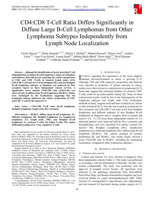
INTERNATIONAL TRENDS IN IMMUNITY VOL.1 NO.2 APRIL 2013ISSN 2326-3121 (Pr<strong>in</strong>t) ISSN 2326-313X (Onl<strong>in</strong>e) http://www.researchpub.org/journal/iti/iti.html<strong>CD4</strong>:<strong>CD8</strong> T-<strong>Cell</strong> <strong>Ratio</strong> <strong>Differs</strong> <strong>Significantly</strong> <strong>in</strong><strong>Diffuse</strong> <strong>Large</strong> B-<strong>Cell</strong> Lymphomas from OtherLymphoma Subtypes Independently fromLymph Node LocalizationCécile Daussy 1,2,3 , Diane Damotte 1,3,4,5 , Thierry J. Mol<strong>in</strong>a 3,4 , Mikael Roussel 6 , Thierry Fest 6,7 , AudreyVar<strong>in</strong> 1,2,3 , Jean-Yves Perrot 8 , Lamia Ouafi 3,4 , Hélène Merle-Béral 9 , Pierre Julia 3,10 , Wolf HermanFridman 1,2,3 , Cather<strong>in</strong>e Sautès-Fridman 1,2,3 , and Sylva<strong>in</strong> Fisson 1,2,3,11,12,13, *Abstract— Although the identification of newly described T-cellsubpopulations <strong>in</strong> human B-cell lymphoma is under <strong>in</strong>vestigation,contradictory data still persist regard<strong>in</strong>g the relative proportionsof <strong>CD4</strong> + and <strong>CD8</strong> + T-cells <strong>in</strong> tumoral lymph nodes (LN).<strong>CD4</strong>:<strong>CD8</strong> T-cell ratio <strong>in</strong> LN from Hodgk<strong>in</strong> (HL) and non Hodgk<strong>in</strong>B-cell lymphoma subtypes at diagnosis was analyzed by flowcytometry based on three <strong>in</strong>dependent cohorts (n=112). Asignificantly lower median <strong>CD4</strong>:<strong>CD8</strong> ratio (0.82±1.06) wasobserved only <strong>in</strong> diffuse large B-cell lymphomas (DLBCL). <strong>Ratio</strong>swere unchanged by the localizations suggest<strong>in</strong>g that thelymphoma subtype <strong>in</strong>fluences the relative proportions of <strong>CD4</strong> +and <strong>CD8</strong> + T-cells <strong>in</strong> the tumoral LN.Index Terms— <strong>CD4</strong>:<strong>CD8</strong> T-cell ratio, B-cell lymphoma,Hodgk<strong>in</strong> lymphoma, lymph node, flow cytometryAbbreviations— DLBCL, diffuse large B-cell lymphoma; FL,follicular lymphoma; HL, Hodgk<strong>in</strong> lymphoma; LL, lymphocyticlymphoma; LN, lymph node; NHL, non Hodgk<strong>in</strong> B-celllymphoma; Tc, cytotoxic T-cells; Th, helper T-cells; TIL, tumor<strong>in</strong>filtrat<strong>in</strong>g lymphocytes; Treg, regulatory T-cells.Paper submitted for review on Feb 24 2103 and accepted March 10, 2013.This work was supported by the Institut National du Cancer (GrantRC013-C06N631-2005), the Association pour la Recherche contre le Cancer,the Institut National de la Santéet de la Recherche Médicale, the UniversityPierre and Marie Curie, the University Paris-Descartes, the pole decompétitivitéIle de France (ImmuCan project). C.D. was recipient of a grantfrom the Institut National du Cancer.1 Institut National de la Santé et de la Recherche Médicale (INSERM),UMRS872, Centre de Recherche des Cordeliers, Paris, F-75006, France;2 UniversitéPierre et Marie Curie-Paris 6, UMRS 872, Paris F-75006, France;3 Université Paris Descartes, UMRS 872, Paris F-75006, France; 4 Serviced’Anatomie Pathologique, Hôtel Dieu, AP-HP, Paris F-75005, France; 5 Serviced’Anatomie Pathologique, Hôpital Européen Georges-Pompidou (HEGP),AP-HP, Paris F-75015, France; 6 Laboratoire d’Hématologie, UniversityHospital Pontchaillou, Rennes, France; 7 INSERM U917, Rennes, France;8 Service d’Hématologie Biologique, Hôtel Dieu, AP-HP, Paris F-75005,France; 9 Service d’Hématologie Biologique, Hôpital de la Pitié-Salpêtrière,AP-HP, Paris F-75013, France; 10 Service de Chirurgie Cardiaque et Vasculaire,Hôpital Européen Georges-Pompidou, AP-HP, Paris F-75015, France.11 Généthon, Evry, France; 12 INSERM, U951, Evry, France; 13 Université d’EvryVal d'Essonne, UMRS 951, Evry, France.* Correspondance: Sylva<strong>in</strong> Fisson. Généthon, INSERM UMRS 951, 1 bisrue de l’Internationale, Evry F-91002, France. Phone: +33 169 473 440 ; Fax :+33 160 778 698 ; e-mail: sylva<strong>in</strong>.fisson@univ-evry.fr45II. INTRODUCTIONNTEREST regard<strong>in</strong>g the importance of the local adaptiveimmune microenvironment <strong>in</strong> cancer is grow<strong>in</strong>g [1-3].Although <strong>CD4</strong> and <strong>CD8</strong> respective proportions and functionwere reported as predictors of patient outcome <strong>in</strong> cancer,studies were often unclear or contradictory <strong>in</strong> lymphomas [4-7].Some data suggest that <strong>in</strong>creased numbers of cytotoxic <strong>CD8</strong> +T-cells could be an unfavourable marker [8]. Some of thesecontroversial results could be due to the heterogeneity of thelymphoma subtypes and lymph node (LN) localization,methods of study, reagents used and time of analysis (ie. beforeor after treatment) [6-7]. Our aim was to analyze extensively byflow cytometry the <strong>CD4</strong>:<strong>CD8</strong> T-cell ratio <strong>in</strong> LN from Hodgk<strong>in</strong>lymphomas and different subtypes of non Hodgk<strong>in</strong> B-celllymphomas at diagnosis and to compare them to normal andreactive LN. 113 LN from three <strong>in</strong>dependent cohorts of 112untreated patients were analyzed both by flow cytometry andhistopathology, and were classified <strong>in</strong> 6 entities: normal (N);reactive (R); Hodgk<strong>in</strong> Lymphoma (HL); lymphocyticlymphoma (LL); follicular lymphoma (FL); diffuse large B-celllymphoma (DLBCL). The cohorts <strong>in</strong>cluded 24 primarymediast<strong>in</strong>al HL and DLBCL. Our results clearly showed alower <strong>CD4</strong>:<strong>CD8</strong> ratio <strong>in</strong> DLBCL, <strong>in</strong>clud<strong>in</strong>g the mediast<strong>in</strong>allocalization, compared to other lymphomas subtypes.II. MATERIALS AND METHODSA. Patients and controlsControl and tumoral LN were selected at diagnosis fromthree <strong>in</strong>dependent cohorts (Table 1). This study was performed<strong>in</strong> accordance with the Declaration of Hels<strong>in</strong>ki. All data weretreated confidentially. The study was performed with theapproval of hospital review boards. All specimens wereanalyzed <strong>in</strong> the pathology department from Paris-HEGP,Paris-Hôtel Dieu, and Rennes-Pontchaillou hospitals and werereviewed bl<strong>in</strong>dly by expert hematopathologists (DD, TJM).Normal LN corresponded to <strong>in</strong>tra-thoracic non <strong>in</strong>flammatoryand non tumoral LN harvested dur<strong>in</strong>g cardiac surgery (n=11,
INTERNATIONAL TRENDS IN IMMUNITY VOL.1 NO.2 APRIL 2013ISSN 2326-3121 (Pr<strong>in</strong>t) ISSN 2326-313X (Onl<strong>in</strong>e) http://www.researchpub.org/journal/iti/iti.htmlcohort #1). Reactive LN were non tumoral but <strong>in</strong>flammatory oradjacent to a lymphomatous LN for one DLBCL patient (n=7,cohort#1). Tumoral LN were HL, LL, FL or DLBCL (n=27,cohort#1; n=22, cohort#2; n=46, cohort#3).TABLE ISUMMARY OF PATIENT CHARACTERISTICS AT DIAGNOSIS FOR COHORTS #1(PARIS-HEGP HOSPITAL), #2 (PARIS-HÔTEL DIEU HOSPITAL), AND #3(RENNES-PONTCHAILLOU HOSPITAL).Cohort#1#2#3Lymph nodeB. <strong>Cell</strong> isolationFresh unfixed LN were m<strong>in</strong>ced with a sterile blade <strong>in</strong> RPMImedium and were either digested with 2mg/mL Collagenase(Worth<strong>in</strong>gton, Roche, Meylan, France) and 0.1mg/mL Dnase I(Roche, Meylan, France) at 37°C for 50m<strong>in</strong>, filtered and r<strong>in</strong>sed<strong>in</strong> PBS with 2mM EDTA and 2% FCS (cohort#1), or weresubjected to a mechanical dissociation <strong>in</strong> petri dishes us<strong>in</strong>g25-gauge needles (cohort#2), or with the Medimach<strong>in</strong>e (BectonDick<strong>in</strong>son, San Jose, CA) (cohort#3).C. Flow cytometry<strong>Cell</strong>s were pre<strong>in</strong>cubated with PBS + 2% human AB serum toblock non-specific b<strong>in</strong>d<strong>in</strong>g to Fc receptors, then 5.10 5 cells perwell were sta<strong>in</strong>ed with ECD anti-CD3 (UCHT1), PE-cyan<strong>in</strong>5anti-<strong>CD4</strong> (13B8.2), PE-cyan<strong>in</strong>7 anti-<strong>CD8</strong> (SFCI21Thy2D3)from Beckman Coulter, or with PE-cyan<strong>in</strong>5 conjugatedanti-CD3 (UCHT1, Dako, Trappes, France) and/or with thefollow<strong>in</strong>g BD-Biosciences (Le Pont-de-Claix, France) mAb:Alexa fluor 700 anti-CD3 (UCHT1), PE anti-<strong>CD4</strong> (SK3),pacific blue anti-<strong>CD4</strong> (SK3), FITC anti-<strong>CD8</strong> (SK1),APC-cyan<strong>in</strong>7 anti-<strong>CD8</strong> (SK1) or the correspond<strong>in</strong>g isotypicmAb controls. Data were acquired us<strong>in</strong>g a n<strong>in</strong>e-colours LSRII(BD-Biosciences) or a four-colors FACS calibur(BD-Biosciences) or a five-colors FC500 (Beckman Coulter),and analyzed with the Diva (BD-Biosciences), <strong>Cell</strong>quest(BD-Biosciences), or CXP softwares (Beckman Coulter) forcohorts #1, #2 and #3, respectively.D. ImmunohistochemistryNumber(% of total)AgeM<strong>in</strong>-max (median)SexM/FNormal 11 (9.7%) 55-81 (72) 9/2Reactive 7 (6.2%) 25-70 (32) 3/4HL :non mediast<strong>in</strong>al localizations7 (6.2%) 21-80 (35) 3/4LL 4 (3.5%) 64-86 (75) 4/0FL 9 (8%) 27-83 (56) 5/4DLBCL : mediast<strong>in</strong>al 2 (1.8%) 25-55 (40) 2/0Other localizations 5 (4.4%) 53-86 (73) 3/2DLBCL : mediast<strong>in</strong>al 10 (9.9%) 18-68 (33) 5/5Other localizations 4 (3.5%) 40-79 (66.5) 4/0HL: mediast<strong>in</strong>al 7 (6.2%) 25-50 (32) 4/3Other localizations 1 (0.9%) 42 (42) 1/0DLBCL : mediast<strong>in</strong>al 4 (3.5%) 20-25 (23.5) 1/3Other localizations 26 (23%) 37-96 (77.5) 15/11HL: mediast<strong>in</strong>al 1 (0.9%) 36 (36) 1/0Other localizations 15 (13.3%) 15-80 (32) 6/9HL, hodgk<strong>in</strong> lymphoma ; LL, lymphocytic lymphoma ; FL, follicularlymphoma ; DLBCL, diffuse large B-cell lymphoma ; M, male ; F, female.Immunohistochemistry sta<strong>in</strong><strong>in</strong>g was performed on paraff<strong>in</strong>embedded sections with the follow<strong>in</strong>g mAb specific to CD3,(clone SP7, Microm Neomarker, Francheville, France), <strong>CD8</strong>(114B, Dako), TiA1 (TiA1, Beckman Coulter, Villep<strong>in</strong>te,France), Granzyme B (GranzB, Tebu, Le Perray-en-Yvel<strong>in</strong>es,France) for the T-cell subtypes. Briefly, immunosta<strong>in</strong><strong>in</strong>g onparaff<strong>in</strong> section was performed after antigen retrieval,avid<strong>in</strong>/biot<strong>in</strong> and Fc receptors block<strong>in</strong>g treatment. Sta<strong>in</strong><strong>in</strong>g wasrevealed with streptavid<strong>in</strong>-peroxidase and DAB.E. Statistical analysisMann-Whitney tests were performed us<strong>in</strong>g StatView (SASInstitute, Cary, NC). P values less than 0.05 were consideredstatistically significant.III. RESULTS AND DISCUSSIONLN from 7 HL and 21 B-NHL subtypes were analyzed byflow cytometry for the presence and the relative distribution of<strong>CD4</strong> + and <strong>CD8</strong> + T-cell subsets. These lymphomas werecompared to 11 normal and 7 reactive LN (Table 1, cohort #1).Non tumoral LN as well as HL, LL and FL samples showed amedian <strong>CD4</strong>:<strong>CD8</strong> ratio > 2 (Fig. 1). Surpris<strong>in</strong>gly, an <strong>in</strong>vertedmedian <strong>CD4</strong>:<strong>CD8</strong> T-cell ratio was specifically observed <strong>in</strong>DLBCL (0.76±0.33; DLBCL vs normal LN: P
INTERNATIONAL TRENDS IN IMMUNITY VOL.1 NO.2 APRIL 2013ISSN 2326-3121 (Pr<strong>in</strong>t) ISSN 2326-313X (Onl<strong>in</strong>e) http://www.researchpub.org/journal/iti/iti.htmlshown <strong>in</strong> Fig. 1 (open and light-blue diamonds, cohorts #2 and#3 respectively) confirmed the low median <strong>CD4</strong>:<strong>CD8</strong> ratio(0.79±0.57 and 1.22±1.27, respectively) without statisticaldifference between the three cohorts (P=0.37). Moreover, <strong>in</strong>two DLBCL cases, tumoral LN from cohorts #1 and #2 weretreated simultaneously but <strong>in</strong>dependently with differenttechniques and the respective <strong>CD4</strong>:<strong>CD8</strong> T-cell ratios werequite similar (data not shown). Interest<strong>in</strong>gly, a tumoral LN andan adjacent non tumoral LN (confirmed by pathologicalexam<strong>in</strong>ation) from one patient were analyzed simultaneouslyand different <strong>CD4</strong>:<strong>CD8</strong> ratios were found (Fig. 1, arrows). Inthis case, the <strong>CD4</strong>:<strong>CD8</strong> ratio from the LN without presence oftumor cells was <strong>in</strong> the range of the set of reactive LN whereasthe tumoral LN displayed the lowest <strong>CD4</strong>:<strong>CD8</strong> ratio (0.11).Therefore, the low <strong>CD4</strong>:<strong>CD8</strong> T-cell ratio for DLBCL samplesseems to be restricted to the tumoral LN and may be <strong>in</strong>duced bythe tumour cells. Moreover, the <strong>CD4</strong>:<strong>CD8</strong> ratio is not l<strong>in</strong>ked tothe T-cell number variation (data not shown).In an extensive study on T cell lymphoma, Gorczyca et coll.noticed a low <strong>CD4</strong>:<strong>CD8</strong> ratio <strong>in</strong> 10 DLBCL cases [5]. Anotherstudy showed that DLBCL patients display<strong>in</strong>g the lowest<strong>CD4</strong>:<strong>CD8</strong> ratios showed a worse overall survival,<strong>in</strong>dependently from the cl<strong>in</strong>ical stage at diagnosis [9]. However,no extensive comparison between normal and tumorallymphoid tissues, nor between LN from HL and non-Hodgk<strong>in</strong>lymphoma subtypes was performed <strong>in</strong> these studies. In thepresent work we analyzed 67 LN from three <strong>in</strong>dependentcohorts classified <strong>in</strong> 6 entities and we demonstrate that DLBCLLN exhibit an <strong>in</strong>verted <strong>CD4</strong>:<strong>CD8</strong> ratio before any treatment.To <strong>in</strong>vestigate the <strong>in</strong>fluence of the mediast<strong>in</strong>al (ie. thymic)localisation on the <strong>CD4</strong>:<strong>CD8</strong> ratio, we compared DLBCL andHL which are the most common lymphomas of themediast<strong>in</strong>um. Identification of these tumors (DLBCL; HL,respectively) was assessed by immunohistochemistry for theexpression of the follow<strong>in</strong>g markers: CD20 + (12/12; 0/7),CD79a + (12/12; 0/7), CD23 + (10/12; 1/7), CD30 + (10/12; 7/7),CD15 + (0/12; 5/7), and LMP-1 + (0/12; 1/7). The median<strong>CD4</strong>:<strong>CD8</strong> ratios are below 1 <strong>in</strong> primary mediast<strong>in</strong>al (0.69±1.15)and non mediast<strong>in</strong>al (0.91±1.03) tumoral LN from DLBCLpatients, and are not significantly different (Fig. 2; P=0.10).Contrast<strong>in</strong>g with DLBCL, the median <strong>CD4</strong>:<strong>CD8</strong> ratio for HLwas over 3, <strong>in</strong>dependently from the mediast<strong>in</strong>al and nonmediast<strong>in</strong>al localisations (3.93±4.29 and 5.61±10.98,respectively; P=0.73). Altogether these results suggest that the<strong>CD4</strong>:<strong>CD8</strong> ratio seems to depend more on the lymphomasubtype than on tumoral LN localization. The <strong>CD4</strong>:<strong>CD8</strong> ratioevaluation <strong>in</strong> LN could be an <strong>in</strong>terest<strong>in</strong>g additional tool for thedifferential diagnosis between some primary mediast<strong>in</strong>alDLBCL and mediast<strong>in</strong>al HL.The <strong>in</strong>verted median ratio <strong>in</strong> DLBCL could be expla<strong>in</strong>ed byan <strong>in</strong>creased number of <strong>CD8</strong> + T-cells, a decreased number of<strong>CD4</strong> + T-cells, or both. Us<strong>in</strong>g immunohistochemistry, we founda high proportion of <strong>CD8</strong> + cells compared to the CD3 sta<strong>in</strong><strong>in</strong>gof T-cells <strong>in</strong> DLBCL (Fig. 3). In contiguous slides, these cellsexpressed cytotoxic prote<strong>in</strong>s like TiA1 and Granzyme B.Moreover, the DLBCL T-cell proportion did not differ from HLand FL, nor reactive and normal LN (data not shown).DLBCLHLCD3<strong>CD8</strong>100nonmediast<strong>in</strong>almediast<strong>in</strong>alnonmediast<strong>in</strong>almediast<strong>in</strong>aliiitioral10e-cT8D:C4 1DCiiiTiA1ivGz-B0.1***********Fig. 2. Analysis of the <strong>CD4</strong>:<strong>CD8</strong> T-cell ratio for different subgroups ofDLBCL patients. <strong>CD4</strong>:<strong>CD8</strong> T-cell ratios are represented on a logarithmicord<strong>in</strong>ate <strong>in</strong> order to respect the proportionality of distribution between valuesover or below 1. The horizontal red bar corresponds to the median value.Statistical analysis was performed us<strong>in</strong>g the Mann-Whitney test.Significativity values are <strong>in</strong>dicated as follow: *: P < 0.05 ; **: P < 0.01 ; ***:P < 0.001.47Fig. 3. Immunosta<strong>in</strong><strong>in</strong>g of (i) CD3, (ii) <strong>CD8</strong>, (iii) TiA-1 and (iv) GranzymeB on a lymph node from a representative mediast<strong>in</strong>al DLBCL patient.Magnification: 200. Bars: 20µm.
INTERNATIONAL TRENDS IN IMMUNITY VOL.1 NO.2 APRIL 2013ISSN 2326-3121 (Pr<strong>in</strong>t) ISSN 2326-313X (Onl<strong>in</strong>e) http://www.researchpub.org/journal/iti/iti.htmlIV. CONCLUSIONAltogether, these data suggest an <strong>in</strong>creased number ofcytotoxic T-lymphocytes, associated to a decreased number of<strong>CD4</strong> + T-cells. S<strong>in</strong>ce this <strong>in</strong>verted median ratio is characteristicof DLBCL, and differs significantly from low grade lymphomasubtypes, the T-cell subset modulations may be <strong>in</strong>volved <strong>in</strong> thelymphomagenesis processes of DLBCL.ACKNOWLEDGMENTThe authors thank the facility core of cellular imag<strong>in</strong>g (CICC,Centre de Recherche des Cordeliers, Paris F-75006, France),the cytometry facility from Rennes Hospital and Sylvie Baudetfor technical assistance. We are grateful to Jo Ann Cahn for hercareful read<strong>in</strong>g of the manuscript.REFERENCES[1] S. S. Dave, G. Wright, B. Tan, A. Rosenwald, R. D. Gascoyne, W. C.Chan, R. I. Fisher, R. M. Braziel, L. M. Rimsza, T. M. Grogan, T. P.Miller, M. LeBlanc, T. C. Gre<strong>in</strong>er, D. D. Weisenburger, J. C. Lynch, J.Vose, J. O. Armitage, E. B. Smeland, S. Kvaloy, H. Holte, J. Delabie, J. M.Connors, P. M. Lansdorp, Q. Ouyang, T. A. Lister, A. J. Davies, A. J.Norton, H. K. Muller-Hermel<strong>in</strong>k, G. Ott, E. Campo, E. Montserrat, W. H.Wilson, E. S. Jaffe, R. Simon, L. Yang, J. Powell, H. Zhao, N.Goldschmidt, M. Chiorazzi, L. M. Staudt, “Prediction of survival <strong>in</strong>follicular lymphoma based on molecular features of tumor-<strong>in</strong>filtrat<strong>in</strong>gimmune cells,” N. Engl. J. Med., vol. 351, pp. 2159-2169, Nov. 2004.[2] F. Pagès, A. Berger, M. Camus, F. Sanchez-Cabo, A. Costes, R. Molidor,B. Mlecnik, A. Kirilovsky, M. Nilsson, D. Damotte, T. Meatchi, P.Bruneval, P. H. Cugnenc, Z. Trajanoski, W. H. Fridman, J. Galon,“Effector memory T cells, early metastasis, and survival <strong>in</strong> colorectalcancer.” N. Engl. J. Med., vol. 353, pp. 2654-2666, Dec. 2005.[3] T. Alvaro, M. Lejeune, M. T. Salvadó, C. Lopez, J. Jaén, R. Bosch, L. E.Pons, “Immunohistochemical patterns of reactive microenvironment areassociated with cl<strong>in</strong>icobiologic behavior <strong>in</strong> follicular lymphomapatients.” J. Cl<strong>in</strong>. Oncol., vol. 24, pp. 5350-5357, Dec. 2006.[4] D. M. Knowles, J. P. Halper, F. A. Jakobiec, “T-lymphocytesubpopulations <strong>in</strong> B-cell-derived non-Hodgk<strong>in</strong>'s lymphomas andHodgk<strong>in</strong>'s disease.” Cancer, vol. 54, pp. 644-651, Aug. 1984.[5] W. Gorczyca, J. Weisberger, Z. Liu, P. Tsang, M. Hosse<strong>in</strong>, C. D. Wu, H.Dong, J. Y. Wong, S. Tugulea, S. Dee, M. R. Melamed, Z. Darzynkiewicz,“An approach to diagnosis of T-cell lymphoproliferative disorders byflow cytometry.” Cytometry, vol. 50, pp. 177-190, Jun. 2002.[6] O. Hernandez, T. Oweity, S. Ibrahim, “Is an <strong>in</strong>crease <strong>in</strong> <strong>CD4</strong>/<strong>CD8</strong> T-cellratio <strong>in</strong> lymph node f<strong>in</strong>e needle aspiration helpful for diagnos<strong>in</strong>g Hodgk<strong>in</strong>lymphoma? A study of 85 lymph node FNAs with <strong>in</strong>creased <strong>CD4</strong>/<strong>CD8</strong>ratio.” Cytojournal, vol. 2, p. 14, Sep. 2005.[7] S. D. Hudnall, E. Betancourt, E. Barnhart, J. Patel, “Comparative flowimmunophenotypic features of the <strong>in</strong>flammatory <strong>in</strong>filtrates of Hodgk<strong>in</strong>lymphoma and lymphoid hyperplasia.” Cytometry B Cl<strong>in</strong>. Cytom., vol. 74,pp. 1-8, Jan. 2008.[8] J. Muris, C. Meijer, S. Cillessen, W. Vos, J. Kummer, B. Bladergroen, M.Bogman, M. Mackenzie, N. Jiwa, L. Siegenbeek Van Heukelom,G.Ossenkoppele, J. Oudejans, “Prognostic significance of activatedcytotoxic T-lymphocytes <strong>in</strong> primary nodal diffuse large B-celllymphomas.” Leukemia, vol. 18, pp. 589-596, Mar. 2004.[9] Y. Xu, S. H. Kroft, R. W. McKenna, D. B. Aqu<strong>in</strong>o, “Prognosticsignificance of tumour-<strong>in</strong>filtrat<strong>in</strong>g T lymphocytes and T-cell subsets <strong>in</strong> denovo diffuse large B-cell lymphoma: a multiparameter flow cytometrystudy.” Br. J. Haematol., vol. 112, pp. 945-949, Mar. 2001.48


