Technical Note - ForteBio
Technical Note - ForteBio
Technical Note - ForteBio

You also want an ePaper? Increase the reach of your titles
YUMPU automatically turns print PDFs into web optimized ePapers that Google loves.
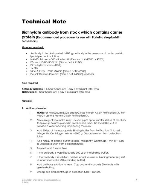
<strong>Technical</strong> <strong>Note</strong>Biotinylate antibody from stock which contains carrierprotein (Recommended procedure for use with FortéBio streptavidinbiosensors)Materials required:• Antibody to be biotinylated (>200µg antibody in the presence of carrier protein;lyophilyzed or in solution)• NAb Protein A or G Purification Kit (Pierce cat # 45200 or 45201)• EZ-Link NHS-LC-LC-Biotin (Pierce cat # 21343)• Dimethylformamide (DMF)• 1x PBS• Slide-A-Lyzer, 10000 MWCO (Pierce cat# 66383)• De-salt Dextran Columns (Pierce cat #43230)- optionalTime required:Antibody isolation: 1.5 hour hands-on; 1 day + overnight total timeBiotinylation: 1 hour hands-on; 1 day + overnight total timeProtocol:1. Antibody isolation1.1. NOTE: For mIgG2a, mIgG2b and IgG3 use Protein A Spin Purification Kit. FormIgG1 use the Protein G Spin Purification Kit.1.2. Mix resin gently to make slurry; use cut pipet tip to transfer 200 µL of the slurryto spin cup column placed in a collection tube. Tip should be cut toprovide a wider opening for pipeting the resin.1.3. Add 300 µL of the appropriate Binding buffer from Purification Kit to resin.Mix gently. Centrifuge 1 min at ~5000 g. Discard solution from collectiontube.1.4. Add 400 µL of Binding buffer to resin. Mix gently. Centrifuge 1 min at ~5000g. Discard solution from collection tube.1.5. Repeat wash 1 more time.1.6. If the antibody is lyophilized, add 300 µL of the binding buffer.1.7. If the antibody is in solution, add an equal volume of binding buffer (eg 250µL of antibody plus 250 µL binding buffer)1.8. Add antibody solution to resin. Cap cup and incubate 30 minute withgentle shaking.1.9. Uncap cup and centrifuge in collection tube 1 minute.Biotinylation when carrier protein present.doc - 1 -K. Witte
1.10. Move cup to a new collection tube. Add 400µL binding buffer and mixbriefly. Centrifuge 1 minute.1.11. Repeat previous wash step 2 more times.1.12. Transfer to a new collection tube. Add 400 µL of Elution Buffer. Cap cupand mix gently for 5 minutes.1.13. Uncap and centrifuge 1 minute. Transfer cup to a new collection tube.1.14. Repeat steps 2 more times for a total of 3 elutions. Elutions should beneutralized with 40µL of 1M sodium phosphate (pH 8).1.15. A denaturing, non-reducing gel should be run of the washes and elutions todetermine efficiency of isolation and location of the antibody (Figure 1).(a)Mol Wt markerStarting materialFlow throughWash 1Wash 2Wash 3Elution 1Elution 2Elution 3Antibody Control (2ug)(b)Mol Wt markerFlow throughWash 1Wash 2Wash 3Elution 1Elution 2Elution 3Starting materialAntibody Control (2ug)Figure 1: Denaturing, non-reducing gels of fractions from antibody isolation. (a) usingProtein G spin purification with mIgG1. (b) using Protein A spin purification with mIgG2a2. Biotinylation2.1. Using a MWCO 10000 slidalyzer, dialyze the elution fractions containing theantibody against PBS at 4C (elution 1 in both examples shown in Figure 1).2.2. NOTE: Since the elution buffer contains primary amines, dialysis must beextensive. Six changes at 1:1000 (> 3 hours between each change) arerecommended. Use of a desalting column is not recommended for thisstep as the procedure is not efficient enough to remove all the reactiveamines from the buffer.2.3. Recover the dialyzed sample from the slidalyzer and transfer to a neweppendorf tube.2.4. Prepare a 10mM Biotin reagent solution: add 2.0 mg NHS-LC-LC-Biotinreagent in 350 µL of DMF. Mix to dissolve.2.5. Calculate the volume of 10 mM biotin reagent needed based on the massof antibody started with prior to antibody isolation. For most antibodies, aBiotinylation when carrier protein present.doc - 2 -K. Witte
5:1 molar coupling ratio (moles NHS-LCLC-biotin: moles antibody at start ofprocedure).mg proteinMW (mg/mmol)X5 mmol biotin1 mmol proteinX1000mL10 mmol biotinX1000 µL1 mL=µL of 10mMsolution of biotin reagent2.6. To each sample, add the appropriate volume (µL) of NHS-LC-LC-Biotinreagent as calculated. Mix immediately2.7. Incubate 30 minutes at Room Temperature.2.8. Stop the reaction by removing the excess biotin reagent by either dialysis(recommended) or desalting column. Dialysis should involve 4 changes ofPBS at 1:1000.2.9. NOTE: Take care to remove all free biotin in order to most efficiently bind thebiotinylated protein to the streptavidin sensor surface.2.10. Biotinylated antibodies in PBS can be stored at 4C. See Figure 2 forexample immobilization of biotinylated antibody onto streptavidin SBCbiosensor.Biotin protein immobilization onto Eight Streptavidin SBC biosensorsnm0.60.50.40.30.20.100 50 100 150 200 250 300time (seconds)Sensor ASensor BSensor CSensor DSensor ESensor FSensor GSensor HFigure 2: Example data for the parallel immobilization of a biotinylated protein onto8 streptavidin SBC biosensors.Biotinylation when carrier protein present.doc - 3 -K. Witte