Review the symposium abstracts (3.19MB PDF) - College of Science
Review the symposium abstracts (3.19MB PDF) - College of Science
Review the symposium abstracts (3.19MB PDF) - College of Science
- No tags were found...

You also want an ePaper? Increase the reach of your titles
YUMPU automatically turns print PDFs into web optimized ePapers that Google loves.
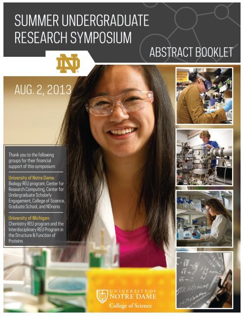
SUMMER UNDERGRADUATERESEARCH SYMPOSIUMABSTRACT BOOKLETAUG. 2, 2013Thank you to <strong>the</strong> followinggroups for <strong>the</strong>ir financialsupport <strong>of</strong> this <strong>symposium</strong>:University <strong>of</strong> Notre Dame:Biology REU program, Center forResearch Computing, Center forUndergraduate ScholarlyEngagement, <strong>College</strong> <strong>of</strong> <strong>Science</strong>,Graduate School, and NDnanoUniversity <strong>of</strong> Michigan:Chemistry REU program and <strong>the</strong>Interdisciplinary REU Program in<strong>the</strong> Structure & Function <strong>of</strong>Proteins
SUMMER UNDERGRADUATE RESEARCH SYMPOSIUMTable <strong>of</strong> ContentsPageOral Presentations ScheduleBiology, Chemistry, and Engineering 4Physics 5Poster Presentations ScheduleSession I 7Session II 10Abstracts 13Index to Abstracts 1783
Oral Presentations ScheduleBiology, Chemistry, and Engineering Jordan Room 105Session I11:00 am - 12:00 noonModerator: Dom Chaloner11:00 Desiree Garcia-Torres - Probing <strong>the</strong> determinants <strong>of</strong> <strong>the</strong> metal binding specificity <strong>of</strong>HDAC811:15 Sean McGee - Incorporation <strong>of</strong> Green Design into Field Iodine Deficiency Test11:30 Clarissa Rogg - Integration <strong>of</strong> Design and Energy Analysis Tools for Cooperative EnergyEfficient Building Design11:45 Kristin Springer - Delineation <strong>of</strong> <strong>the</strong> role for Notch signaling during zebrafish kidneyregenerationSession II2:00 pm – 3:00 pmModerator: Dom Chaloner2:00 Frances Acevedo Mariani - Polymerized HDLs for vaccine delivery2:15 Jordan Campbell - Field emission-driven Townsend discharges for silver nanoparticlesyn<strong>the</strong>sis2:30 Bryce Jones - Syn<strong>the</strong>ses and SAR Analysis <strong>of</strong> Thiophene Aldehyde Derivatives2:45 Akash Kannegulla - Optical modulation <strong>of</strong> continuous terahertz waves towardsreconfigurable quasi-optical terahertz components4
Physics Jordan Room 322Session I9:00 am - 10:15 amModerator: Umesh Garg9:00 Edward Kielb, Taylor Corpuz, and Michelle Berg - The World’s First Diffraction LimitedDoppler Spectrometer: iLocater9:15 Shanel Deal - Using Gamma Ray Burst to Estimate Luminosity Distances9:30 Sarah Dietz - Detangling <strong>the</strong> Cosmic Web: Computational Models <strong>of</strong> Galaxies inFilaments9:45 María Muñoz López - Analytical model <strong>of</strong> galactic feedback processes10:00 Jared Johnson, Aaron Sawyer, and Adrien Saremi - Formation <strong>of</strong> relativistic jets along<strong>the</strong> axis <strong>of</strong> rotation <strong>of</strong> a black holeSession II10:30 am - 12:00 noonModerator: Umesh Garg10:30 Liza Mulder - Electronic Properties <strong>of</strong> Lead Telluride Quantum Wells10:45 Kevin Lee - Exploring <strong>the</strong> Growth Mechanisms <strong>of</strong> GaAs Nanowires Grown by MBE11:00 Elissa Canseco - Damage <strong>of</strong> DNA in DNA-Gold Nanoparticle Mixture by AtmosphericPressure Plasma Jet11:15 Diana Gutierrez Zedano - Understanding Strontium’s Role in <strong>the</strong> Stability <strong>of</strong> SrAu3Ge11:30 Christine Kuryla - Ma<strong>the</strong>matical Modeling <strong>of</strong> Dynamic Instability in Microtubules5
Physics Jordan Room 322Session III1:30 pm - 2:45 pmModerator: Umesh Garg1:30 Bryce Frentz - New Targets for New accelerators1:45 Zach Tully - Exploring <strong>the</strong> Collective Properties <strong>of</strong> 160-Gd2:00 Benedict Pinyero - Double Folding Analysis <strong>of</strong> 6Li Elastic and Inelastic Scattering to lowlying states on 208Pb2:15 Hua Zhang - Constructing and Testing a Beam line for 5U Accelerator and GermaniumGamma Ray Detector Tests2:30 Trevor Satterfield - Beam Monitor Prototype Using Gas DischargeSession IV3:00 pm - 4:30 pmModerator: Umesh Garg3:00 Ruiyang Zhao - Accelerator Mass Spectrometry (AMS) applied in <strong>the</strong> measurement <strong>of</strong>93Zr3:15 Kirby Hermansen - Carbon 14 AMS: Establishing a Carbon Dating Procedure at NotreDame3:30 Alissa Murray - Vaccine Mandates and HPV3:45 Hanyi Yi - Summary <strong>of</strong> CMS Minimum Bias Pythia 8 Tunes4:00 Edward Varty - CMS Upgrade Simulations4:15 Concluding Remarks6
Poster Presentations ScheduleJordan GalleriaSession I12:00 noon - 1:00 pmEphraim Acevedo - EspN and FL-EC7 interact with o<strong>the</strong>r proteins in <strong>the</strong> mycobacterial ESX-1secretion systemMaria Agostini - Understanding <strong>the</strong> role <strong>of</strong> <strong>the</strong> CCR4-NOT1 complex in <strong>the</strong> microRNA pathwayusing single particle techniquesGregory M Alberding - Combustion Syn<strong>the</strong>sis <strong>of</strong> Ni and Co catalysts on CeO2, for <strong>the</strong>production H2 from EthanolJonathan Alvarez - What Does Time Matter? The Impact <strong>of</strong> Temporal Sampling in Home RangeArea CalculationsMegan Baker and Alexander Medvedeff - A first step in evaluating sNLP-12b as an antagonist tored flour beetle sulfakinin signalingOmari Baruti - Identifying In Vivo Coactivator Targets <strong>of</strong> Transcriptional Activator Gal4 byPhotocrosslinking with an Unnatural Amino AcidSteven Boggess - Ratiometric fluorescent sensors <strong>of</strong> cysteine sulfenylationRachael Bridgman - Determining SERS cross-section <strong>of</strong> amino acids for protein characterizationAlexa Carollo - Investigating <strong>the</strong> Photodissociation <strong>of</strong> Hydroxocobalamin using Steady State andTime Resolved UV-Visible SpectroscopyKatelyn Caro<strong>the</strong>rs - Knockdown <strong>of</strong> G-protein coupled receptors in adult female Anophelesgambiae using RNAiGonzalo Cazes-Nasitiqui - Integer Solutions to Diophantine EquationsRachel Choi - TcpP Diffusion Rate and Concentration in a Chemical Gradient Using Single-Molecule Fluorescence Imaging <strong>of</strong> Vibrio choleraeSarah Cox - A New Computational Approach to Gelator DiscoveryRandolph David II - Modeling <strong>the</strong> Formation and Propagation <strong>of</strong> Thermal Bar in Lake Ontariousing Climatological ParametersAllison Dianis - Inverse Islands: Comparing Matrilineal Structure Using mtDNA BetweenSingapore and Bali Long-Tailed MacaquesSimone Dozier - Synonymous Codon UsageNnamdi Edokobi - Investigation <strong>of</strong> changes in cardiac innervation in a mouse model <strong>of</strong> DravetSyndromeNichole Etienne - Ma<strong>the</strong>matical modeling and control <strong>of</strong> co-transmitting soil-transmittedhelmin<strong>the</strong>s7
Hea<strong>the</strong>r Fick, Jennifer Parra, Kirin Kuar, Stephanie Sesse, Vera Marcello, Arika Haines andJames K. Campbell - Design <strong>of</strong> a Fast-Acting Coliphage Biosensor DeviceAmy Fraley - Characterization <strong>of</strong> Halogenases in <strong>the</strong> Malbrancheamide PathwayBurke Gao - Visualizing Mismatch Repair in Live Bacillus subtilis using Single-MoleculeFluorescence MicroscopyJohn Gensic - Making Gasoline from Grass-Using heterogeneous catalysts to convert biomassinto bi<strong>of</strong>uelDaniel Gomez - PrivacyRangerAndrea Hakaj - Approaches to Identify Novel Phosphorylation Sites in <strong>the</strong> Checkpoint ProteinZW10Andrew Hamilton - Exploring Plasmon-Exciton Interactions between Gold Nanoparticles andGold NanoclustersJoel Hlavaty - Biomechanical Engineering and a Model Of Phenotypic Plasticity In DevelopingMammalsPeter H<strong>of</strong>fman - Polymer-Templated Rainbow Solar Cells & Implementing Photovoltaics into<strong>the</strong> Chemistry ClassroomShayna Hu - The Role <strong>of</strong> GRP78 Expression in Pancreatic Cancer ChemoresistanceMichael Hunckler - Feasibility <strong>of</strong> gel electrophoresis to determine <strong>the</strong> permeability properties <strong>of</strong>tendonsJonathan Jou - The Role <strong>of</strong> Fgf Signaling During Regeneration <strong>of</strong> <strong>the</strong> Zebrafish MesonephrosPatrick Jung - Microcontact Printing <strong>of</strong> DNA Origami onto Silicon SubstratesAkash Kannegulla - Optical modulation <strong>of</strong> continuous terahertz waves towards reconfigurablequasi-optical terahertz componentsMicah Katz - Investigating In Vivo Binding Partners <strong>of</strong> Transcriptional Activator VP16 through<strong>the</strong> Genetic Incorporation <strong>of</strong> Unnatural, Photocrosslinking Amino AcidsTaylor Kelson - Thermosensitive acrylamide-based hydrogel nanoparticles as a novel targeteddrug delivery vehicle for anti-cancer agentsKyle Kimler - The Relationship <strong>of</strong> Vision and Circadian Rhythm in Aedes aegyptiChristine Kuryla - Ma<strong>the</strong>matical Modeling <strong>of</strong> Dynamic Instability in MicrotubulesDaniel Kwasnieski - Characterization <strong>of</strong> Ag SERS-active microelectrodesJudy Long - Data Git: Provenance Extraction Tool Using Version ControlRui Yan Ma - Evaluating <strong>the</strong> Effects <strong>of</strong> Spatial Scale on <strong>the</strong> Performance <strong>of</strong> Climate EnvelopeModelingAziz Mamur - Syn<strong>the</strong>sis <strong>of</strong> a Selective Kinase Inhibitor for <strong>the</strong> M.tb. Ser/Thr Kinase - PknBRebecca Marton - Interkinetic Nuclear Migration in <strong>the</strong> Regenerating Zebrafish RetinaGrace McKenna - An Enantioselective Total Syn<strong>the</strong>sis <strong>of</strong> Cis-2,5-Disubstituted Pyrrolidines8
Christine Mei - Roles and Interaction <strong>of</strong> MRPP1 and MRPP2 in Reconstitution <strong>of</strong> HumanMitochondrial RNase PMax Miller - Performance Evaluation <strong>of</strong> Amazon EC2 and Google Compute EngineCiaran Mooney - Mechanisms Governing Müller Glial Interkinetic Nuclear Migration in <strong>the</strong>Regenerating Zebrafish RetinaVaishnav Murthy - Analysis <strong>of</strong> holes in graphene monolayersAnsel Nalin - Progress Towards <strong>the</strong> Total Syn<strong>the</strong>sis <strong>of</strong> Ambruticin JHamim Nigena - Syn<strong>the</strong>sis <strong>of</strong> IRMOF-3 and Conversion <strong>of</strong> Dihydroxyacetone to Ethyl LactateJoshua Ostrander - The Structure and Dynamics <strong>of</strong> CORM-3 in Solution Studied Using FTIRand 2D-IR SpectroscopyAakash Patel - Determining Heart Rate from VideoJaqueline-Mae Picache - Characterization <strong>of</strong> APC-induced microtubule branchingJulian Pilate-Hutcherson - Thiolated gold and silver nanoclusters absorption <strong>of</strong> visible lightJessica Rafson - Crystalline InSb from Simple Electrodeposition <strong>of</strong> Sb onto <strong>the</strong> ReactiveInterface <strong>of</strong> In Electrodes under Aqueous Benchtop SettingsJulia Resil - Population genetic structure <strong>of</strong> Balinese long-tailed macaques (Macaca fascicularis)Olaf Rodríguez Gutiérrez - Functional characterization <strong>of</strong> <strong>the</strong> carbon dioxide receptor genesin Culex quinquefasciatus and Culex stigmatosomaSarah Semanek - Role <strong>of</strong> cyclins in Müller glia and neuronal progenitor cell proliferation in <strong>the</strong>zebrafish retinaLynda Smith - Hey! Can Someone Turn <strong>the</strong> Light On? SOS! (Sustainably Off Sunlight)Jordan Stern - Improved Efficiency <strong>of</strong> Negative Ion Electron Capture Dissociation (niECD)Through Charge State Reduction and Laser ActivationDaniel Steyer - Analysis <strong>of</strong> SIRT1 Activity by Capillary ElectrophoresisDonghyuk Suh - Ultrafast 2DIR probe on an organic polymer for a model HFL system.Joseph Thomaz - Does Visible Light Photolyze Hydroxocobalamin?Thibault Twahirwa - Experimental Studies on Field Emission-Driven MicrodischargesValerie Verdun - Analysis <strong>of</strong> lunatic fringe in Renal Progenitor Patterning During ZebrafishKidney DevelopmentAlyse Volino - Stasis <strong>of</strong> Saltwater Tolerance in Anopheles farauti FAR1 MosquitoesJustin Waller - Syn<strong>the</strong>tic processes <strong>of</strong> gold nano-sphere (NS) & nano-rod (NR) particles for <strong>the</strong>enhancement <strong>of</strong> solar cellsNicholas Weidner - Hurricane and Storm Surge Modeling at Notre DameRussell Williams III - Receptor Interacting Protein Mediated Cell Death in ECM-detached BreastCancer Cells9
Nathaniel Wirgau - Syn<strong>the</strong>sis and Characterization <strong>of</strong> Five- and Six-Coordinate Ferrous Heme-Nitrosyl ComplexesYing Zhang - Chiral Guest Recognition by Lanthanide Metallacrown Hosts with ThreoninebasedLigandsSession II1:00 - 2:00 pmThomas C. Adams - Thermal Rectifiers - The Effects <strong>of</strong> Thermal ConductivityMichael Ahlers - Syn<strong>the</strong>sis <strong>of</strong> a GEX1A Analogue for Niemann-Pick Type C Disease ResearchPauline Alokolaro - Light It Up: High Performance Backside Solar Cell FabricationArnaud Bacye - External modulation <strong>of</strong> mid-infrared quantum cascade lasers with integratedMach-Zehnder interferometric modulatorElijah Barstis - Creating a Device to Detect Substandard Tagamet HB 200 ® PillsOmar Beleh - Development <strong>of</strong> a TR-FRET Assay to Screen for Allosteric Kinase InhibitorsBuchanan Bourdon - Electrostatic Doping <strong>of</strong> 2D materials via Polymer Electrolytes: UsingCOMSOL to Simulate Ion-electron TransportKrista Briedis - Biological Characterization <strong>of</strong> Novel Mcl-1 InhibitorsSarah Caruso - α3DH3-GSGA-F31A, A De Novo Designed MetallopeptideBrianna Chamberlin - Exploring <strong>the</strong> polymorphism <strong>of</strong> piperineKasey Clear - A Hybrid Optical Detection MethodPhillip Cook - The New Digital Playground: Applications <strong>of</strong> 3D Printing within a K-12 SettingWill Crosby - Syn<strong>the</strong>sis <strong>of</strong> Nickel Diimine Complexes as Possible Catalysts for <strong>the</strong>Polymerization <strong>of</strong> Electron Deficient MonomersChristine Cuthbertson - Syn<strong>the</strong>sis and Analysis <strong>of</strong> 2,5,6-trifluoro-3,4-dihydroxybenzoate as aPotential Inhibitor <strong>of</strong> Wild-type Class 1A Dihydroorotate DehydrogenaseWilliams Dixon - Examining <strong>the</strong> Conformational Dynamics <strong>of</strong> Proteins and snRNAs duringSplicing using smFRETKevin Durand - Plasmon-Enhanced Fluorescence Imaging <strong>of</strong> <strong>the</strong> Intrinsic Emission <strong>of</strong> SingleCoenzyme B12 MoleculesOsama El-Sayed - Characterization <strong>of</strong> J-Binding Protein 1Taylor Evans - Syn<strong>the</strong>sis <strong>of</strong> α-SnWO 4Nathan Foje - Non-Invasive Optical Imaging <strong>of</strong> Near Infrared Electrospun NanoparticlesJessica Freeman - An Investigation into <strong>the</strong> Factors that Affect Compartment Size in DrosophilaEpidermal Larval Segments during EmbryogenesisDesiree Garcia-Torres - Probing <strong>the</strong> determinants <strong>of</strong> <strong>the</strong> metal binding specificity <strong>of</strong> HDAC810
Jeremy Gerick - Green’s Functions in Chemistry; Optimization and Implementation <strong>of</strong> PT2 Self-EnergyBrandon Gutierrez - Development <strong>of</strong> Visually Impaired Transgenic MosquitoesJosh Halpern - Enzymatic Syn<strong>the</strong>sis <strong>of</strong> 3-deoxy-D-manno-octulosonic acid from in situ D-Arabinose-5-Arsenate as a Substrate AnalogueJohn Hargett - Visualizing Imitation Through Motion Capture TechnologySara Hockney - Role <strong>of</strong> pax6 in neuronal progenitor cell proliferation during zebrafishphotoreceptor regenerationGwendolyn Hooley - Identification <strong>of</strong> a novel gene required for ESX-1 secretion and virulence inMycobacterium marinumMeghan Hudak - Evolution <strong>of</strong> Mycobacterium marinum for loss <strong>of</strong> ESX-1 associated hemolyticactivityElizabeth Huschke - On-Chip Optical Diagnosis Using Capillary ElectrophoresisNirbhay Jain - A Simple Method <strong>of</strong> Minimizing Ionic Strength to Reduce Chemical ShiftVariance in NMR-based Metabolomics <strong>of</strong> Urine SpecimenKipchumba Kaitany - Determining Hepatitis C NS3 Helicase’s Active Oligomeric StateGordon Kane - Assay Development Monitoring VirF Binding to VirB PromoterMilan Kaushik - Nanomaterial Induced Changes in Cell Membrane Conductivity: A Whole CellPatch Clamp StudyDaniel Kestell - Simulation <strong>of</strong> Nanomagnets under Dynamic ConditionsAnthony Krenselewski - Manipulation <strong>of</strong> Graphene Morphology Compounds via MetalNanoparticle Mediated Hydroxyl Radical AttackChristopher Kubitskey - Biosyn<strong>the</strong>tic Production <strong>of</strong> Novel SubstratesAlexa Kutch - 3D Printing: Rapid Prototyping using Additive ManufacturingMegan Leander - DMS Ligand-Receptor Interactions Reflect SAR DataNevin Longenecker - Possible Student Investigations Related to Engineering a More SustainableEnergy FutureAmber Lott - Membranes with Multiple Porous Crystals for Chemical SeparationsMichael Manning - Examining Interprotein Interactions Using Coarse Grained MolecularDynamics SimulationsRobert Mat<strong>the</strong>ws - Raman Spectroscopic Monitoring <strong>of</strong> Integration <strong>of</strong> a Tissue-Engineered OralMucosa ConstructJill McNabnay - Changing Climate; Changing LifeRachel Miceli - Genetic analysis <strong>of</strong> podocyte developmentAlannah Miranda - Low glucose exposure favors <strong>the</strong> appearance <strong>of</strong> fast vs. slow oscillations inisolated mouse pancreatic islets11
Cameron Moore - Optimizing Paper Tests for Anti-Tuberculosis MedicationsBrenda Mueller - Carbon Dioxide Capture in Ionic LiquidsSarah Neville - Syn<strong>the</strong>sis <strong>of</strong> New Non-Heme Iron NitrosylsHea<strong>the</strong>r M. Nimon - Employing <strong>the</strong> Energy <strong>of</strong> Light to Advance TechnologyClarence Pascual - Identifying <strong>the</strong> significance <strong>of</strong> <strong>the</strong> dual Peroxisomal Targeting Signals inArabidopsis thaliana LACS7Nathaniel Pelz - The Geppetto ProjectSamantha Piekos - Generation and characterization <strong>of</strong> tnfa and nr2e3 transgenic zebrafish linesKenneth Poling - Dye Sensitized Solar CellsSashary Ramos - Characterization <strong>of</strong> fluticasone propionate microcrystalline drug product usingquantitative polarization microscopyGrayson Ritch - Structure and Catalytic Activity <strong>of</strong> Iron 6,6’Dihydroxy Terpyridine ComplexesAnn Ru<strong>the</strong>rford - Development <strong>of</strong> s-block Organometallic Catalysts for co-polymerization <strong>of</strong>PolycarbonatesDillon Skeehan - Opportunistic Grid Computation for High Energy PhysicsEmmanuel Sosa - Revolutions <strong>of</strong> 1848: History <strong>of</strong> Contingency?India Stewart - Data Git: Provenance Extraction Tool Using Version ControlJerone Stoner - Determining <strong>the</strong> Significance <strong>of</strong> <strong>the</strong> ZNF217-PKM2 Interaction in <strong>the</strong> MetabolicRegulation <strong>of</strong> Breast Cancer ProgressionJoseph Terranova - Effects <strong>of</strong> pH on Pore Size <strong>of</strong> poly(isoprene-b-styrene-b-dimethylacrylimade)MembraneSara Tweedy - MD Simulations Reveal <strong>the</strong> Impact <strong>of</strong> Crystal Packing on <strong>the</strong> Post-SET Loop <strong>of</strong>NSD1Ca<strong>the</strong>rine Eve Vogt - Investigation <strong>of</strong> Small Molecule Binding to <strong>the</strong> Exon Splicing Silencer inHuman Immunodeficiency Virus Type 1 Tat Exon 3Ian VonWald - S-Nitroso-N-hexanylpenicillamine (SNHP)-Doped Polymers: Syn<strong>the</strong>sis,Leaching Stability, and Characterization <strong>of</strong> Nitric Oxide ReleaseJiarui Wang - Structure probing <strong>of</strong> <strong>the</strong> transcriptionally acting preQ 1 riboswitch using singlemolecule FRET (smFRET)Milena Westarb - Methods for Cocrystal Syn<strong>the</strong>sisMark Wilson - Kinetics <strong>of</strong> Reaction <strong>of</strong> (MeClamp)Ti with HydrazobenzeneCharles Cong Yang Xu - Genetic Differentiation among Populations <strong>of</strong> Echinolittorina radiataacross East Asia12
ABSTRACTS13
Poster PresentationEspN and FL-EC7 interact with o<strong>the</strong>r proteinsin <strong>the</strong> mycobacterial ESX-1 secretion systemEphraim AcevedoMarika Kuspa, Dept. <strong>of</strong> Biological <strong>Science</strong>s, University <strong>of</strong> Notre DameAdvisor: Patricia Champion, Dept. <strong>of</strong> Biological <strong>Science</strong>s, University <strong>of</strong> Notre DameTuberculosis is one <strong>of</strong> <strong>the</strong> most deadly diseases known, killing 1.4 million people worldwideeach year (Who, 2012). The virulence <strong>of</strong> Mycobacterium tuberculosis is dependent upon <strong>the</strong>ESX-1 secretion system. This system is conserved in pathogenic mycobacteria and certain Grampositivebacteria. One <strong>of</strong> <strong>the</strong> functions <strong>of</strong> <strong>the</strong> ESX-1 secretion system is to manipulate <strong>the</strong> hostresponse to infection. The goal <strong>of</strong> my project is to study protein-protein interactions within <strong>the</strong>ESX-1 secretion system. To do so I am focusing on how two ESX-1 associated proteins, EspNand FL-EC7, interact with components <strong>of</strong> <strong>the</strong> secretion system. EspN is a novel protein that isinvolved in <strong>the</strong> ESX-1 secretion system. FL-EC7 is a construct <strong>of</strong> <strong>the</strong> substrates EspC and CFP-10 that shows interactions with two ESX-1 ATPases. We are performing this study by using ayeast two-hybrid screen in order to see which proteins from a M. tuberculosis cDNA libraryinteract with EspN and FL-EC7. Using this approach, we found that EspN interacted withproteins that are found in <strong>the</strong> membrane, cell wall and <strong>the</strong> cytosol. Some <strong>of</strong> <strong>the</strong>se proteins arefound when M. tuberculosis is under stress. This stress may be associated to <strong>the</strong> conditions thatare found when M. tuberculosis is trapped within <strong>the</strong> phagosome <strong>of</strong> <strong>the</strong> macrophages, where M.tuberculosis resides after infection. This screen will identify novel ESX-1 components, as well asto facilitate studies focused on how <strong>the</strong>se components interact with existing ESX-1 proteins.14
Oral PresentationPolymerized HDLs for vaccine deliveryFrances Acevedo MarianiGwangseong Kim, Dept <strong>of</strong> Pharmaceutical <strong>Science</strong>s, University <strong>of</strong> MichiganAdvisor: Anna Schwendeman, Dept. <strong>of</strong> Medicinal Chemistry, University <strong>of</strong> MichiganHigh-density lipoprotein (HDL) is a natural nanoparticle composed phospholipid bilayer andApolipoprotein A-I (ApoA-I). The main functions <strong>of</strong> HDL are to efflux <strong>the</strong> excess <strong>of</strong> cholesterolfrom endo<strong>the</strong>lial cells and transport it to <strong>the</strong> liver for elimination. Due to <strong>the</strong>ir size (8-10 nm) andlong circulation half-life (1-3 days) <strong>the</strong>y could be used for intravenous delivery <strong>of</strong> peptideantigens covalently bound to HDL phospholipids. Unfortunately HDL undergoes rapidremodeling in plasma by exchanging phospholipids with o<strong>the</strong>r membranes, and potentiallyloosing phospholipid bound surface antigens. Polymerization <strong>of</strong> HDL lipid bilayer will likelyprevent plasma remodeling. ApoAI mimetic peptide (5A) was complexed with polymerizablelipid, 1,2-bis(10,12-tricosadiynoyl)-sn-glycero-3-phosphocholine (Diyne PC), andsphingomyelin (SM) to form sHDL nanoparticles. Diyne PC, SM and 5A were dissolved inorganic solvent, combined at 2:0:1, 1:1:1, 0.5:1.5:1 and 0.25:1.75:1 weight ratios andlyophilized. The powders were hydrated to form HDLs and polymerized by UV light exposurefor 2 hours. The resulting sHDLs were analyzed to determine size (DLS), purity (gelchromatography – GPC), presence <strong>of</strong> liposome impurity (turbidity) and stability after cooling to4°C. Only polymerized HDLs composed exclusively <strong>of</strong> Diyne PC (2:0:1) remained intact, while<strong>the</strong> increase in impurities was observed for all o<strong>the</strong>r preparations. To increase purity andoptimize size, HDL were prepared at 1:0.5, 1:0.75, 1:1, 1:1.5 and 1:2 wt/wt ratios <strong>of</strong> 5A to DiynePC. The HDL purity increased with decrease <strong>of</strong> lipid content and stability was improved bypolymerization. The polymerized HDLs were prepared and <strong>the</strong>ir composition was optimized toachieve size characteristic size <strong>of</strong> 8-10 nm and physical stability. The polymerized HDL could bepotentially used for vaccine antigen delivery.15
Poster PresentationThermal Rectifiers – The Effects <strong>of</strong> Thermal ConductivityThomas C. AdamsKy-Quan NguyenAdvisor: Tengfei Luo, Dept. <strong>of</strong> Aerospace and Mechanical Engineering,University <strong>of</strong> Notre DameWhile still in progress, a bi-layer junction could be constructed by using <strong>the</strong> properties <strong>of</strong>hexadecane to create a <strong>the</strong>rmal rectifier – one-way heat flux transfer mechanism. Interestingly,<strong>the</strong>se predications and calculations are made via simple secondary level algebra and statisticsmethods with regards to introductory physics applications.16
Poster PresentationUnderstanding <strong>the</strong> role <strong>of</strong> <strong>the</strong> CCR4-NOT1 complex in <strong>the</strong> microRNA pathwayusing single particle techniquesMaria AgostiniAdvisor: Nils Walter, Dept. <strong>of</strong> Chemistry, University <strong>of</strong> MichiganMicroRNAs (miRNA) are short, non-coding RNAs approximately 22 nucleotides in length thatregulate post-transcriptional gene expression. They function by assembling on mRNA targetsand associating with components <strong>of</strong> <strong>the</strong> RNA-induced silencing complex (RISC). The majorcomponents <strong>of</strong> RISC are an Argonaute protein (AGO), GW182, and <strong>the</strong> CCR4-NOT1 complex.This study focuses primarily on <strong>the</strong> role <strong>of</strong> <strong>the</strong> CCR4-NOT1 complex by means <strong>of</strong> an importantsubunit involved in deadenylation <strong>of</strong> <strong>the</strong> mRNA target transcripts, CNOT1. Here, we verifysiRNA knockdown <strong>of</strong> CNOT1 by Western Blot before analyzing differences in distribution <strong>of</strong>processing bodies (P bodies) that are <strong>of</strong>ten <strong>the</strong> site <strong>of</strong> mRNA degradation in U2OS cells stablyexpressing DCP1a labeled with GFP. In addition to this observation, using a method previouslydescribed by members <strong>of</strong> our group called intracellular single-molecule, high resolutionlocalization and counting (iSHiRLoC), we aim to discover <strong>the</strong> different localization anddiffusion coefficients <strong>of</strong> let-7-a1 miRNAs that result in CNOT1 knockdown U2OS cells. Thisprocess includes microinjecting double-stranded let-7-a1 miRNA labeled with Cy5 on <strong>the</strong> 3’ end<strong>of</strong> <strong>the</strong> guide strand. Previously, using this method, members <strong>of</strong> our group discovered thatmiRNA exhibit specific localization and diffusion coefficients when microinjected into <strong>the</strong> cell.Specifically, <strong>the</strong>y observed two kinetically distinct populations. The goal <strong>of</strong> this study is todetermine <strong>the</strong> effects <strong>of</strong> <strong>the</strong> CCR4-NOT1 complex on <strong>the</strong>se values by observing shifts in <strong>the</strong>sebehaviors when <strong>the</strong> cell is depleted <strong>of</strong> CNOT1. These observations may serve to ei<strong>the</strong>r verify oralter our current understanding <strong>of</strong> <strong>the</strong> role <strong>of</strong> <strong>the</strong> CCR4-NOT1 complex in this pathway.17
Poster PresentationSyn<strong>the</strong>sis <strong>of</strong> a GEX1A Analogue for Niemann-Pick Type C Disease ResearchMichael AhlersEve Granatosky and Jarred Pickering, Dept. <strong>of</strong> Chemistry and Biochemistry,University <strong>of</strong> Notre DameAdvisor: Richard Taylor, Dept. <strong>of</strong> Chemistry and Biochemistry,University <strong>of</strong> Notre DameNiemann-Pick Type C disease (NPC) is a rare and fatal lysosomal storage disease that typicallypresents before <strong>the</strong> age <strong>of</strong> 10. Specifically, NPC is characterized by mutations to ei<strong>the</strong>r <strong>the</strong> NPC1or NPC2 proteins that result in defective cholesterol trafficking. Although hydroxypropyl β-cyclodextrin (HPβCD) and histone deacetylase inhibitors (HDACi) such as trichostatin A arecurrent <strong>the</strong>rapeutic candidates for NPC, <strong>the</strong>re is currently no FDA approved treatment. We haverecently demonstrated <strong>the</strong> ability <strong>of</strong> GEX1A, a type I polyketide natural product isolated fromStreptomyces chrom<strong>of</strong>uscus, to restore cholesterol trafficking in NPC1 mutant cell lines (Figure1). Interestingly, herboxidiene is not an HDACi. Given rapid access to GEX1A throughfermentation processes, our attention has shifted towards <strong>the</strong> syn<strong>the</strong>sis <strong>of</strong> non-natural analogues<strong>of</strong> <strong>the</strong> parent compound such as 1. Thus, an efficient route to fragment A has been realizedthrough crotylation chemistry reported by Leighton and co-workers. Once completed, thisanalogue will be used in NPC1 and NPC2 mutant cell line assays for comparison with GEX1A.18
Poster PresentationCombustion Syn<strong>the</strong>sis <strong>of</strong> Ni and Co catalysts on CeO2, for <strong>the</strong> production H2 from EthanolGregory M AlberdingAllison Cross, Dept. <strong>of</strong> Chemical and Biomolecular EngineeringAdvisor: Eduardo Wolf, Dept. <strong>of</strong> Chemical and Biomolecular Engineering,University <strong>of</strong> Notre DameThis research looks at catalysts for <strong>the</strong> production <strong>of</strong> hydrogen gas from ethanol. Hydrogen gasproduction is an important step for <strong>the</strong> functioning <strong>of</strong> fuel cells. We created six different CeO2catalysts, some containing Ni, Co, or both. We are using combustion syn<strong>the</strong>sis techniques tocreate <strong>the</strong>se different metal-containing catalysts. We <strong>the</strong>n characterized <strong>the</strong> catalysts, looking atcomposition, surface area, pore size, and particle size. Finally we used each catalyst in anethanol reaction to look for selectivity <strong>of</strong> each catalyst in <strong>the</strong> production <strong>of</strong> hydrogen.19
Poster PresentationLight It Up: High Performance Backside Solar Cell FabricationPauline AlokolaroYuning Zhao, Dept. <strong>of</strong> Electrical Engineering, University <strong>of</strong> Notre DameAdvisor: Patrick Fay, Dept. <strong>of</strong> Electrical Engineering, University <strong>of</strong> Notre DameConsiderations for meeting future worldwide energy needs must include a serious discussionregarding practical and efficient processes for harnessing solar energy. Although o<strong>the</strong>r forms <strong>of</strong>renewable energy should be a part <strong>of</strong> a balanced, clean, renewable energy economy, only solarenergy <strong>of</strong>fers <strong>the</strong> quantities <strong>of</strong> energy that can meet <strong>the</strong> world’s increasing demands.Theoretically, up to 86.7% <strong>of</strong> sunlight can be harnessed and stored, but current solartechnologies cap out at less than half that efficiency. Urban solar power plants will not becomeviable options until solar cells are small enough and efficient enough to fit within a denselypopulated area.Fabrication <strong>of</strong> compound-semiconductor based multi-junction high-performance solar cellsinvolves <strong>the</strong> development <strong>of</strong> “back-side” solar cells in which all <strong>of</strong> <strong>the</strong> contacts andinterconnections can be made on <strong>the</strong> back side <strong>of</strong> <strong>the</strong> wafer. Multi-junction backside solar cells<strong>of</strong>fer <strong>the</strong> additional benefit <strong>of</strong> strong absorption <strong>of</strong> multiple regions <strong>of</strong> <strong>the</strong> solar spectrum.Current work focuses on packaging <strong>the</strong> solar cells so that <strong>the</strong>y are practical for commercial use.A thin layer <strong>of</strong> tin will be applied to <strong>the</strong> bottom <strong>of</strong> <strong>the</strong> solar cell for packaging purposes.The experience will be incorporated into a high school chemistry class during a unit involving<strong>the</strong> electromagnetic spectrum and quantum chemistry. The unit will include a discussion <strong>of</strong> <strong>the</strong>different wavelengths <strong>of</strong> sunlight and methods <strong>of</strong> capturing <strong>the</strong> most energy from <strong>the</strong> sunlight aswell as a laboratory activity involving <strong>the</strong> construction <strong>of</strong> a solar cell and measurements <strong>of</strong> itspower.20
Poster PresentationWhat Does Time Matter? The Impact <strong>of</strong> Temporal Samplingin Home Range Area CalculationsJonathan AlvarezAmy Klegarth, Dept. <strong>of</strong> Biological <strong>Science</strong>s, University <strong>of</strong> Notre DameAdvisors: Hope Hollocher, Dept. <strong>of</strong> Biological <strong>Science</strong>s,and Agustin Fuentes, Dept. <strong>of</strong> Anthropology, University <strong>of</strong> Notre DameGlobal Positioning System (GPS) are currently widely used to study larger mammals, includingcarnivores and ungulates. Only recently have GPS collars been a viable method to studyprimates, as size restrictions hindered potential studies. The constant locational updates as wellas <strong>the</strong>ir noninvasive nature after deployment provide great utility for managers by helping <strong>the</strong>mmodel future ranging patterns based on past land use. GPS collars remove a potential bias <strong>of</strong>researches closely following primate groups and provide a simple method <strong>of</strong> acquiring data fromanywhere in <strong>the</strong> world.Results from a previous study in Singapore, which used GPS collars to track daily and homeranges for 3 monkeys, show that home range sizes calculated using Minimum Convex Polygons(MCPs) display a significant increasing trend with <strong>the</strong> iterative addition <strong>of</strong> sample points. Recentstatistical analyses show that when <strong>the</strong> same home range areas are calculated using KernelDensity Estimations (KDEs), <strong>the</strong> significance between sample sizes is no longer present.However, linear regressions <strong>of</strong> <strong>the</strong> data suggest that <strong>the</strong> difference between sample sizesbecomes significantly smaller as more points are added.This project aims to apply <strong>the</strong> methods from <strong>the</strong> Singapore study to six different monkeyslocated in Gibraltar. The Gibraltar monkeys have been collared for five months as opposed totwo months for <strong>the</strong> Singapore monkeys. This increased data size allows <strong>the</strong> project to examine<strong>the</strong> temporal element <strong>of</strong> <strong>the</strong> iterative analysis.Ultimately, <strong>the</strong> results from this project will inform future analytic methods for a larger collarproject in Singapore. The information about home range size and general movement behaviorwill be incorporated within a landscape genetic Geographic Information Systems model.Specifically, this project will help parameterize migration ability based on recorded home rangeareas as well as distance and movement rates.21
Poster PresentationExternal modulation <strong>of</strong> mid-infrared quantum cascade lasers withintegrated Mach-Zehnder interferometric modulatorArnaud BacyeMichael Harter and Galen Harden, Dept. <strong>of</strong> Electrical Engineering, University <strong>of</strong> Notre DameAdvisor: Anthony H<strong>of</strong>fman, Dept. <strong>of</strong> Electrical Engineering, University <strong>of</strong> Notre DameAbstract: We present a quantum cascade laser (QCL) with an integrated Mach-Zehnderinterferometer that function as an electro-optic modulator. The laser and interferometer arefabricated from <strong>the</strong> same QC wafer that consists <strong>of</strong> two core sections: <strong>the</strong> active region forproviding optical gain and a super-lattice <strong>of</strong> asymmetric coupled quantum wells (ACQWs) forcontrolling <strong>the</strong> phase difference between <strong>the</strong> arms <strong>of</strong> <strong>the</strong> interferometer. Amplitude modulation isachieved by tuning <strong>the</strong> intersubband absorption <strong>of</strong> <strong>the</strong> ACQWs via an applied voltage over <strong>the</strong>super-lattice. The intersubband absorption <strong>of</strong> <strong>the</strong> ACQWs was designed using a two-band solverthat includes energy band nonparabolicity. Our design has an intersubband transitional energythat tunes from 78.9 meV at a field <strong>of</strong> -75 kV/cm to 115.6 meV at 75 kV/cm. The waveguidewas designed by including <strong>the</strong> voltage-dependent index <strong>of</strong> <strong>the</strong> ACQWs in a commercial 2-Dfinite element solver to calculate <strong>the</strong> propagating mode and <strong>the</strong> effective modal index. Theoverlap <strong>of</strong> <strong>the</strong> optical mode with <strong>the</strong> ACQWs, approximately 1%, was engineered to enablesufficient control <strong>of</strong> <strong>the</strong> phase for optical modulation while minimizing optical loss from <strong>the</strong>contact layer. Such a device has application in mid infrared photo-acoustic spectroscopy, atechnique used for trace gas detection in <strong>the</strong> medical, environmental, and homeland defensefields.22
Poster PresentationA first step in evaluating sNLP-12b as an antagonist to red flour beetle sulfakinin signalingMegan BakerAlexander MedvedeffBenjamin Maynard and Ruthann Nichols Dept. <strong>of</strong> Biological Chemistry, University <strong>of</strong> MichiganAdvisor: Ruthann Nichols, Dept. <strong>of</strong> Biological Chemistry, University <strong>of</strong> MichiganSulfakinins (SKs) are insect neuropeptides which regulate gut and heart contractions and inhibitfood intake by signaling through family A G protein-coupled receptors. Identifying SK peptidereceptorcontact sites is important to understanding how <strong>the</strong>se peptides act. In a previous study,<strong>the</strong> nematode peptide sNLP-12b behaved as a putative antagonist to SK receptors in red flourbeetle as measured by a food intake assay. Docking SK ligands as well as sNLP-12b to red flourbeetle SK receptors (TcSKRs) was a first step to determine whe<strong>the</strong>r <strong>the</strong> putative antagonist actsthrough <strong>the</strong> beetle SK pathway or through a different signaling pathway. Our work includednovel receptor modeling and SK ligand docking to TcSKR1 and to TcSKR2, a protein previouslyunexplored in <strong>the</strong> signaling pathway. Our results suggest that sNLP-12b makes contacts withTcSKRs and may act as an antagonist to <strong>the</strong> SK signaling pathway.23
Poster PresentationCreating a Device to Detect Substandard Tagamet HB 200® PillsElijah BarstisToni Barstis, Dept <strong>of</strong> Chemistry & Physics, Saint Mary's <strong>College</strong>Advisor: Toni Barstis, Dept <strong>of</strong> Chemistry & Physics, Saint Mary's <strong>College</strong>This research focused on <strong>the</strong> creation <strong>of</strong> a Paper Analytical Device (PAD) to screen forsubstandard Tagamet HB 200® pills. Different colorimetric reagent tests are used on <strong>the</strong> PAD todetect <strong>the</strong> API Cimetidine and select excipients found in genuine pills and select substituentsfound in substandard pills. The colorimetric reagent tests and <strong>the</strong> results <strong>of</strong> a small field test willbe presented.1. “Essential Drug List.” Web accessed July 22, 2013.www.who.int/selection_medicines/country_lists/phl_edl_2007.pdf2. “Screening for Counterfeit Drugs Using Near-Infrared Spectroscopy.”Web accessed July 22, 2013. http://www.pharmainfo.net/pharmaceutical-packagingarticles/screening-counterfeit-drugs-using-near-infrared-spectroscopy24
Poster PresentationIdentifying In Vivo Coactivator Targets <strong>of</strong> Transcriptional Activator Gal4 byPhotocrosslinking with an Unnatural Amino AcidOmari BarutiAmanda Dugan Rachel Pricer, Cassie Joiner, Dept. <strong>of</strong> Department <strong>of</strong> Chemistry,University <strong>of</strong> MichiganAdvisor: Anna Mapp, Dept. <strong>of</strong> Department <strong>of</strong> Chemistry, University <strong>of</strong> MichiganTranscriptional activators are modular proteins that are required to initiate transcription, <strong>the</strong>essential cellular process wherein DNA is transcribed into mRNA. Activators have a DNAbinding domain (DBD) that specifies localization at a gene, and transcriptional activation domain(TAD) that regulates expression <strong>of</strong> gene by recruiting <strong>the</strong> transcriptional machinery, includingRNA Polymerase II, to <strong>the</strong> gene promoter. Although activators are known to recruit multiplecoactivator complexes, identifying <strong>the</strong> individual subunits within <strong>the</strong>se coactivators and <strong>the</strong>transcriptional machinery, and thus understanding <strong>the</strong>ir individual role in transcription, hasproven challenging. Here we use in vivo photocrosslinking with a genetically incorporatedphoto-crosslinker amino acid, p-benzoyl-L-phenylalanine (Bpa) to capture <strong>the</strong> direct targets <strong>of</strong><strong>the</strong> model yeast activator Gal4 in Saccharomyces cerevisiae. Specifically, we examine itsinteractions with <strong>the</strong> chromatin modifying complex SAGA. To do this, we used molecularcloning techniques to generate myc-tagged constructs <strong>of</strong> SAGA subunits, Tra1, Ada2, and Taf12,believed to be putative targets <strong>of</strong> Gal4. We co-transform <strong>the</strong>se tagged coactivator proteinsalongside a Gal4 construct that bears a LexA DBD and <strong>the</strong> Gal4 TAD and <strong>the</strong>n grow <strong>the</strong> yeast in<strong>the</strong> presence <strong>of</strong> Bpa. Live yeast are <strong>the</strong>n irradiated with UV light to form a covalent crosslinkbetween <strong>the</strong> Bpa in <strong>the</strong> Gal4 TAD and its protein binding partners. We <strong>the</strong>n immunoprecipitate<strong>the</strong> crosslinked activator complexes from yeast lysate and probe for <strong>the</strong> presence <strong>of</strong> covalentlybound tagged co activator using Western blotting techniques. In this way, we can identify <strong>the</strong>coactivators that are directly recruited by Gal4 in living cells.25
Poster PresentationDevelopment <strong>of</strong> a TR-FRET Assay to Screen for Allosteric Kinase InhibitorsOmar BelehAdvisor: Mat<strong>the</strong>w Soellner, Dept. <strong>of</strong> Medicinal Chemistry, University <strong>of</strong> MichiganProtein kinases have become important drug targets due to <strong>the</strong>ir involvement in many biologicalprocesses, including cell division, motility, and overall survival. Dysregulation <strong>of</strong> <strong>the</strong>se signalingmolecules has been shown to lead to disease, particularly in a number <strong>of</strong> different cancers.Previous research has led to <strong>the</strong> development <strong>of</strong> ATP-competitive inhibitors that target multiplekinases to achieve <strong>the</strong>ir desired effect. However, since <strong>the</strong>re are over 500 protein kinases in <strong>the</strong>human body with similar ATP-binding domains, selectivity and characterization <strong>of</strong> specificsignaling pathways can be difficult when designing an inhibitor. One approach to increaseselectivity and limit potential toxicity is to develop allosteric kinase inhibitors; that is, aninhibitor that targets a nonconserved binding site specific to a given kinase. One such allostericinhibitor <strong>of</strong> c-Abl kinase is GNF-2, which binds to its myristate binding pocket. Here we report<strong>the</strong> syn<strong>the</strong>sis <strong>of</strong> a derivative <strong>of</strong> GNF-2 labeled with <strong>the</strong> fluorescent dye Cy5. This GNF-2 Cy5derivative is used in <strong>the</strong> development <strong>of</strong> a TR-FRET assay to screen for allosteric inhibitors <strong>of</strong> c-Abl. Initial hits resulting from <strong>the</strong> screen will be subjected to fur<strong>the</strong>r optimization.26
Poster PresentationRatiometric fluorescent sensors <strong>of</strong> cysteine sulfenylationSteven BoggessChris Tom, Aaron Konopko, and Matt Stone, Dept. <strong>of</strong> Chemistry, University <strong>of</strong> MichiganAdvisor: Brent Martin, Dept. <strong>of</strong> Chemistry, University <strong>of</strong> MichiganOxidation <strong>of</strong> cysteine is an important post-translational modification that regulates manyimportant signaling pathways in cells by blocking <strong>the</strong> activity <strong>of</strong> protein phosphatases andenhancing growth factor receptor activation. Upon epidermal growth factor stimulation, NADPHoxidase is stimulated to produce hydrogen peroxide (H2O2), which acts as a second messengerby oxidizing reactive cysteines to <strong>the</strong> sulfenic acids. Recently, functionalized derivatives <strong>of</strong> <strong>the</strong>active methylene compound dimedone have been used to capture sites <strong>of</strong> sulfenylation forbiochemical and proteomic analysis. Dimedone exists in its enolate form at physiological pH(pKa 4.3), where <strong>the</strong> α-carbon reactivity is tuned to selectively react with weakly electrophilicsulfenic acids. While <strong>the</strong>se probes are valuable for biochemical studies, fluorophore conjugatesare useless for live cell imaging. In order to overcome this limitation, we present <strong>the</strong> syn<strong>the</strong>sisand activity <strong>of</strong> a family <strong>of</strong> small-molecule probes that combines dimedone with a fluorescentnaphthalene, linking <strong>the</strong> fluorescent properties to <strong>the</strong> reactive α-carbon.. These DiNaps(Dimedone Napthalene) retain <strong>the</strong> reactivity and selectivity <strong>of</strong> dimedone, and conjugate tonumerous known sulfenylated proteins. In order to induce a fluorescence change, we substituteda single α-proton with a methyl or fluorine blocking group. Blocking <strong>the</strong> α-position prevents asecond deprotonation <strong>of</strong> <strong>the</strong> dimedone upon conjugation to protein which locks it into itsdiketone form, <strong>the</strong>reby changing <strong>the</strong> electronics <strong>of</strong> <strong>the</strong> ring in order to provide a change in <strong>the</strong>excitation spectrum <strong>of</strong> <strong>the</strong> probe. Ratiometric imaging <strong>of</strong> H2O2 stimulated immature B-cellsdemonstrates <strong>the</strong> first presentation <strong>of</strong> dynamic sulfenylation in live cells.27
Poster PresentationElectrostatic Doping <strong>of</strong> 2D materials via Polymer Electrolytes:Using COMSOL to Simulate Ion-electron TransportBuchanan BourdonJoshua Vahala, Electrical Engineering, University <strong>of</strong> Notre DameAdvisors: Alan Seabaugh, Dept. <strong>of</strong> Electrical Engineering, University <strong>of</strong> Notre Dame and SusanFullerton, Dept. <strong>of</strong> Electrical Engineering, University <strong>of</strong> Notre DameTo reduce power dissipation in electronics, we must reduce <strong>the</strong> operating voltages <strong>of</strong> <strong>the</strong>individual components, including memory. Recent ion-based memory concepts have focused on<strong>the</strong> formation and destruction <strong>of</strong> a conductive filament formed by <strong>the</strong> migration <strong>of</strong> ions;however, one drawback to this approach is that it requires a high-voltage forming-step. The longtermgoal <strong>of</strong> this project is to develop a low-voltage nanoionic memory based on electrostaticdoping that implements graphene and a solid polymer-electrolyte. Graphene is used because ithas a high electrical conductivity in which electrons move at 1/300th <strong>the</strong> speed <strong>of</strong> light, a surfaceat which ions will not react, and it is a two-dimensional material that is only one atomic layerthick, allowing us to approach <strong>the</strong> limits <strong>of</strong> scaling. The memory will operate by moving ions in<strong>the</strong> polymer towards and away from <strong>the</strong> graphene surface, <strong>the</strong>reby modulating its conductivity,which will be sensed to indicate <strong>the</strong> memory state. The goal <strong>of</strong> this summer’s research was to useCOMSOL Multiphysics s<strong>of</strong>tware to simulate ion transport in a simplified architecturecomprising a source, drain, and backgate to model how <strong>the</strong> ions respond to an electric potential.We simulated systems with a continuous graphene channel and ones that had a gap. Thebackgate controls <strong>the</strong> channel electric potential between <strong>the</strong> source and drain. Using materialsproperties derived from experimentation, we determined that <strong>the</strong> ions require five seconds toreach a homogeneous concentration pr<strong>of</strong>ile for a backgate voltage <strong>of</strong> -5V – a timescale longerthan preconceived. Specifically, a p-n-p junction is formed in <strong>the</strong> channel when <strong>the</strong> backgatevoltage is positive, and an n-p-n junction is formed when it is negative. When relaxing <strong>the</strong>system by switching <strong>the</strong> backgate voltage from -5V to 0V, we determined that it can take up to20 and 35 seconds for <strong>the</strong> anions and cations, respectively, to return to a homogeneousconcentration pr<strong>of</strong>ile. These results will inform decisions on how to gate <strong>the</strong> deviceexperimentally. Future work will include adding a topgate to generate a uniform electricpotential, and accounting for non-idealities such as electrochemistry to more accurately represent<strong>the</strong> experimental system.28
Poster PresentationDetermining SERS cross-section <strong>of</strong> amino acids for protein characterizationRachael BridgmanZachary Schultz, Dept. <strong>of</strong> Chemistry and Biochemistry, University <strong>of</strong> Notre DameAdvisor: Zachary Schultz, Dept. <strong>of</strong> Chemistry and Biochemistry, University <strong>of</strong> Notre DameProteins are <strong>the</strong> building blocks <strong>of</strong> all life forms and are composed <strong>of</strong> amino acids. The chemicalstructure <strong>of</strong> proteins is important for understanding <strong>the</strong>ir function. The amino acid sequencedictates protein structure. Raman is a chemically specific method that can be used to identify <strong>the</strong>amino acids in a protein. Raman is a low signal experiment; however, <strong>the</strong> signals can beenhanced using gold and silver nanostructures, commonly referred to as surface enhancedRaman spectroscopy (SERS). The signal intensities observed in SERS are <strong>of</strong>ten different thanregular Raman. Determining each amino acid’s SERS cross-section provides a measure <strong>of</strong> howlikely an amino acid will scatter compared to ano<strong>the</strong>r. However, it has been difficult to develop areliable process for determining <strong>the</strong> SERS cross-section <strong>of</strong> molecules. The goal <strong>of</strong> this projectwas to determine <strong>the</strong> SERS cross-section <strong>of</strong> all 22 amino acids and to use this information tospecify amino acid contributions to <strong>the</strong> SERS spectrum <strong>of</strong> proteins like Bovine Serum Albumin(BSA). Toward this goal, we quantified <strong>the</strong> regular Raman cross section <strong>of</strong> each amino acid as asolid and in aqueous solution. These results serve as a reference upon which to analyze <strong>the</strong>bands observed in SERS. The empirical data was used to dissect BSA’s spectra and determinewhich amino acids were detected in <strong>the</strong> protein. This analysis suggested that <strong>the</strong> amino acidresidues histidine, serine and phenylalanine are present in BSA. Future work quantifying <strong>the</strong>SERS response <strong>of</strong> each amino acid will help understand differences in <strong>the</strong> conventional Ramanand SERS spectrum <strong>of</strong> proteins.29
Poster PresentationBiological Characterization <strong>of</strong> Novel Mcl-1 InhibitorsKrista BriedisAhmed Mady, Lei Miao, Dept. <strong>of</strong> Pathology, University <strong>of</strong> MichiganAdvisor: Zaneta Nikolovska-Coleska, Dept. <strong>of</strong> Pathology, University <strong>of</strong> MichiganMcl-1, myeloid cell leukemia protein, is an anti-apoptotic member <strong>of</strong> <strong>the</strong> Bcl-2 protein familythat regulates <strong>the</strong> intrinsic pathway <strong>of</strong> apoptosis. Mcl-1 binds and sequesters activators <strong>of</strong> proapoptoticproteins, <strong>the</strong>reby blocking apoptosis. Mcl-1 is overexpressed in many human cancerscausing disease progression and resistance to chemo<strong>the</strong>rapies. Previous studies indicate thatspecific down-regulation <strong>of</strong> Mcl-1 induces apoptosis and overcomes Mcl-1-mediated resistanceto apoptosis, emerging Mcl-1 as an attractive molecular target for developing small moleculeinhibitors as novel anticancer <strong>the</strong>rapies. The goal <strong>of</strong> this study was to accomplish biochemicaland cell-based characterization <strong>of</strong> several small molecule inhibitors <strong>of</strong> Mcl-1. Competitivefluorescence polarization based assay was used to determine <strong>the</strong> binding affinity <strong>of</strong> testedcompounds using a fluorescein-labeled 21mer Bid BH3 peptide and recombinant Mcl-1 protein.Using CellTiter-Glo luminescent cell viability assay, <strong>the</strong> potency <strong>of</strong> <strong>the</strong> small-molecule Mcl-1inhibitors to inhibit <strong>the</strong> growth <strong>of</strong> cancer cells was determined in a panel <strong>of</strong> acute myeloidleukemia (AML) cell lines. Fur<strong>the</strong>rmore, <strong>the</strong> compounds were tested in murine embryonicfibroblasts (MEFs), wild type and deficient in both Bax and Bak (double knock-out). Thesemodel cell lines were used to determine if <strong>the</strong> mechanism <strong>of</strong> cell death induced by <strong>the</strong> testedcompounds was through <strong>the</strong> Bak/Bax-dependent intrinsic apoptotic pathway. The resultsobtained from this study will provide useful information for design <strong>of</strong> novel small moleculeinhibitors with improved potency, as well as for fur<strong>the</strong>r understanding <strong>the</strong> role <strong>of</strong> Mcl-1 proteinin AML.30
Oral PresentationField emission-driven Townsend discharges for silver nanoparticle syn<strong>the</strong>sisJordan CampbellPaul Rumbach and Mat<strong>the</strong>w Kerins, Dept. <strong>of</strong> Aerospace and Mechanical Engineering,University <strong>of</strong> Notre DameAdvisor: David Go, Dept. <strong>of</strong> Aerospace and Mechanical Engineering, University <strong>of</strong> Notre DameElectron driven reactions have motivated research into alternatives to lithographic processes. Ourfocus has been to scale down discharges to lower potential differences by increasing <strong>the</strong> electricfield by using smaller gap sizes between electrodes, still maintaining a comparable magnitude <strong>of</strong>current. Field emission-driven Townsend discharges were identified as a viable type <strong>of</strong> dischargeand pro<strong>of</strong> <strong>of</strong> concept experiments were conducted to demonstrate atmospheric – pressuresyn<strong>the</strong>sis <strong>of</strong> silver nanoparticles in polymer films. Polymer films were spincoated onto SiO2wafers sputtered with tungsten and were made from an aqueous mixture <strong>of</strong> 0.1 – 0.001 MAgNO3 and 0.5 – 1 wt % polyvinyl alcohol. A tungsten needle with a tip radius <strong>of</strong> ei<strong>the</strong>r 1 or 5µm was positioned 2 µm above PVA/AgNO3 film and an electric potential <strong>of</strong> 300 V was appliedyielding field emission currents on <strong>the</strong> order <strong>of</strong> 1 nA. The field emitted electrons from <strong>the</strong> needlewere used to electrochemically reduce silver ions to produce silver nanoparticles. SEM and EDXanalysis was conducted on each sample before and after field emission trials and confirmed <strong>the</strong>formation <strong>of</strong> nanoparticles on <strong>the</strong> order <strong>of</strong> 5 – 50 nm silver metal nanoparticles. Future work willfocus on repeatability and parametric studies to fully understand and control <strong>the</strong> syn<strong>the</strong>sisprocess.31
Oral PresentationDamage <strong>of</strong> DNA in DNA-Gold Nanoparticle Mixture by Atmospheric Pressure Plasma JetElissa CansecoSylwia Ptasinska, Dept. <strong>of</strong> Physics, University <strong>of</strong> Notre DameAdvisor: Sylwia Ptasinska, Dept. <strong>of</strong> Physics, University <strong>of</strong> Notre DamePlasma healthcare has made pronounced scientific encounters that are revealed clearly in manyareas such as medicine, food safely, environmental hygiene, and cosmetics [1]. In medicine, <strong>the</strong>plasma synergy with gold nanoparticles (GNPs) can possibly yield in greater DNA damage intumor cells than that inflicted solely by <strong>the</strong> plasma. Our previous research that utilizedAtmospheric Pressure Plasma Jet (APPJ) showed a high increase <strong>of</strong> strand breaks in DNAtreated by APPJ [2]. It is well known that <strong>the</strong> addition <strong>of</strong> GNPs, which can penetrate biologicaltissue, has a significant potential as a treatment for infectious diseases and cancers. In <strong>the</strong> presentwork our attention is on <strong>the</strong> use <strong>of</strong> APPJ and GNP, we compare qualitatively and quantitativelydamage to DNA during APPJ exposure with and without GNPs in samples. The conclusion camethrough qualitative analyses via a standard gel electrophoresis technique for identifying damagein DNA samples. We are focused on <strong>the</strong> effects <strong>of</strong> <strong>the</strong> APPJ with <strong>the</strong> addition <strong>of</strong> goldnanoparticles at different times <strong>of</strong> plasma exposure, GNP concentrations, and distances from <strong>the</strong>plasma source. We have found that using both technologies, i.e., plasma and GNPs, <strong>the</strong> DNAmolecule experiences more breaks in its sugar-phosphate backbone.References:[1] G E Morfill et al 2009 New J. Phys. 11 115011 DOI:10.1088/1367-2630/11/11/115011[2] S Ptasinska et al. 2010 Phys Chem Chem Phys. 12 7779 DOI: 10.1039/C001188F32
Poster PresentationInvestigating <strong>the</strong> Photodissociation <strong>of</strong> Hydroxocobalamin using Steady Stateand Time Resolved UV-Visible SpectroscopyAlexa CarolloJoseph ThomazBrenden Arruda and Theodore Wiley, Dept. <strong>of</strong> Chemistry, University <strong>of</strong> MichiganAdvisor: Roseanne Sension, Dept. <strong>of</strong> Chemistry, University <strong>of</strong> MichiganIt is known that alkylcobalamins, such as methylcobalamin, photolyze and producecob(II)alamin and an alkyl ligand radical. The photochemistry <strong>of</strong> hydroxocobalamin (HOCbl), anon-alkylcobalamin, is <strong>of</strong> interest since <strong>the</strong>oretical calculations simulating photolysis havesuggested that photodissociation <strong>of</strong> <strong>the</strong> cobalt-oxygen (Co-OH) bond does occur with excitementat <strong>the</strong> correct wavelength. The photophysics <strong>of</strong> HOCbl is also <strong>of</strong> interest due to its suggested usein <strong>the</strong> temporal control <strong>of</strong> DNA cleavage. Previous experimental data suggests that <strong>the</strong> hydroxylradical formed from <strong>the</strong> photolysis <strong>of</strong> HOCbl causes significant DNA cleavage that can becontrolled by visible light illumination with a mercury lamp. In <strong>the</strong> work presented, we studied<strong>the</strong> photochemistry <strong>of</strong> HOCbl using steady state UV-visible and ultrafast transient absorptionspectroscopy. In an attempt to reproduce past experiments, steady state spectroscopy was usedto characterize <strong>the</strong> photoproducts following <strong>the</strong> illumination <strong>of</strong> HOCbl in solvents <strong>of</strong> differentpH measurements and with different radical scavengers. UV-visible absorption spectra wereobtained for illumination times up to ninety minutes. Difference spectra from <strong>the</strong> initial scanprior to illumination showed no significant change in absorption, giving inconclusiveinformation about <strong>the</strong> photoproducts <strong>of</strong> HOCbl. Femtosecond transient absorption spectroscopywas used to study <strong>the</strong> excited state intermediate <strong>of</strong> HOCbl following excitation at 266 nm.Average difference spectra were obtained, and <strong>the</strong> excited state lifetime <strong>of</strong> HOCbl in solutionwas determined to be 2.9 ps. The total ground state recovery suggests a low quantum yield <strong>of</strong>cob(II)alamin. Fur<strong>the</strong>r research includes studying <strong>the</strong> wavelength dependence <strong>of</strong> <strong>the</strong>photoproducts by exciting HOCbl at additional wavelengths.33
Poster PresentationKnockdown <strong>of</strong> G-protein coupled receptors in adult female Anopheles gambiae using RNAiKatelyn Caro<strong>the</strong>rsDouglas Shoue, Mary McDowell Dept. <strong>of</strong> Biological <strong>Science</strong>s, University <strong>of</strong> Notre DameAdvisor: Mary McDowell, Dept. <strong>of</strong> Biological <strong>Science</strong>s, University <strong>of</strong> Notre DameMalaria, yellow fever, and o<strong>the</strong>r insect transmitted diseases are a growing worldwide concern,responsible for millions <strong>of</strong> human deaths every year. Increased human population andurbanization <strong>of</strong> tropical areas contribute to <strong>the</strong> continued spread <strong>of</strong> <strong>the</strong> diseases, as well asgrowing insect resistance to current insecticides and concerns <strong>of</strong> <strong>the</strong>ir effect on <strong>the</strong> environmentand on human health. G-protein coupled receptors, already a common drug target in humans,contribute to a variety <strong>of</strong> physiological processes in eukaryotes, and are under consideration asnovel targets for insecticides. The targets <strong>of</strong> this study included AgOAR45B, an octopaminereceptor, and AgNPF10626 and AgNPF2733, <strong>the</strong> respective receptor and ligand <strong>of</strong> neuropeptideF in Anopheles gambiae. We attempted to knock down <strong>the</strong> production <strong>of</strong> AgOAR45B in adultfemale Anopheles gambiae via infection with a recombinant Sindbis virus containing <strong>the</strong>antisense sequence to <strong>the</strong> AgOAR45B, in order to induce RNA interference in <strong>the</strong> cells. We alsotranscribed dsRNA for AgNPF10626 and AgNPF2733 for direct injection into <strong>the</strong> mosquito asan alternative to <strong>the</strong> viral pathway <strong>of</strong> gene knockdown. We <strong>the</strong>n quantified <strong>the</strong> expression <strong>of</strong> <strong>the</strong>genes in infected and control mosquitoes using qRT-PCR. Knockdown <strong>of</strong> gene expression byRNAi can contribute to greater knowledge <strong>of</strong> <strong>the</strong> target gene’s role in <strong>the</strong> organism. Futurestudies may include screening <strong>of</strong> agonists and antagonists for AgOAR45B and AgNPF10626,and <strong>the</strong>ir development as components in future insecticides.34
Poster Presentationα3DH3-GSGA-F31A, A De Novo Designed MetallopeptideSarah CarusoVirginia Cangelosi, Dept. <strong>of</strong> Chemistry, University <strong>of</strong> MichiganAdvisor: Vincent Pecoraro, Dept. <strong>of</strong> Chemistry, University <strong>of</strong> MichiganDe novo protein design is a powerful method for modeling biological systems. Using thistechnique, it should be possible to design receptors, enzymes, and ion channels from scratch.However, <strong>the</strong>re are many challenges to using this technique, and <strong>the</strong> field <strong>of</strong> de novo proteindesign is just reaching <strong>the</strong> point where researchers can model natural enzymes with rates that arewithin several orders <strong>of</strong> magnitude. The Pecoraro lab recently published a carbonic anhydrasemodel (CAII) with rates <strong>of</strong> CO2 hydration that are only 550-fold slower than <strong>the</strong> natural enzyme.One drawback to this current model is that it has been constructed in a highly symmetricenvironment, making substitutions to fur<strong>the</strong>r improve catalysis difficult. Using de novo proteindesign, my goal is to model CAII within a single-stranded three-helix construct, α3DH3, whichcontains three histidine residues at <strong>the</strong> c-terminus for binding Zn(II) and will allow easy pointmutations.35
Poster PresentationInteger Solutions to Diophantine EquationsGonzalo Cazes-NasitiquiAdvisor: Samuel Evens, Dept. <strong>of</strong> Ma<strong>the</strong>matics, University <strong>of</strong> Notre DameDiophantine equations are polynomial equations with integer coefficients, whose solutions aregiven in integers. Among <strong>the</strong> many Diophantine equations are <strong>the</strong> ones known as Bachet'sequations. These are equations <strong>of</strong> <strong>the</strong> form y[^2^]= x[^3^] + k, where k is some integer. For anyinteger, k, <strong>the</strong>re are only finitely many solutions to <strong>the</strong> equation, if any exist at all. There aremany conditions on k that determine whe<strong>the</strong>r or not a solution exists, and how to find it. I willdiscuss certain conditions that guarantee no solutions, as well as some that do give solutions, aswell as proving a general formula for finding <strong>the</strong> solutions for certain values <strong>of</strong> k.36
Poster PresentationExploring <strong>the</strong> polymorphism <strong>of</strong> piperineBrianna ChamberlinAdvisor: Adam Matzger, Dept. <strong>of</strong> Department <strong>of</strong> Chemistry, University <strong>of</strong> MichiganPiperine, a component <strong>of</strong> Piper Longum, was studied to determine if <strong>the</strong> compound exhibitedpolymorphism, <strong>the</strong> ability <strong>of</strong> a crystalline material to form multiple unique packing structures.The discovery <strong>of</strong> novel crystalline forms is <strong>of</strong> great importance to <strong>the</strong> pharmaceutical industrydue to <strong>the</strong> effects unique molecular packing can have on <strong>the</strong> physiochemical properties <strong>of</strong> acompound. In addition, each form is a unique composition <strong>of</strong> matter leading to intellectualproperty implications. Polymorphic studies <strong>of</strong> piperine are especially intriguing because itexhibits low solubility in water and also qualities <strong>of</strong> a biomodulator. Therefore piperine can vary<strong>the</strong> bioavailability <strong>of</strong> ano<strong>the</strong>r compound when administered simultaneously. Using polymerinducedheteronucleation (PIHn) and o<strong>the</strong>r crystallization techniques, three novel polymorphshave been discovered and one crystal structure has been determined. The crystal forms <strong>of</strong>piperine have been characterized by Raman spectroscopy, X-ray diffraction, and differentialscanning calorimetry.37
Poster PresentationTcpP Diffusion Rate and Concentration in a Chemical GradientUsing Single-Molecule Fluorescence Imaging <strong>of</strong> Vibrio choleraeRachel ChoiDavid Rowland, Dept <strong>of</strong> Biophysics, University <strong>of</strong> MichiganAdvisor: Julie Biteen, Dept. <strong>of</strong> Chemistry, University <strong>of</strong> MichiganThe bacterium, Vibrio cholerae, is responsible for <strong>the</strong> disease cholera that afflicts many peoplewho lack access to good sanitation and clean water. Since this bacterium has a polar flagellum, itis very motile. Cholera toxin (CT) is a protein complex that causes water and chloride to besecreted in <strong>the</strong> intestines, leading to <strong>the</strong> dehydration affiliated with <strong>the</strong> disease. TcpP, atranscription activator <strong>of</strong> toxT, regulates <strong>the</strong> production <strong>of</strong> CT. TcpP works with ToxR to activatetoxT transcription while <strong>the</strong> pathway <strong>of</strong> V. cholerae regulating ToxT expression is turned on;during transcription, TcpP should move slowly. When <strong>the</strong> transcription pathway is turned <strong>of</strong>f,TcpP should move more quickly. We <strong>the</strong>refore propose to study <strong>the</strong> motion <strong>of</strong> TcpP under <strong>the</strong>effects <strong>of</strong> chemicals that can turn <strong>the</strong> pathways on and <strong>of</strong>f, and to use gradients <strong>of</strong> <strong>the</strong>se inducersto access multiple concentration points. In <strong>the</strong> experiment, an agar pad is used as a medium forimmobilized cells and a concentration gradient <strong>of</strong> auto-inducer, pH, or spent media. To observe<strong>the</strong> cells, <strong>the</strong> agar is put on a microscope slide and imaged by single-molecule super-resolutionmicroscopy. From a few recent experiments, a dye, fluorescein, was used to trace <strong>the</strong> movement<strong>of</strong> <strong>the</strong> gradient. Through <strong>the</strong> microscope, a heavy flow <strong>of</strong> cells could be seen moving, which wasreduced by <strong>the</strong> addition <strong>of</strong> M9 solution. This imaging will show with nanometer-resolution howTcpP moves in <strong>the</strong> cells when <strong>the</strong>y are at different concentration levels in <strong>the</strong> chemical gradient.Single-molecule fluorescence imaging <strong>of</strong> V. cholerae cells should show a direct correlationbetween <strong>the</strong> TcpP diffusion rate and <strong>the</strong> concentration in a chemical gradient.38
Poster PresentationA Hybrid Optical Detection MethodKasey ClearSarah StroudAdvisor: Bradley Smith, Dept. <strong>of</strong> Chemistry and Biochemistry, University <strong>of</strong> Notre DameSensing <strong>of</strong> biologically relevant anions is an important <strong>the</strong>me in supramolecular and analyticalchemistry. Both ‘naked-eye’ and luminescence methods are commonly used in sensor design.While easy to use and interpret, naked-eye sensing methods are limited by relatively lowsensitivity and detection limit for analytes. Conversely, fluorescent and o<strong>the</strong>r luminescentsensors have far better sensitivity, but require special instrumentation to detect. The presentstudy demonstrates a method for simultaneous determination <strong>of</strong> anions by incorporation <strong>of</strong> bothnaked-eye detection and turn-on luminescence strategies, allowing rapid qualitative andquantitative analyte detection. In <strong>the</strong> system, a complexometric indicator is bound to a lanthanidein aqueous solution. Upon addition <strong>of</strong> <strong>the</strong> analyte, <strong>the</strong> indicator is displaced, leading to adetectable color change and a concomitant increase in luminescence by energy transfer from <strong>the</strong>analyte.39
Poster PresentationThe New Digital Playground: Applications <strong>of</strong> 3D Printing within a K-12 SettingPhillip CookAlexa KutchAdvisor: Paul McGinn, Dept. <strong>of</strong> Chemical and Biomolecular Engineering,University <strong>of</strong> Notre Dame3D printing <strong>of</strong>fers novel means by which to design and fabricate custom prototypes out <strong>of</strong> avariety <strong>of</strong> materials. As <strong>the</strong> cost <strong>of</strong> entry into 3D printing continues to decrease, <strong>the</strong> technologybecomes increasingly accessible to K-12 schools. 3D printers <strong>of</strong>fer a myriad <strong>of</strong> uses within a K-12 educational framework; from <strong>the</strong> creation <strong>of</strong> “maker” spaces to integration within existingscience, ma<strong>the</strong>matics and engineering courses. Over <strong>the</strong> course <strong>of</strong> a seven-week ResearchExperience for Teachers program, a locally manufactured and sourced 3D printer wasconstructed from scratch. The printer was <strong>the</strong>n calibrated and utilized to develop curriculum inma<strong>the</strong>matics and science. The culminating experience from this project sheds insight on what ittakes to create a “maker” environment within a school setting, as well as raises challenges toensuring <strong>the</strong> thoughtful and careful integration <strong>of</strong> <strong>the</strong> printer into existing educationalenvironments.40
Poster PresentationA New Computational Approach to Gelator DiscoverySarah CoxKesley King, Dept. <strong>of</strong> Chemistry, University <strong>of</strong> Michigan,Advisor: Anne McNeil, Dept. <strong>of</strong> Chemistry, University <strong>of</strong> MichiganMolecular gels are materials that exhibit solid-like properties despite being comprised <strong>of</strong> mostlyliquid. Gelators can be applied to a variety <strong>of</strong> different uses such as sensing, tissue regeneration,and drug delivery. Small molecule gels self-assemble into long entangled cross linking fibers thatcan entrap solvent. To predict gelators, which are normally found by chance, a method wasdeveloped to examine <strong>the</strong> solid-state packing structures <strong>of</strong> lead-containing compounds through<strong>the</strong> use <strong>of</strong> crystal morphology prediction s<strong>of</strong>tware. Compounds with needle-like predictedmorphologies were specifically targeted due to <strong>the</strong>ir similarity to <strong>the</strong> fibers in a gel network.Lead-containing compounds were selected for initial screening to develop a gel-based sensor todetect lead in paint. From <strong>the</strong> total compounds screened through <strong>the</strong> s<strong>of</strong>tware, 5% <strong>of</strong> <strong>the</strong> largestpredicted aspect ratios were scrutinized for possible syn<strong>the</strong>sis and gel screening. Aftersyn<strong>the</strong>sizing 7 derivatives <strong>of</strong> <strong>the</strong> first selected compound, a new molecular gel was found! Thispresentation will describe <strong>the</strong> efforts to predict new gelators using computational methods and<strong>the</strong> efforts that lead to syn<strong>the</strong>sizing a new lead containing gelator.41
Poster PresentationSyn<strong>the</strong>sis <strong>of</strong> Nickel Diimine Complexes as Possible Catalysts for <strong>the</strong> Polymerization <strong>of</strong>Electron Deficient MonomersWill CrosbyEd Palermo and Mitchell Smith, Dept. <strong>of</strong> Chemistry, University <strong>of</strong> MichiganAdvisor: Anne McNeil, Dept. <strong>of</strong> Chemistry, University <strong>of</strong> MichiganConjugated polymers are promising materials for optical and electronic devices such as solarcells and organic light emitting diodes due <strong>the</strong>ir ability to absorb/emit light and conduct chargealong <strong>the</strong> π-backbone. These properties are different from most organic polymers, which arecolorless insulators. In <strong>the</strong> past, syn<strong>the</strong>sis <strong>of</strong> conjugated polymers was only done through stepgrowthpolymerization, which produces polymers <strong>of</strong> various molecular weights giving someundesirable properties. This technique has changed within <strong>the</strong> last 10 years with <strong>the</strong> discovery <strong>of</strong>living chain-growth polymerization techniques leading to more regular and easily controlledstructures and, <strong>the</strong>refore, properties, leading conjugated polymers to be more useful and efficientin <strong>the</strong>ir applications. To date, one limitation <strong>of</strong> chain-growth syn<strong>the</strong>sis <strong>of</strong> π-conjugated polymersis that only electron rich monomers can be polymerized with <strong>the</strong> current catalysts. Nickeldiimine catalysts have been used in <strong>the</strong> chain-growth polymerization <strong>of</strong> olefins and 3-hexylthiophene, so <strong>the</strong>ir possible use as catalysts for electron deficient conjugated monomers,such as thiazoles, was investigated. Two diimine ligands and <strong>the</strong>ir resulting nickel complexeswere successfully syn<strong>the</strong>sized. These catalysts were <strong>the</strong>n used in <strong>the</strong> attempted polymerization<strong>of</strong> (5-bromo-4-hexylthiazol-2-yl)magnesium chloride. No high molecular weight polymer wasobtained, only oligomers, similar to <strong>the</strong> behavior <strong>of</strong> more traditional catalysts for chain-growthpolymerizations <strong>of</strong> 4-hexylthiazole, so it does not appear to be a suitable candidate for a catalystfor <strong>the</strong> polymerization <strong>of</strong> (5-bromo-4-hexylthiazol-2-yl)magnesium chloride.42
Poster PresentationSyn<strong>the</strong>sis <strong>of</strong> a Hydroxybenzoate Ligand for a Class 1A Dihydroorotate DehydrogenaseChristine CuthbertsonAdvisor: Bruce Palfey, Dept. <strong>of</strong> Biological Chemistry, University <strong>of</strong> MichiganDihydroorotate dehydrogenases (DHODs) catalyze <strong>the</strong> only redox reaction in pyrimidinebiosyn<strong>the</strong>sis—<strong>the</strong> oxidation <strong>of</strong> dihydroorotate to orotate. Class 1A DHODs are found in somedisease-causing protozoans, which make <strong>the</strong>m possible drug targets in <strong>the</strong> treatment <strong>of</strong>Leishmaniasis, Chagas’ disease, and African sleeping sickness. An enzymatic route forsyn<strong>the</strong>sizing ligands has been established utilizing <strong>the</strong> enzyme p-hydroxybenzoate hydroxylase(PHBH). PHBH was overexpressed using <strong>the</strong> plasmid pIE-130 in Escherichia coli strain JM105and purified by salting-out some impurities with ammonium sulfate, dialyzing, treating withheat, followed by affinity chromatography using a Red-Dye column. A ligand was made by <strong>the</strong>hydroxylation <strong>of</strong> hydroxybenzoate-precursor by PHBH. Purification was completed by HPLCand <strong>the</strong> ligand was verified by UV-vis spectrophotometry and NMR spectroscopy.43
Poster PresentationModeling <strong>the</strong> Formation and Propagation <strong>of</strong> Thermal Bar in Lake Ontariousing Climatological ParametersRandolph David IIRumana Reaz Arifin, Dept. <strong>of</strong> Civil, Environmental Engineering and Earth <strong>Science</strong>s,University <strong>of</strong> Notre DameDilkushi Pitts, Center for Research Computing, University <strong>of</strong> Notre DameAshish Sharma, Dept. <strong>of</strong> Civil, Environmental Engineering and Earth <strong>Science</strong>s,University <strong>of</strong> Notre DameScott C. James, Exponent, Los Angeles, CAHarindra J. Fernando, Dept. <strong>of</strong> Civil, Environmental Engineering and Earth <strong>Science</strong>s,University <strong>of</strong> Notre DameAndika Suhardjo, Center for Research Computing, University <strong>of</strong> Notre DameAdvisor: Dilkushi Pitts, Center for Research Computing, University <strong>of</strong> Notre DameThe formation <strong>of</strong> <strong>the</strong>rmal bars during Spring turnover is an important phenomenon in largetemperate lakes. This study is motivated by concerns on nearshore lake health caused bydownwelling <strong>of</strong> dense water at <strong>the</strong> <strong>the</strong>rmal bar, which acts as a barrier to horizontal mixing.Decreased horizontal mixing reduces <strong>the</strong> exchange <strong>of</strong> nutrients, alters habitats <strong>of</strong> aquatic species,and may intensify eutrophication (algal blooms). Thermal bar formation and <strong>the</strong>ir propagationthrough Lake Ontario are simulated using hourly climatological input parameters from wea<strong>the</strong>rstations around Lake Ontario: wind speed and direction, atmospheric pressure, air temperature,water temperature, relative humidity, precipitation, solar radiation and cloud cover factor. The3D hydrodynamic model – Environmental Fluid Dynamics Code (EFDC) – is used to simulateyear 2011, February to August, on a curvilinear grid at ~2-km resolution. Simulation <strong>of</strong> <strong>the</strong><strong>the</strong>rmal bar formation will be continued for subsequent years 2010-2005 and results will becompared with remotely sensed surface-temperature data to help understand <strong>the</strong> climatic impacton <strong>the</strong>rmal stratification in Lake Ontario.The mission for <strong>the</strong> summer REU project was to find an efficient way to ga<strong>the</strong>r <strong>the</strong> wea<strong>the</strong>r datafor <strong>the</strong> years 2012-2005, format, and critically analyze <strong>the</strong> data sheets for <strong>the</strong> hydrodynamicmodel. In order to format <strong>the</strong> wea<strong>the</strong>r station data efficiently, python programming was used t<strong>of</strong>ormat and do data analyze by using plotting python libraries and examining any possibleoutliers.44
Oral PresentationUsing Gamma Ray Burst to Estimate Luminosity DistancesShanel DealAdvisor: Peter Garnavich, Dept. <strong>of</strong> Physics, University <strong>of</strong> Notre DameGamma ray bursts (GRBs) are short flashes <strong>of</strong> gamma-rays that occur in star forming regions <strong>of</strong>galaxies. In <strong>the</strong> early 2000s, a correlation between <strong>the</strong> isotropic equivalent energy radiated by <strong>the</strong>gamma ray burst and <strong>the</strong> spectral peak energy was found. This is now known as <strong>the</strong> AmatiRelation. With this correlation GRBs could be used as cosmic standard candles. GRBs distancescan be estimated by using <strong>the</strong> calculated isotropic energy and <strong>the</strong> observed fluence with <strong>the</strong>inverse-square law. We used <strong>the</strong> NASA Swift database to identify approximately 150 gammaray bursts with known redshifts. From <strong>the</strong> database, we collected <strong>the</strong> duration, fluence, and <strong>the</strong>Band Function parameters such as alpha, beta, and <strong>the</strong> peak energy. We fitted a linear relationbetween <strong>the</strong> spectral peak energy and <strong>the</strong> isotropic energy to test <strong>the</strong> Amati Relation. We see arough correlation with scatter and many GRBs fall far from <strong>the</strong> Amati Relation. We also applied<strong>the</strong> inverse-square law test. Our results suggest that <strong>the</strong> Amati Relation will not provide reliabledistances to GRBs but o<strong>the</strong>r methods <strong>of</strong> estimating luminosity distance merit fur<strong>the</strong>r research.45
Poster PresentationInverse Islands: Comparing Matrilineal Structure Using mtDNA Between Singaporeand Bali Long-Tailed MacaquesAllison DianisAmy Klegarth, Dept. <strong>of</strong> Biological <strong>Science</strong>s, University <strong>of</strong> Notre DameAdvisors: Hope Hollocher, Dept. <strong>of</strong> Biological <strong>Science</strong>s, University <strong>of</strong> Notre Dame and AgustinFuentes, Dept. <strong>of</strong> Anthropology, University <strong>of</strong> Notre DameLandscapes genetically structure populations by restricting gene flow between groups. Longtailedmacaques, Macaca fascicularis, have an overall range spanning mainland Asia downthrough Indonesia, which presents numerous opportunities for populations to be shaped byhuman altered landscapes. In previous analyses <strong>of</strong> mitochondrial DNA (mtDNA) <strong>of</strong> <strong>the</strong> longtailedmacaque, two distinct monophyletic lineages are observed – <strong>the</strong> ‘insular’ and ‘continental.’It has been found that individuals sampled from Bali are grouped within a single ‘insular’ clade<strong>of</strong> M. fascicularis. A preliminary haplotype network constructed using <strong>the</strong> mtDNA D-LOOPhypervariable region I revealed structure indicative <strong>of</strong> high site fidelity - leading to <strong>the</strong> belief that<strong>the</strong> matrilineal structure <strong>of</strong> M. fascicularis populations has fostered this structure and maintainedvariation between groups in Bali. In contrast, a preliminary haplotype network constructed usingmtDNA D-LOOP hypervariable region I and II from individuals sampled across Singaporeindicated minimal population structure across <strong>the</strong> island. Here I propose a research plan fordetermining how matrilines vary across M. fascicularis populations in Singapore because <strong>of</strong> <strong>the</strong>landscape differences between Singapore and Bali. While Bali is a large island with anagricultural landscape and a population who provisions macaques, Singapore is a small islandwith an urban landscape where macaques are less revered by <strong>the</strong> local culture. This difference inlandscape could reflect in <strong>the</strong> difference in mitochondrial DNA in Singapore and Balipopulations. I also seek to answer how landscape influences <strong>the</strong> number <strong>of</strong> matrilines present in apopulation because long-tailed macaques live in matrilineal based groups; <strong>the</strong>refore, <strong>the</strong>matrilines present could be linked to <strong>the</strong> relative isolation <strong>of</strong> particular populations. Likewise,this information could correlate to <strong>the</strong> presence <strong>of</strong> specific haplotypes within matrilines, showingdispersal trends, or to <strong>the</strong> landscape <strong>of</strong> <strong>the</strong> island, indicating <strong>the</strong> influence on <strong>the</strong> maintenance <strong>of</strong>matrilines.46
Oral PresentationDetangling <strong>the</strong> Cosmic Web: Computational Models <strong>of</strong> Galaxies in FilamentsSarah DietzAdvisor: Lara Arielle Phillips, Dept. <strong>of</strong> Physics, University <strong>of</strong> Notre DameSheets, filaments, and voids are Large Scale Structures (LSS) that describe patterns <strong>of</strong> galaxyorganization in what is known as <strong>the</strong> Cosmic Web. Filaments contain galaxies, gas and darkmatter, and it is <strong>of</strong> interest to study how galaxies interact with <strong>the</strong> filaments <strong>the</strong>y are embeddedin. We modify an existing fluid dynamics program to create a two-dimensional computationalmodel <strong>of</strong> a galaxy in a filament. We studied both galaxies with spins parallel to <strong>the</strong> flow <strong>of</strong> <strong>the</strong>irhost filaments and galaxies with spins perpendicular to <strong>the</strong>ir filaments, in order to observe how<strong>the</strong> exchange <strong>of</strong> material between galaxy and filament differed for each case. The program solves<strong>the</strong> incompressible Navier-Stokes Equation allowing us to model <strong>the</strong> exchange <strong>of</strong> energybetween galaxies and filaments. However it does not allow for variable density and <strong>the</strong> modelingmatter exchange between galaxies and filaments. We <strong>the</strong>refore turn to programs that providedsolutions to <strong>the</strong> compressible Navier-Stokes Equation to study <strong>the</strong> models.47
Poster PresentationExamining <strong>the</strong> Conformational Dynamics <strong>of</strong> Proteins and snRNAsduring Splicing using smFRETWilliams DixonMat<strong>the</strong>w Kahlscheuer, Dept. <strong>of</strong> Chemistry, University <strong>of</strong> MichiganAdvisor: Nils Walter, Dept. <strong>of</strong> Chemistry, University <strong>of</strong> MichiganThe spliceosome is a complex cellular machine responsible for catalyzing <strong>the</strong> two step process inwhich noncoding introns are removed from precursor messenger RNA (pre-mRNA) transcriptsfollowed by ligation <strong>of</strong> <strong>the</strong> flanking coding exon sequences to produce mature messenger RNA(mRNA) through a process termed splicing. Spliceosome assembly upon a pre-mRNA substraterequires <strong>the</strong> stepwise coordination <strong>of</strong> five small nuclear RNAs (snRNAs) and numerous proteinswith <strong>the</strong> purpose <strong>of</strong> creating mRNA that can be correctly transcribed by <strong>the</strong> ribosome. Given <strong>the</strong>highly dynamic nature <strong>of</strong> <strong>the</strong> spliceosome and <strong>the</strong> lack <strong>of</strong> ideal tools available to study splicing,many specific steps <strong>of</strong> splicing and spliceosome assembly are not well understood. Singlemoleculefluorescence microscopy tools have recently been developed in <strong>the</strong> Walter Laboratorythat allow for a more comprehensive study <strong>of</strong> splicing. In an effort to expand on <strong>the</strong>setechniques, this study has two components. The first objective was to investigate whe<strong>the</strong>r a vitalheat resistant first step protein, Cwc25, undergoes conformational changes prior to or during <strong>the</strong>first step <strong>of</strong> chemistry. This required recombinant expression and purification <strong>of</strong> a modifiedCwc25 protein containing cysteine residues at <strong>the</strong> N and C-termini for incorporation <strong>of</strong> FRETpairfluorescent dyes onto <strong>the</strong> protein. The second objective was to observe <strong>the</strong> relationshipbetween <strong>the</strong> U6 and U2 snRNA during catalysis. Endogenous U6 and U2 snRNAs weredepleted from yeast cell extract using DNA oligonucleotides D1 and SRU2 which anneal to U6and U2, respectively, allowing for recognition and digestion by RNaseH. This depleted extractcan <strong>the</strong>n be reconstituted with in vitro transcribed, 3’ labeled (Cy3) U6 snRNA and 5’ labeled(Cy5) U2 snRNA. Using a spliceosome stalled immediately prior to <strong>the</strong> first step <strong>of</strong> splicing, <strong>the</strong>relationship between <strong>the</strong> U6 and U2 snRNA can be monitored using single-molecule FRET.48
Poster PresentationSynonymous Codon UsageSimone DozierAdvisor: Scott Emrich, Dept. <strong>of</strong> Computer <strong>Science</strong> and Engineering, University <strong>of</strong> Notre DameCodons are nucleotide triplets that code for amino acids. A set <strong>of</strong> codons that correspond to <strong>the</strong>same amino acid are “synonymous”. Surprisingly, which synonym is used can have an effect ontranslation speed and <strong>the</strong> final structure <strong>of</strong> <strong>the</strong> folded protein. I generated visuals for existingprojects, and investigated whe<strong>the</strong>r codon functionality extended to viruses.49
Poster PresentationPlasmon-Enhanced Fluorescence Imaging <strong>of</strong> <strong>the</strong> Intrinsic Emission<strong>of</strong> Single Coenzyme B12 MoleculesKevin DurandMou-Chi Cheng, Dept. <strong>of</strong> Chemistry, University <strong>of</strong> MichiganAdvisor: Julie Biteen, Dept. <strong>of</strong> Chemistry, University <strong>of</strong> MichiganThe inability <strong>of</strong> traditional optical microscopy to visualize biomolecular events has hindered <strong>the</strong>ability <strong>of</strong> researchers to fur<strong>the</strong>r comprehend biological processes. The benefits <strong>of</strong> fluorescencemicroscopy are two-fold: biomolecular activity can be studied in vivo and specific biomoleculescan be labeled, but this method is limited to <strong>the</strong> standard diffraction limit <strong>of</strong> light, approximately0.5 µm. With <strong>the</strong> introduction <strong>of</strong> single-molecule fluorescence (SMF) microscopy, however,resolutions far below <strong>the</strong> standard diffraction limit can now be attained. [1,2] Unfortunately,SMF requires bright emission. As such, SMF does not typically resolve intrinsic emission;ra<strong>the</strong>r, biomolecules are generally tagged with artificial markers such as fluorescent proteins ororganic dyes. [3]In <strong>the</strong> hope <strong>of</strong> eradicating false reporting by extrinsic markers, here we utilize intrinsicfluorescent reporters to study B12-dependent enzymes. Our goal was to use a c<strong>of</strong>actor directlyinvolved with <strong>the</strong> biomolecule’s function, allowing us to avoid artifacts from artificial labels. For<strong>the</strong>se SMF experiments, <strong>the</strong> compound hydroxocobalamin, a derivative <strong>of</strong> coenzyme B12, hasbeen studied in vitro. Coenzyme B12 is a valuable reporter molecule because <strong>of</strong> its intrinsicfluorescence. Fur<strong>the</strong>rmore, <strong>the</strong> activity <strong>of</strong> coenzyme B12 has been extensively studied. Using areporter molecule for which thorough information is readily available makes this c<strong>of</strong>actorenzymepair <strong>the</strong> perfect springboard for <strong>the</strong> advancement <strong>of</strong> <strong>the</strong> study <strong>of</strong> B12-dependentcoenzymes.Although using hydroxocobalamin to assess enzymatic activity has clear and significantadvantages over reporter-engineered molecules, its inherent fluorescence is very weak. In orderto enhance its fluorescence, this compound was coupled to silver nanoparticles, which cansupport a localized plasmon (charge-oscillation) mode and generate electromagnetic fields toenhance fluorescence. We aim to achieve plasmonic enhancement <strong>of</strong> hydroxocobalamin on <strong>the</strong>silver nanoparticles. We have grown silver nanoparticles via <strong>the</strong> rapid deposition techniquesdeveloped by <strong>the</strong> Geddes group [4] and immobilized coenzyme B12 in a network <strong>of</strong>polyelectrolyte layers constructed on top <strong>of</strong> <strong>the</strong> silver nan<strong>of</strong>ilm. [5] In <strong>the</strong> future, we hope tostudy adenosylcobalamin in vivo to report on <strong>the</strong> activity <strong>of</strong> glutamate mutase. As coenzymeB12 in glutamate mutase approaches <strong>the</strong> nanoparticles, <strong>the</strong> protein-coenzyme complex shouldreside at <strong>the</strong> optimal distance corresponding to maximum fluorescent enhancement.50
Poster PresentationInvestigation <strong>of</strong> changes in cardiac innervation in a mouse model <strong>of</strong> Dravet SyndromeNnamdi EdokobiCharles Anumonwo, David Auerbach, and Hea<strong>the</strong>r O’Malley, Dept. <strong>of</strong> Pharmacology,University <strong>of</strong> MichiganAdvisor: Lori Isom, Depts. <strong>of</strong> Pharmacology and Molecular and Integrative Physiology,University <strong>of</strong> MichiganDravet Syndrome (DS, also known as Severe Myoclonic Epilepsy <strong>of</strong> Infancy) is a catastrophicpediatric epileptic encephalopathy caused by heterozygous loss <strong>of</strong> function mutations in SCN1A,encoding <strong>the</strong> α subunit <strong>of</strong> <strong>the</strong> voltage-gated sodium channel Na v 1.1, which is expressed in brainand heart. Individuals with DS experience severe seizures, behavioral and developmental delays,ataxia, growth and nutrition issues, chronic infections, sensory integration disorder, anddisruptions <strong>of</strong> <strong>the</strong> autonomic nervous system. They also face higher incidences <strong>of</strong> SuddenUnexpected Death in Epilepsy (SUDEP) than individuals with general forms <strong>of</strong> epilepsy. Themechanism(s) underlying SUDEP are not understood, however, cardiac arrhythmias andautonomic imbalance are thought to play a major role. For example, aberrant cardiac innervationby autonomic neurons during development may lead to fatal cardiac arrhythmias. We are using aDS mouse model expressing a human SCN1A DS mutation to test for differences in cardiacinnervation. We prepared cardiac whole mounts from wild-type and DS mice to compare <strong>the</strong>localization <strong>of</strong> cholinergic, adrenergic, and peptidergic neural components usingimmunohistochemical labeling followed by fluorescence microscopy. We propose that, throughunderstanding <strong>the</strong> mechanism <strong>of</strong> SUDEP in mouse models, we may be able to translate ourresults to <strong>the</strong> future development <strong>of</strong> novel <strong>the</strong>rapeutic agents to treat this devastating disease inchildren.51
Poster PresentationCharacterization <strong>of</strong> J-Binding Protein 1Osama El-SayedAdvisor: Raymond Trievel, Dept. <strong>of</strong> Biological Chemistry, University <strong>of</strong> Michiganβ-D-glucopyranosylomethyluracil, or base J, is <strong>the</strong> first hypermodified base found in eukaryoticDNA. It is used in organisms to switch nucleotide sequences in DNA in order to change <strong>the</strong>variant surface glycoprotein that is expressed. As a result, <strong>the</strong> expressed glycoproteins are able tochange <strong>the</strong> amino acid sequence as well as <strong>the</strong> attached sugar. This unique ability is used by<strong>the</strong>se organisms to avoid being destroyed by <strong>the</strong> adaptive immune system. Very little is stillknown about <strong>the</strong> exact function <strong>of</strong> base J.JBP1 is a protein involved in <strong>the</strong> propagation <strong>of</strong> base J. It has two main domains: J-Binding(JBD) domain and thymidine hydroxylase (TH) domain. The JBD domain is <strong>the</strong> domaininvolved in binding <strong>the</strong> base J while <strong>the</strong> TH domain is <strong>the</strong> catalytic domain involved in <strong>the</strong>reaction. In my project we looked into characterizing <strong>the</strong> TH domain via x-ray crystallographyand limited proteolysis to learn more about its structure and function.52
Poster PresentationMa<strong>the</strong>matical modeling and control <strong>of</strong> co-transmitting soil-transmitted helmin<strong>the</strong>sNichole EtienneAdvisors: Brajendra Singh and Edwin Michael, Dept. <strong>of</strong> Biological <strong>Science</strong>s,University <strong>of</strong> Notre DameSoil Transmitted Helmin<strong>the</strong>s (STH) refers to a group <strong>of</strong> nematode worms causing humaninfection through <strong>the</strong> contact <strong>of</strong> parasite eggs or larvae which usually thrive in tropical orsubtropical areas <strong>of</strong> <strong>the</strong> world. These worms usually enter <strong>the</strong> human body via direct or indirecttransmission, sometimes even via skin penetration. Infection by such worms may result inimpairment in physical, intellectual or cognitive development. Over <strong>the</strong> past few years <strong>the</strong>numbers for individuals being infected by such parasite has greatly increased, raising awarenessworldwide. With this in mind <strong>the</strong>re is a need for a system that controls <strong>the</strong> number <strong>of</strong> infection inorder to reduce morbidity, hence <strong>the</strong> use <strong>of</strong> ma<strong>the</strong>matical modeling. Ma<strong>the</strong>matical modelingrefers to a system <strong>of</strong> concept and languages. This research mainly looks mainly at eliminationand control. Ma<strong>the</strong>matical modeling has <strong>the</strong> ability to help in <strong>the</strong> understanding <strong>of</strong> <strong>the</strong> parasitepopulation biology and <strong>the</strong> effects <strong>of</strong> control intervention as well as <strong>the</strong> exploration <strong>of</strong> differentparameters and <strong>the</strong> likely outcomes. The STH model used in this research is based on a basicmodel derived from Anderson and Medley. In this, <strong>the</strong> host population is taken intoconsideration as well as worm population and <strong>the</strong> different parameters <strong>of</strong> transmission andcontact. All <strong>of</strong> which are based on differential equations and all tested.53
Poster PresentationSyn<strong>the</strong>sis <strong>of</strong> α-SnWO 4Taylor EvansKayla Pyper, Dept. <strong>of</strong> Chemistry, University <strong>of</strong> MichiganAdvisors: Bart Bartlett and Kayla Pyper, Dept. <strong>of</strong> Chemistry, University <strong>of</strong> MichiganIf we look at <strong>the</strong> energy consumption today in terms <strong>of</strong> <strong>the</strong> amount consumed in one year we areusing 13 TW. This number is projected to increase to 30 TW by <strong>the</strong> year 2050. Using <strong>the</strong> sun tosplit water by converting solar energy into chemical energy in order to produce hydrogen as analternative fuel is <strong>the</strong> goal. There are many challenges in trying to produce a material that will beefficient in producing both hydrogen and oxygen. The rate limiting step <strong>of</strong> this reaction is <strong>the</strong>four electron redox process needed for water oxidation and is <strong>the</strong> focus <strong>of</strong> my research.α-SnWO 4 is projected to be a promising photocatalyst for <strong>the</strong> purpose <strong>of</strong> water oxidation basedon <strong>the</strong> experimental and <strong>the</strong>oretical calculations <strong>of</strong> its band structure and absorption properties.The material α-SnWO 4 if syn<strong>the</strong>sized in air. Common syn<strong>the</strong>ses utilize vacuum or inertatmospheric conditions, which make it difficult to produce a thin film or uniform powdersamples. We have employed a new low temperature hydro<strong>the</strong>rmal method to convert thincrystalline WO 3 films and nanopowders to α-SnWO 4 , which will be used to study <strong>the</strong>photoelectrochemical properties.54
Poster PresentationDesign <strong>of</strong> a Fast-Acting Coliphage Biosensor DeviceHea<strong>the</strong>r Fick, Jennifer Parra, Kirin Kuar, Stephanie Sesse, Vera Marcello,Arika Haines, and James K. CampbellAdvisor: George Twaddle, Dept. <strong>of</strong> Biotechnology, Ivy Tech Community <strong>College</strong> <strong>of</strong> IndianaThe American Public Health Assay (APHA) for human waste contaminated water is hamperedby a dependency on culturing coliform bacteria with a turn-around time exceeding one day. Anassay for coliphage detection, as surrogate for coliforms, has been developed which employs <strong>the</strong>release <strong>of</strong> <strong>the</strong> host indicator cell β-galactosidase enzyme (β-gal) activity by lytic coliphage and<strong>the</strong> conversion <strong>of</strong> a colorimetric substrate. While significantly shortening <strong>the</strong> APHA time to 3hrs., this assay requires a laboratory equipment to separate phage-induced released β-gal enzymefrom intact cells before addition <strong>of</strong> <strong>the</strong> substrate. To optimize this assay we have takenadvantage <strong>of</strong> <strong>the</strong> α-complementation feature <strong>of</strong> β-gal. We are engineering cell lines containingei<strong>the</strong>r <strong>the</strong> gene fragments <strong>of</strong> LacZα or LacZΩ <strong>of</strong> β-gal on separate plasmids. We predict uponlysis <strong>of</strong> a mix population <strong>of</strong> cells by coliphage in <strong>the</strong> presence <strong>of</strong> substrate, <strong>the</strong> gene products willcomplement in trans reconstituting <strong>the</strong> β-gal activity thus eliminating a separation step in <strong>the</strong>assay. In addition we are testing <strong>the</strong> introduction <strong>of</strong> a cassette <strong>of</strong> coliphage cell lysis genes onboth plasmids under <strong>the</strong> control <strong>of</strong> a coliphage promoter to accelerate <strong>the</strong> enzyme fragmentrelease thus fur<strong>the</strong>r reducing test time. Our goal is to design a fast-acting hand-held coliphagebiosensor device that can be used by someone without any special technical expertise. Weanticipate our device being useful in rapidly reporting <strong>the</strong> contamination <strong>of</strong> recreational waters in<strong>the</strong> U.S. or assuring potable water in <strong>the</strong> water sources in <strong>the</strong> developing World.55
Poster PresentationNon-Invasive Optical Imaging <strong>of</strong> Near Infrared Electrospun NanoparticlesNathan FojeRaghuraman Kannan, University <strong>of</strong> Missouri-ColumbiaSarah Chapman and W. Mat<strong>the</strong>w Leevy, Dept. <strong>of</strong> Chemistry and Biochemistry,University <strong>of</strong> Notre DameAdvisor: W Mat<strong>the</strong>w Leevy, Dept. <strong>of</strong> Chemistry and Biochemistry, University <strong>of</strong> Notre DameNanoparticles are part <strong>of</strong> a growing, powerful wave <strong>of</strong> new technology that focuses on <strong>the</strong>special properties that come from extremely small size. Nanoparticles possess great potential inmedicine and biology due to <strong>the</strong>ir unique properties that allow <strong>the</strong>m to be selectively taken up byspecific organs based on <strong>the</strong>ir size and chemical properties. Electrospun nanoparticles are madeby creating an electric field between <strong>the</strong> tip <strong>of</strong> a syringe containing liquid nanoparticle materialand a pool <strong>of</strong> water. As <strong>the</strong> syringe is depressed small droplets <strong>of</strong> charged nanoparticle materialare expelled and follow <strong>the</strong> electric field into <strong>the</strong> water where <strong>the</strong>y are collected. Thesenanoparticles are especially important in that <strong>the</strong>y can be tuned to selectively collect in specificorgans for imaging or potential drug delivery. Near infrared nanoparticles emit light whenexcited at a certain wavelength which allows researchers to determine <strong>the</strong> location <strong>of</strong> <strong>the</strong>nanoparticles within <strong>the</strong> animal using high-tech optical imaging equipment. Due to <strong>the</strong>ir noninvasivenature nanoparticles show great promise for use in longitudinal imaging studies. Herewe discuss <strong>the</strong> methodology behind <strong>the</strong> testing <strong>of</strong> near infrared electrospun nanoparticles, <strong>the</strong>importance <strong>of</strong> <strong>the</strong>se nanoparticles, and <strong>the</strong> results <strong>of</strong> several pilot studies.56
Poster PresentationCharacterization <strong>of</strong> Halogenases in <strong>the</strong> Malbrancheamide PathwayAmy FraleyHong Tran, Dept. <strong>of</strong> Medicinal Chemistry, University <strong>of</strong> MichiganAdvisor: David Sherman, Dept. <strong>of</strong> Medicinal Chemistry, University <strong>of</strong> MichiganMalbrancheamide is a compound isolated from a fungus, Malbranchea aurantiaca, that hasdemonstrated chemo<strong>the</strong>rapeutic potential through its use as a calmodulin antagonist. The halogensubstituents located on <strong>the</strong> indole ring differentiate Malbrancheamide from <strong>the</strong> rest <strong>of</strong> <strong>the</strong>bicyclo[2.2.2]diazaoctane family, and <strong>the</strong>y are critical to its biological activity. Consequently,this investigation was aimed at determining and characterizing <strong>the</strong> enzymes involved in <strong>the</strong>halogenation step <strong>of</strong> <strong>the</strong> Malbrancheamide biosyn<strong>the</strong>tic pathway. It has been hypo<strong>the</strong>sized that aflavin-dependent halogenase, as well as a flavin-reductase are required for <strong>the</strong> dichlorination <strong>of</strong>Premalbrancheamide in two sequential steps to form Malbrancheamide. MalA, a halogenasefrom <strong>the</strong> fungal gene cluster consisting <strong>of</strong> seven genes that are responsible for <strong>the</strong> production <strong>of</strong>Malbrancheamide, was expressed alongside previously identified putative halogenase, MalHal,and reductase, MalRed. Additionally, phaC, which codes for a known flavin-reductase fromPseudomonas aeruginosa, was also expressed and tested in <strong>the</strong>se reactions in order to determine<strong>the</strong> halogenase specificity for <strong>the</strong> reductase that it interacts with. Through testing <strong>the</strong> isolatedenzymes on various substrates (Tryptophan, Premalbrancheamide, Malbrancheamide B, andIsomalbrancheamide B), <strong>the</strong> details behind <strong>the</strong> chlorinations in <strong>the</strong> pathway may be revealed.The product formation was monitored through LC-MS analysis <strong>of</strong> <strong>the</strong> reaction solution andfur<strong>the</strong>r studies will be performed to elucidate <strong>the</strong> identities <strong>of</strong> potential products being formed.57
Poster PresentationAn Investigation into <strong>the</strong> Factors that Affect Compartment Sizein Drosophila Epidermal Larval Segments during EmbryogenesisJessica FreemanCody Narciso, Dept. <strong>of</strong> Chemical and Biomolecular Engineering,University <strong>of</strong> Notre DameAdvisor: Jeremiah Zartman, Dept. <strong>of</strong> Chemical and Biomolecular Engineering,University <strong>of</strong> Notre DameThe epidermal growth factor receptor (EGFR) signaling pathway is a prominent signalingpathway involved in cell proliferation that has been linked to tumor development. This projectuses Drosophila melanogaster, <strong>the</strong> common fruit fly, as a model organism to study <strong>the</strong> effectEGFR has on <strong>the</strong> patterning <strong>of</strong> apoptosis during Drosophila embryogenesis. In Joseph Parker’spaper, Control <strong>of</strong> Compartment Size by an EGF Ligand from Neighboring Cells, he proposes <strong>the</strong>primary EGFR signaling ligand, Spitz, is <strong>the</strong> main control <strong>of</strong> compartment size within <strong>the</strong>developing Drosophila embryo. Parker found that Spitz, a ligand secreted from <strong>the</strong> neighboringanterior (A) compartment, regulates <strong>the</strong> posterior (P) compartment size in <strong>the</strong> Drosophilaembryo through a balance <strong>of</strong> cell proliferation and apoptosis. The aim <strong>of</strong> this project is to fur<strong>the</strong>rtest his conclusions as well as quantify a concentration gradient across <strong>the</strong> developing epidermalsegments for several main ligands associated with <strong>the</strong> EGFR signaling pathway. Apoptoticactivity in Drosophila embryos between stages 10-12 is examined to investigate links betweenEGFR signaling and apoptotic activity. A long-term goal <strong>of</strong> this project is to monitor <strong>the</strong> stages<strong>of</strong> development using live imaging techniques to visualize <strong>the</strong> patterns <strong>of</strong> apoptosis. Thesepatterns can be compared to concentration gradient data to determine whe<strong>the</strong>r <strong>the</strong>re arecorrelations between which cells undergo apoptosis and <strong>the</strong>ir relative concentrations <strong>of</strong> importantmorphogens. Immunohistochemical techniques on genetically modified Drosophila embryos areused to visualize and quantify morphogen concentrations. Staining for TUNEL and cleaved-Caspase-3 mark apoptotic cells. This project is particularly important to cancer research as <strong>the</strong>EGFR signaling pathway is one <strong>of</strong> <strong>the</strong> main pathways implicated in tumor development. Somecancer treatments are specifically targeting this pathway in cancer cells to stop proliferation.Understanding <strong>the</strong> main ligands involved and <strong>the</strong>ir effect on cellular decision-making is a crucialstep toward better understanding tumor development.58
Oral PresentationNew Targets for New AcceleratorsBryce FrentzAdvisors: Khachatur Manukyan and Ani Aprahamian, Dept. <strong>of</strong> Physics,University <strong>of</strong> Notre DameNew accelerators, such as <strong>the</strong> 5MV St. ANA accelerator, at <strong>the</strong> University <strong>of</strong> Notre Dame, willproduce more powerful beams up to 100’s <strong>of</strong> μAmps. These accelerators require a completerethinking <strong>of</strong> target preparation since <strong>the</strong> high intensity <strong>of</strong> such beams would melt conventionaltargets. Traditionally, accelerator targets are made with a tantalum backing. Tantalum, whilebeing a successful material to stop hadron beams, is also brittle, a poor conductor, and, ifproduced commercially, <strong>of</strong>ten contains impurities (e.g. fluorine) that produce undesirablebackground signals and reaction products. Despite tungsten’s brittle structure and poorconductivity, its high proton number, allowing it to stop hadron beams, and lack <strong>of</strong> impuritiesmake tungsten a more desirable backing than tantalum. In conjunction with tungsten’s properties,copper is robust and a far superior conductor <strong>of</strong> heat. We describe a new method <strong>of</strong> reactivejoining, which we developed for creating targets by using <strong>the</strong> advantageous properties <strong>of</strong> bothtungsten and copper. This process involved placing a reactive mixture (<strong>the</strong>rmite, Ti+C+Ni, orTi+C+Si) between tungsten and copper and applying a load force. The mixture is <strong>the</strong>n ignited,and while under pressure, <strong>the</strong> system produces conditions to join <strong>the</strong> materials. We present ourinvestigation to optimize <strong>the</strong> process <strong>of</strong> reactive joining, as well as some <strong>of</strong> <strong>the</strong> final target’sproperties.59
Poster PresentationVisualizing Mismatch Repair in Live Bacillus subtilisusing Single-Molecule Fluorescence MicroscopyBurke GaoYi Liao, Dept. <strong>of</strong> Chemistry, University <strong>of</strong> MichiganAdvisor: Julie Biteen, Dept. <strong>of</strong> Chemistry, University <strong>of</strong> MichiganDuring DNA replication, <strong>the</strong> mismatch repair (MMR) system corrects base-paring errors madeby DNA polymerase, and increases <strong>the</strong> fidelity <strong>of</strong> DNA replication several thousand-fold. Thefidelity <strong>of</strong> DNA replication is important as <strong>the</strong> consequences <strong>of</strong> replication errors include <strong>the</strong>development <strong>of</strong> cancer in humans and antibiotic resistance in bacteria. Of particular importanceto MMR machinery is <strong>the</strong> highly conserved protein MutS, which exists in both prokaryotes andeukaryotes, and which is responsible for mismatch detection and recruitment <strong>of</strong> downstreamMMR proteins. However, <strong>the</strong> exact mechanisms by which MutS initiates MMR remains highlydebated. A number <strong>of</strong> studies have shown that MutS can slide along DNA strands untilencountering a mismatch-containing site. These findings support <strong>the</strong> “scanning-model,” whichargues that MutS binds to DNA and scans it independently in search for mismatched base pairs.Meanwhile, however, a second competing model known as <strong>the</strong> “replisome-associated model,”proposes that MutS proteins are positioned near <strong>the</strong> replisomes subunit DnaN prior to mismatchbinding, which increases <strong>the</strong> efficiency <strong>of</strong> mismatch detection in comparison to <strong>the</strong> scanningmodelby allowing MutS to stay near <strong>the</strong> replication fork where it can target newly formed DNA,ra<strong>the</strong>r than having to scan <strong>the</strong> entire DNA for mismatches.In order to discover exactly how MutS detects mismatches, we used single-moleculefluorescence microscopy to directly monitor how MutS and DnaN move and interact with eacho<strong>the</strong>r in live cells <strong>of</strong> <strong>the</strong> model organism Bacillus subtilis. Because it has superior geneticcompetence and MMR repair pathways that are highly homologous with correspondingpathways in humans, B. subtilis serves as an ideal platform to carry out such an investigation.Our results have shown enrichment <strong>of</strong> MutS in <strong>the</strong> vicinities <strong>of</strong> DnaN, and have identified threesub-populations <strong>of</strong> MutS, each with its distinct diffusivity, and thus provide preliminary supportfor <strong>the</strong> replisome-associated model.60
Oral and Poster PresentationProbing <strong>the</strong> determinants <strong>of</strong> <strong>the</strong> metal binding specificity <strong>of</strong> HDAC8Desiree Garcia-TorresByung Chul Kim, Dept. <strong>of</strong> Chemistry, University <strong>of</strong> MichiganAdvisor: Carol Fierke, Depts. <strong>of</strong> Chemistry and Biological Chemistry, University <strong>of</strong> MichiganHistone deacetylases (HDAC) play a key role in regulating transcription and o<strong>the</strong>r cellularprocesses by catalyzing <strong>the</strong> hydrolysis <strong>of</strong> ɛ-acetyl-lysine residues. Class I, II and IV HDACs andmany o<strong>the</strong>r enzymes incorporate a metal c<strong>of</strong>actor that directly participates in <strong>the</strong> chemicalreaction catalyzed by <strong>the</strong> enzyme. Under in vitro conditions HDAC8 is activated by Co(II),Zn(II) and Fe(II) while <strong>the</strong> metal ion that activates <strong>the</strong> enzyme in vivo is likely Zn(II) or Fe(II).Determination <strong>of</strong> <strong>the</strong> metal-dependent activity and metal affinity in vitro provides insight into <strong>the</strong>selective metal incorporation in <strong>the</strong> cell. The ability <strong>of</strong> this enzyme to selectively bind Zn(II) orFe(II) depends on both <strong>the</strong> relative free metal ion concentrations in cells and <strong>the</strong> relative K Dvalues for <strong>the</strong> metal-enzyme complexes. Metal binding sites typically contain amino acids thatdirectly coordinate <strong>the</strong> metal ion and amino acids that form hydrogen bonds with <strong>the</strong> direct metalligands, called “second shell ligands”, that orient <strong>the</strong> ligands. However, in HDAC8 <strong>the</strong> secondshell ligands are mainly hydrophobic and this may contribute to <strong>the</strong> relaxed metal selectivity.The goal <strong>of</strong> this project is to understand <strong>the</strong> functional role <strong>of</strong> <strong>the</strong> second shell ligands <strong>of</strong>HDAC8 by determining <strong>the</strong> effects <strong>of</strong> varying <strong>the</strong> structure <strong>of</strong> <strong>the</strong> second shell ligand at position273 (Met substituted with Glu) on metal binding affinity and catalysis. The M273E mutationdecreases <strong>the</strong> Co(II) affinity by 5-fold and may have similar effects on Zn(II) and Fe(II) affinity.However, <strong>the</strong> metal dissociation rate constants, k <strong>of</strong>f , for <strong>the</strong> mutant enzyme is comparable to thatfor <strong>the</strong> wild type, suggesting an alteration in <strong>the</strong> apparent association rate constant. Additionally,this mutation decreases <strong>the</strong> catalytic efficiency <strong>of</strong> HDAC8; <strong>the</strong> k cat / K M value decreases 1000-fold compared to <strong>the</strong> wild-type enzyme. These experiments provide insight into <strong>the</strong> determinants<strong>of</strong> metal selectivity <strong>of</strong> HDAC8 in vitro and in vivo.61
Poster PresentationMaking Gasoline from Grass-Using heterogeneous catalysts to convert biomass into bi<strong>of</strong>uelJohn GensicGregory Neumann, Dept. <strong>of</strong> Chemical and Biomolecular Engineering,University <strong>of</strong> Notre DameAdvisor: Jason Hicks, Dept. <strong>of</strong> Chemical and Biomolecular Engineering,University <strong>of</strong> Notre DameCurrent use patterns and evidence lead us to <strong>the</strong> inevitable depletion <strong>of</strong> fossil fuels. While plantbasedalternatives to fossil fuels can slow this depletion, it would be ideal to use non-foodbiomass for fuel. Lignin, a highly oxygenated and indigestible energy containing plant molecule,provides a possible starting point to make gasoline range hydrocarbon chains. In order toconvert biomass into gasoline range hydrocarbon chains, special catalysts are needed to reducecoke and deoxygenate <strong>the</strong> biomass molecules. Catalysts widely used for various applications arezeolites. Zeolites have pores and surfaces that led to high surface are area to allow morereactions to happen. This catalyst also has solid acidic properties due to <strong>the</strong> way Al-O-Si bondwith each o<strong>the</strong>r in <strong>the</strong> zeolite crystal. The Hicks lab is experimenting with different metals and<strong>the</strong> zeolite surfaces. There are a large variety <strong>of</strong> zeolite architectures and <strong>the</strong> Hicks group islooking at MFI (mordenite framework inverted), BEA and FAU. The goal <strong>of</strong> <strong>the</strong> lab I workedin is to use and characterize zeolite catalysts that can be used in reactions related to <strong>the</strong>conversion <strong>of</strong> biomass into bi<strong>of</strong>uels. Zeolites are porous minerals (predominately Aluminum,Silicon, and Oxygen) that can hold various cations. These zeolites can act as solid acids andhave unique mesoporous and reactive properties.62
Poster PresentationGreen’s Functions in Chemistry; Optimization and Implementation <strong>of</strong> PT2 Self-EnergyJeremy GerickAdvisor: Dominika Zgid, Dept. <strong>of</strong> Chemistry, University <strong>of</strong> MichiganThere are many materials that merit study in <strong>the</strong> field <strong>of</strong> solid-state physics, but <strong>of</strong>ten <strong>the</strong>se arecomposed <strong>of</strong> layers <strong>of</strong> metals and o<strong>the</strong>r solids which can be prohibitively expensive to produce.It is our goal to develop and improve upon <strong>the</strong>oretical methods to study <strong>the</strong> properties <strong>of</strong> socalled“strongly-correlated” materials, materials that display high degrees <strong>of</strong> electron correlation.Our methods make use <strong>of</strong> a Green's function, a ma<strong>the</strong>matical object analogous to a wavefunctionor density matrix. The advantage <strong>of</strong> a Green's function, however, is that it gives direct access to<strong>the</strong> material's photoelectron spectrum, which is an important quantity that relates <strong>the</strong>oreticalresults to experimental data. The Green's function is a function <strong>of</strong> <strong>the</strong> self-energy, a measure <strong>of</strong><strong>the</strong> interaction between particles and <strong>the</strong> surrounding system. It is calculation <strong>of</strong> this self-energythat limits Green's function <strong>the</strong>ory; a first approximation (using second-order perturbation<strong>the</strong>ory) has K 5 computational scaling, where K is <strong>the</strong> number <strong>of</strong> spatial orbitals. Our lab hasadapted and optimized a method that reduces <strong>the</strong> cost <strong>of</strong> using perturbation <strong>the</strong>ory to calculatethis quantity, and implemented it into our base code. On top <strong>of</strong> basic Green's functioncalculations, our lab is working on adapting Dynamical Mean Field Theory (DMFT) for use onstrongly-correlated materials. It is our main goal to use <strong>the</strong>se methods to calculate <strong>the</strong>temperature <strong>of</strong> <strong>the</strong> Kondo effect, an experimentally-discovered phenomenon in metals withmagnetic impurities. The knowledge <strong>of</strong> <strong>the</strong> Kondo temperature may have interesting uses inmolecular electronics and nanotechnology.63
Poster PresentationPrivacyRangerDaniel GomezAdvisor: Timothy Wright, Dept. <strong>of</strong> Computer <strong>Science</strong> and Engineering,University <strong>of</strong> Notre DamePrivacyRanger aims to enhance <strong>the</strong> security <strong>of</strong> mobile devices, such as iPhones and Androidphones, by visualizing privacy risks in an ongoing manner for <strong>the</strong> user. Security for mobiledevices has been somewhat limited until now because user interaction with <strong>the</strong> underlyingsystem is greatly limited and <strong>the</strong> devices <strong>the</strong>mselves do not have quite as much processing poweras standard desktops and laptops. Therefore, ordinary security measures that work on desktopsand laptops, such as firewalls, are not always readily ported to mobile devices. Likewise, a largenumber <strong>of</strong> <strong>the</strong> people who use mobile devices do not have a good deal <strong>of</strong> technical knowledge,creating a need for easily understood security solutions. In order to satisfy both <strong>of</strong> <strong>the</strong>se needs,<strong>the</strong> PrivacyRanger app takes a passive approach to securing mobile devices and protecting users’data. PrivacyRanger monitors <strong>the</strong> mobile device and alerts users when privacy risks exceed auser-defined threshold. As a result <strong>of</strong> this process, PrivacyRanger calculates a near-real time riskrating in three different areas pertaining to a device’s data: confidentiality, integrity, andavailability. These ratings can <strong>the</strong>n be presented to <strong>the</strong> user in a format that even those without atechnical background will be able to understand. In addition, PrivacyRanger <strong>of</strong>fers suggestions to<strong>the</strong> user as to how he can mitigate <strong>the</strong> risk <strong>of</strong> data disclosure and o<strong>the</strong>r potential threats. The finaldecision, however, is left up to <strong>the</strong> user. After all, <strong>the</strong> user knows best <strong>the</strong> value <strong>of</strong> his personalinformation. Therefore, <strong>the</strong> app takes no action <strong>of</strong> its own, o<strong>the</strong>r than to alert <strong>the</strong> user so aninformed decision can be made to better protect <strong>the</strong> data on <strong>the</strong> mobile device.64
Poster PresentationDevelopment <strong>of</strong> Visually Impaired Transgenic MosquitoesBrandon GutierrezAdvisors: Joseph O’Tousa and Michelle Whaley, Dept. <strong>of</strong> Biological <strong>Science</strong>s,University <strong>of</strong> Notre DameAedes aegypti is <strong>the</strong> primary vector for transmission <strong>of</strong> yellow fever and dengue fever. Treatment<strong>of</strong> <strong>the</strong>se diseases is costly and not effective, causing vector control to become a promisingmethod for disease prevention. The visual systems <strong>of</strong> mosquitoes likely provides sensory inputdriving behavioral responses. This study is designed to fur<strong>the</strong>r understand <strong>the</strong> role <strong>of</strong> visualsystems in vector behaviors such as blood-feeding, oviposition, flying, and mating. Rhodopsinsare a protein family responsible for initiation <strong>of</strong> <strong>the</strong> phototransduction cascade. The role <strong>of</strong> amajor rhodopsin, Aaop1, as well as vision as a whole in vector behavior will be evaluated bydeveloping visually impaired mosquitoes. Two transgenic strategies are being utilized to create<strong>the</strong>se animals. The first strategy is to develop a transgenic line <strong>of</strong> mosquito in which Aaop1 willbe knocked out through <strong>the</strong> use <strong>of</strong> transcription activator-like effector nucleases (TALENs). Thesecond transgenic approach is targeted ablation <strong>of</strong> photoreceptor cells by using <strong>the</strong> Aaop1promoter to drive retinal expression <strong>of</strong> michelob_x, a cell death gene. A construct containing <strong>the</strong>promoter-driven death gene will <strong>the</strong>n be inserted into <strong>the</strong> genome <strong>of</strong> Ae. aegypti throughutilization <strong>of</strong> a transgenic line containing a recombination site, or through use <strong>of</strong> <strong>the</strong> piggyBacmethod to insert <strong>the</strong> construct into <strong>the</strong> genome at a random location. The construct will beflanked by a gypsy insulator sequence which will serve to prevent any repression caused by <strong>the</strong>localization <strong>of</strong> <strong>the</strong> insert. Upon successful generation <strong>of</strong> <strong>the</strong>se transgenic lines, behavioral assayswill be used to determine <strong>the</strong> effect <strong>of</strong> visual impairment on vector behaviors using wild typemosquitoes as a control group.65
Oral PresentationUnderstanding Strontium’s Role in <strong>the</strong> Stability <strong>of</strong> SrAu 3 GeDiana Gutierrez-ZedanoAdvisors: Kathie Newman and Danielle McDermott, Dept. <strong>of</strong> Physics, University <strong>of</strong> Notre DameWe study <strong>the</strong> effect <strong>of</strong> valence charge on <strong>the</strong> material properties in compounds with <strong>the</strong>geometry <strong>of</strong> experimentally syn<strong>the</strong>sized SrAu 3 Ge. By replacing strontium with barium, calcium,sodium, and magnesium, we examine <strong>the</strong> geometry and bonding <strong>of</strong> a compound formed <strong>of</strong> goldgermaniumoctahedra. Using Density Functional Theory (DFT), we calculate <strong>the</strong> energy,geometry, and electronic structure for each compound. Initial calculations show <strong>the</strong> structure <strong>of</strong>BaAu 3 Ge, CaAu 3 Ge and MgAu 3 Ge are similar to that <strong>of</strong> SrAu 3 Ge while cerium and sodium,which have a different valence charge than strontium, undergo structural rearrangements. Acomparison between <strong>the</strong> compounds can help us understand <strong>the</strong> bonds between gold andgermanium. These bonds call tell us whe<strong>the</strong>r <strong>the</strong> number <strong>of</strong> valence electrons contributes to <strong>the</strong>stability <strong>of</strong> gold-germanium bonds.66
Poster PresentationApproaches to Identify Novel Phosphorylation Sites in <strong>the</strong> Checkpoint Protein ZW10Andrea HakajAdvisor: Kevin Vaughan, Dept. <strong>of</strong> Biological <strong>Science</strong>s, University <strong>of</strong> Notre DameMitosis is a highly regulated process where cells duplicate <strong>the</strong>ir chromosomes and <strong>the</strong>n segregate<strong>the</strong>se duplicated chromosomes into two new daughter cells. Because each stage <strong>of</strong> mitosisrequires <strong>the</strong> completion <strong>of</strong> specific molecular events, an elaborate series <strong>of</strong> “checkpoints” hasevolved to couple completion <strong>of</strong> critical events with transition to <strong>the</strong> next stage <strong>of</strong> mitosis. One<strong>of</strong> <strong>the</strong>se checkpoints is mediated by <strong>the</strong> Rod-Zw10-Zwilch (RZZ) complex, a highly-conservedprotein complex that recruits and regulates multiple essential proteins at <strong>the</strong> kinetochores <strong>of</strong>mitotic chromosomes. At least five distinct kinetochore proteins are dependent on <strong>the</strong> RZZcomplex, including dynein, dynactin, spindly, Mad1/2 and BubR1. How RZZ recruits andregulates <strong>the</strong>se proteins is not known. One possibility, suggested by previous work in <strong>the</strong>Vaughan lab, is that phospho-regulation <strong>of</strong> RZZ proteins controls recruitment and regulation <strong>of</strong><strong>the</strong> o<strong>the</strong>r components. Zw10, in particular, is a likely target for this phospho-regulation becauseit has been shown to bind dynein, spindly and Mad1/2. However, Zw10 has been difficult toanalyze because it is bound tightly to kinetochores and because limited reagents are available toisolate <strong>the</strong> protein. In this project, we propose to use novel protocols to: 1) release Zw10 intocytosolic extracts <strong>the</strong>reby making it more accessible, and 2) to evaluate methods to purify thisreleased Zw10. Building on previous work in <strong>the</strong> Vaughan lab that identifies Zwint-1 as a novelsubstrate for AurB (Kasuboski et al., 2011), we will treat NRK2 cells with ZM447439 to blockrecruitment <strong>of</strong> <strong>the</strong> RZZ complex to kinetochores <strong>the</strong>reby enhancing <strong>the</strong> cytoplasmic pool <strong>of</strong>Zw10. After evaluating <strong>the</strong> impact <strong>of</strong> ZM treatment, we will test immunoprecipitation with anti-Zw10 antibodies and purification with recombinant zwint-1 as methods to isolate Zw10. Usingwhich ever method provides biochemical quantities <strong>of</strong> Zw10, we will subject purified Zw10 toMS/MS analysis to identify phosphorylation sites. These sites will be evaluated usingmutagenesis and transfection assays. Toge<strong>the</strong>r this work will test <strong>the</strong> hypo<strong>the</strong>sis that Zw10phosphorylation is crucial to regulation <strong>of</strong> progression through mitosis.67
Poster PresentationEnzymatic Syn<strong>the</strong>sis <strong>of</strong> 3-deoxy-D-manno-octulosonic acid from in situ D-Arabinose-5-Arsenate as a Substrate AnalogueJosh HalpernAndrew Pratt and Christopher Holt, David Cech, Dept. <strong>of</strong> Medicinal Chemistry,University <strong>of</strong> MichiganAdvisor: Ronald Woodard, Dept. <strong>of</strong> Medicinal Chemistry, University <strong>of</strong> MichiganKdo (3-deoxy-D-manno-octulosonic acid) is an acidic monosaccharide found in <strong>the</strong> inner core <strong>of</strong>lipopolysaccharide (LPS), <strong>the</strong> main component <strong>of</strong> <strong>the</strong> outer leaflet <strong>of</strong> <strong>the</strong> outer membrane criticalfor survival and growth <strong>of</strong> Gram-negative bacteria. These bacteria are human and plantpathogens that are responsible for many bacterial infections and understanding <strong>the</strong> biologicalroles Kdo can help us create novel antibiotics. Kdo serves as a linker between <strong>the</strong> coreoligosaccharides and lipid A <strong>of</strong> LPS. Absence <strong>of</strong> Kdo in WT E. coli results in stagnant bacterialgrowth and increased sensitivity to antibiotics. The syn<strong>the</strong>sis <strong>of</strong> Kdo using in situ D-arabinose-5-arsenate as a substrate mimic in place <strong>of</strong> <strong>the</strong> endogenous substrate, D-arabinose-5-phosphate,is a more facile, direct, and inexpensive in vitro syn<strong>the</strong>sis that requires <strong>the</strong> enzyme, KdsA (3-deoxy-D-manno-octulosonate 8-phosphate synthase) which fur<strong>the</strong>r lacks <strong>the</strong> need for <strong>the</strong>phosphatase activity <strong>of</strong> KdsB due to water mediated hydrolysis <strong>of</strong> <strong>the</strong> arsenate linkage. Largequantities <strong>of</strong> Kdo are needed in order to prepare analogues that will provide a deeperunderstanding <strong>of</strong> <strong>the</strong> chemical and biophysical properties <strong>of</strong> <strong>the</strong> enzymes involved in Kdobiosyn<strong>the</strong>sis and incorporation into LPS with potential to discover novel inhibitors. KdsA fromthree species were overexpressed and purified with <strong>the</strong> KdsA from Acintobacter baumanii beingfound to provide <strong>the</strong> maximal amount <strong>of</strong> Kdo. The concentrations <strong>of</strong> starting materials, D-arabinose, phosphoenolpyruvate, sodium arsenate, and protein concentrations were optimized toprovide us with <strong>the</strong> highest yield <strong>of</strong> Kdo. Fur<strong>the</strong>r optimization occurred by varying <strong>the</strong> reactiontime and volumes. Kdo produced in <strong>the</strong>se reactions was quantified via <strong>the</strong> Amin<strong>of</strong>f assay.Various purification methods <strong>of</strong> <strong>the</strong> Kdo produced via this method are currently being explored.68
Poster PresentationExploring Plasmon-Exciton Interactionsbetween Gold Nanoparticles and Gold NanoclustersAndrew HamiltonYong Siou Chen, Dept. <strong>of</strong> Chemistry and Biochemistry, University <strong>of</strong> Notre DameAdvisor: Prashant Kamat, Dept. <strong>of</strong> Chemistry and Biochemistry, University <strong>of</strong> Notre DameNanoparticles (NPs) are structures <strong>of</strong> materials whose size is below 100 nm that have distinctphysical and optical properties. These NPs interact with a specific frequency <strong>of</strong> incomingradiation (depending on size) by <strong>the</strong> collective oscillation <strong>of</strong> conduction band electrons on <strong>the</strong>irsurface, an effect known as localized surface plasmon resonance (LSPR). When <strong>the</strong> size <strong>of</strong>particles falls below approximately 2 nm, different properties are observed, and we term define<strong>the</strong> substances as nanoclusters (NCs). A discrete electronic structure is found due to quantumconfinement effects, resulting in molecular-like properties. The optical transition between <strong>the</strong>sediscrete electronic levels leads to bound electron-hole pairs (excitons), resulting in uniquephotoluminescence (PL) properties. When two such structures are brought into close proximity,interactions can occur between <strong>the</strong> LSPR and excitons, resulting in well-known phenomena suchas increased absorption and photoluminescence <strong>of</strong> <strong>the</strong> nanoclusters, increased radiative rates, andexciton-plasmon energy transfer, all <strong>of</strong> which can be controlled due to <strong>the</strong>ir distance-dependence.This research intended to investigate <strong>the</strong> existence <strong>of</strong> plasmon-exciton interaction between AuNPs and Au NCs by linking <strong>the</strong> two structures. To do so, we syn<strong>the</strong>sized gold nanoclusters andgold nanoparticle solutions by known method. Additionally, we capped some <strong>of</strong> <strong>the</strong> particleswith a silica case – that was functionalized later – <strong>of</strong> varying thickness in order to investigate anydistance-dependence <strong>of</strong> <strong>the</strong> aforementioned plasmon-exciton interaction. To evaluate <strong>the</strong> success<strong>of</strong> this experiment, various techniques such as UV-Vis absorption, emission spectroscopy, andtransmission electron microscopy (TEM) were used. The characteristic properties <strong>of</strong> <strong>the</strong> goldNCs include a broad absorption range that onsets at 525nm and shoulders at approximately400nm before continuing strongly into <strong>the</strong> UV region, orange fluorescence with a maximumemission <strong>of</strong> 610nm, and a diameter around 1.5nm. The syn<strong>the</strong>sized NPs had a particle sizearound 15nm and a characteristic plasmonic peak at 520nm (that varies with <strong>the</strong> size <strong>of</strong> <strong>the</strong>particles). This experiment is ongoing and we intend to fur<strong>the</strong>r study any interaction using avariety <strong>of</strong> o<strong>the</strong>r techniques such as fluorescence lifetime and nanosecond flash-photolysis.69
Poster PresentationVisualizing Imitation Through Motion Capture TechnologyJohn HargettAdvisors: Charles Crowell and Michael Villano, Dept. <strong>of</strong> Psychology, University <strong>of</strong> Notre DameMotion capture technology has been used over <strong>the</strong> years for a variety <strong>of</strong> purposes includingstudies <strong>of</strong> how humans execute full body movements, as well as methods to animate non-humanmotion so that it closely mimics human movement. The latter objective has become a verycommon means for creating realistic characters in animated movies. The goal <strong>of</strong> this project wasto develop an application that will be able to represent data visually that has been ga<strong>the</strong>redduring episodes in which human motion has been captured using <strong>the</strong> low-cost Micros<strong>of</strong>t Kinecttechnology.I used Visual Studio to develop a visualization and filtering application for <strong>the</strong> eMotion andeCognition lab. Using code based on <strong>the</strong> Kinect SDK that was developed by BenjaminBockstege from <strong>the</strong> Computer <strong>Science</strong> department for data collection, I am able to capture andstore a multimodal data stream over time derived from how <strong>the</strong> Kinect’s joint-based anatomicalmodel represents gross body movements.The application I developed accesses this stored multimodal data for visual playback andfiltering. My application supports visualization <strong>of</strong> two separate data streams recorded fromKinect as skeletal stick-figure images that reproduce <strong>the</strong> original recorded movements. For eachdata stream, my application also produces a graph <strong>of</strong> joint location in time or a graph <strong>of</strong> <strong>the</strong>height <strong>of</strong> a joint over time. Filtering is supported in <strong>the</strong> application by allowing users to select ajoint to visualize from among all those <strong>the</strong> recorded in <strong>the</strong> multimodal input stream. Theapplication also writes information to a text file, specifically <strong>the</strong> time <strong>the</strong> movement was startedand at what time a joint reaches a certain specified location.One use <strong>of</strong> <strong>the</strong> application developed in this project will be in <strong>the</strong> study <strong>of</strong> imitation. This type<strong>of</strong> study commonly involves an “actor” who executes a movement (e.g., an arm motion) that an“imitator” is <strong>the</strong>m asked to observe and reproduce. If both <strong>the</strong> actor’s and imitator’s actions arerecorded with Kinect, <strong>the</strong>y both can be visualized simultaneously using my application. Thiswill greatly facilitate efforts to compare <strong>the</strong> motions <strong>of</strong> actor and imitator.70
Oral PresentationCarbon 14 AMS: Establishing a Carbon Dating Procedure at Notre DameKirby HermansenAdvisor: Philippe Collon, Dept. <strong>of</strong> Physics, University <strong>of</strong> Notre DameAccelerator mass spectrometry (AMS) is a method <strong>of</strong> separating isotopes and isobars <strong>of</strong> a certainelement through a variety <strong>of</strong> beam-analysis techniques. One <strong>of</strong> <strong>the</strong> applications <strong>of</strong> this tool is inradiocarbon dating. By determining <strong>the</strong> isotopic ratio 14C/C in a specific sample, it is possible todetermine <strong>the</strong> age <strong>of</strong> <strong>the</strong> sample. In this way, AMS is similar to standard mass spectrometry, but<strong>the</strong> inclusion <strong>of</strong> an accelerator allows for analysis <strong>of</strong> <strong>the</strong> particles at a higher energy, which for anumber <strong>of</strong> reasons affords smaller sample sizes and shorter experiment times. However, 14CAMS is complicated by <strong>the</strong> presence <strong>of</strong> many contaminants, notably (7Li)2 and 14N, whichmaintain <strong>the</strong> same mass-to-charge ratio as 14C and thus swamp <strong>the</strong> detector, clouding out <strong>the</strong>wanted 14C. Data collected in a short July run on <strong>the</strong> 10 MV FN Tandem accelerator in <strong>the</strong>Nuclear <strong>Science</strong> Laboratory (NSL) confirm <strong>the</strong>se problem-ions, but a lack <strong>of</strong> sufficient beamtime prevented <strong>the</strong> application <strong>of</strong> solutions. Noting <strong>the</strong> past problems and <strong>the</strong>ir possiblesolutions, new settings will be ready for full experimentation in <strong>the</strong> fall. Optimizing thisprocedure will <strong>the</strong>n allow for <strong>the</strong> NSL to work in conjunction with o<strong>the</strong>r departments at NotreDame on precise dating <strong>of</strong> artifacts and samples.71
Poster PresentationBiomechanical Engineering and a Model Of Phenotypic Plasticity In Developing MammalsJoel HlavatyAdvisor: Mat<strong>the</strong>w Ravosa, Dept. <strong>of</strong> Biological <strong>Science</strong>s, University <strong>of</strong> Notre DameAdaptive plasticity is an organism’s ability to respond to its environment during ontogenesis.While <strong>the</strong> evolutionary significance <strong>of</strong> mammalian crani<strong>of</strong>acial form can be attributed to naturalselection, adaptive plasticity also plays an unappreciated role. Employing a naturalistic approachto studying plasticity in an experimental setting, <strong>the</strong> Ravosa lab (Biological <strong>Science</strong>s) isexploring craniomandibular development from weaning to adulthood in white rabbits. Bydividing 40 subjects into dietary cohorts to model masticatory over use and dietary seasonality,we are examining <strong>the</strong> fine-tuning effects <strong>of</strong> adaptive plasticity in <strong>the</strong> feeding apparatus and o<strong>the</strong>rcranial elements. Covering <strong>the</strong> entire duration <strong>of</strong> rabbit growth facilitates a novel perspective onmusculoskeletal plasticity, especially given <strong>the</strong> multiple anatomical, behavioral and functionalsimilarities between rabbits and higher primates.We employed microCT imaging <strong>of</strong> rabbit postnatal development to document diet-inducedchanges in skull dimensions. To evaluate <strong>the</strong> functional significance <strong>of</strong> such bony variation, I amexamining <strong>the</strong> mechanical properties <strong>of</strong> rabbit mandibular corpus explants during simulated jawloadingparameters. Collaborating with <strong>the</strong> Niebur lab (Aerospace and Mechanical Engineering),we developed a 3-point bending model <strong>of</strong> <strong>the</strong> mandible. To be properly loaded using <strong>the</strong> testsystem, each dissected hemi-mandible is embedded in epoxy at both ends. Subsequently, <strong>the</strong>embedded portion is polished, which allows for testing in both mediolateral and superoinferiorbending, modeling <strong>the</strong> same jaw-loading regimes encountered during in vivo postcanine bitingand chewing. By utilizing initial tests with relatively smaller loading parameters, <strong>the</strong> force can beplotted against <strong>the</strong> displacement to characterize <strong>the</strong> elastic modulus or stiffness. With subsequentincreases in applied forces, we can also determine <strong>the</strong> load-to-ultimate-failure, both <strong>of</strong> whichassess <strong>the</strong> mechanical properties <strong>of</strong> <strong>the</strong> mandible between dietary cohorts.We have developed a successful loading protocol for testing jaw strength in opportunisticallyobtained rabbit samples. We are now collecting preliminary data to test <strong>the</strong> hypo<strong>the</strong>sis that,versus rabbits raised on a control diet <strong>of</strong> pellets, rabbits subject to annual dietary overloading(hay feeding) exhibit increased mandibular osteogenesis and correspondingly stiffer and strongermasticatory elements. Such findings will have implications for understanding <strong>the</strong> biomechanicalcorrelates <strong>of</strong> morphological variation in closely related mammals that vary in diet.72
Poster PresentationRole <strong>of</strong> pax6 in neuronal progenitor cell proliferationduring zebrafish photoreceptor regenerationSara HockneyAdvisor: David Hyde, Dept. <strong>of</strong> Biological <strong>Science</strong>s, University <strong>of</strong> Notre DameMacular degeneration is <strong>the</strong> leading cause <strong>of</strong> vision loss in North America (Stell 2012).Zebrafish (Danio rerio) are a valuable model for studying retinal degeneration because <strong>the</strong>zebrafish retina is structurally and functionally similar to <strong>the</strong> human eye. Unlike humans,however, zebrafish naturally possess <strong>the</strong> ability to regenerate retinal neurons after injury. Upondamage to <strong>the</strong> zebrafish retina, an increased number <strong>of</strong> Müller glial cells reenter <strong>the</strong> cell cycleand produce neuronal progenitor cells that can replenish any <strong>of</strong> <strong>the</strong> lost retinal cell types,restoring <strong>the</strong> visual response (Vihtelic et al 2006). The mechanism that induces thistransformation <strong>of</strong> Müller glial cells and <strong>the</strong> subsequent regeneration process is not fullycharacterized. It has been shown that <strong>the</strong> two pax6 genes, pax6a and pax6, are likely required forprogenitor cells’ continued proliferation and localization (Thummel et al 2008). To study <strong>the</strong>transcriptional regulation <strong>of</strong> pax6, transgenic constructs were generated using transposonmediatedBAC transgenesis for both pax6a, and pax6b. pax6b was designed to express GFP as areporter gene, but pax6a will express mCherry so that <strong>the</strong> two can be differentially visualized infuture studies. Although many methods <strong>of</strong> transgenesis are available, this method is particularlyvaluable because <strong>the</strong> BAC can hold a large genomic fragment <strong>of</strong> over 1000 kb that includes <strong>the</strong>long-range cis-regulatory elements required for correct cell type–specific and temporalexpression <strong>of</strong> <strong>the</strong> gene (Suster et al 2011). Microinjection <strong>of</strong> single-cell embryos with <strong>the</strong>recombineered BAC constructs allows for <strong>the</strong> localized expression pattern <strong>of</strong> <strong>the</strong> pax6a andpax6b reporter transgenes in <strong>the</strong> separate lines. To verify that <strong>the</strong> expression <strong>of</strong> <strong>the</strong> pax6a andpax6b reporter transgenes mimics <strong>the</strong> endogenous spatial and temporal expression, in situhybridization was performed with DIG-labeled probes specifically designed to localize <strong>the</strong> RNAexpression <strong>of</strong> pax6a or pax6b mRNA. This allows for verification that <strong>the</strong> reporter transgene isexpressed in <strong>the</strong> same spatial and temporal pattern as <strong>the</strong> endogenous gene, signifying that allnecessary cis¬-regulatory elements required for normal expression <strong>of</strong> <strong>the</strong> protein are present.73
Poster PresentationPolymer-Templated Rainbow Solar Cells & Implementing Photovoltaics into <strong>the</strong>Chemistry ClassroomPeter H<strong>of</strong>fmanAdvisor: Ian Lightcap, Center for Sustainable Energy, University <strong>of</strong> Notre DameQuantum dot solar cells promise high efficiency and at relatively low costs. O<strong>the</strong>r advantages tousing semiconductor quantum dots are <strong>the</strong> ability to tune <strong>the</strong> band gap and generate multiplecharge carriers with a single photon. A multi-layer, multi-sized cadmium-sulfide quantum dotsolar cell, or a rainbow solar cell, has <strong>the</strong> promise to capture a wide range <strong>of</strong> <strong>the</strong> solar spectrum.However, one reason <strong>the</strong> <strong>the</strong>oretical efficiency has not been approached is due to difficulty infabricating <strong>the</strong> “rainbow” layering. We propose a method that will successfully deposit each <strong>of</strong><strong>the</strong> three quantum dot layers, each layer with its own unique band gap, using successive ioniclayer adsorption and reaction (SILAR) onto <strong>the</strong> optically transparent electrode (OTE) coatedwith TiO 2 coupled with a polymethyl methacrylate (PMMA) backfill to control <strong>the</strong> diffusion anddeposition <strong>of</strong> CdS particles.74
Poster PresentationIdentification <strong>of</strong> a novel gene required for ESX-1 secretionand virulence in Mycobacterium marinumGwendolyn HooleyAdvisors: Patricia Champion and George Kennedy, Dept. <strong>of</strong> Biological <strong>Science</strong>s,University <strong>of</strong> Notre DameThe bacteria that cause Tuberculosis, Mycobacterium tuberculosis, use protein secretion tomediate <strong>the</strong> response to infection. One system responsible for transporting proteins from <strong>the</strong>bacterial cell into <strong>the</strong> host cell during infection is <strong>the</strong> ESX-1 secretion system. Disruption <strong>of</strong> <strong>the</strong>ESX-1 system results in attenuation <strong>of</strong> M. tuberculosis and o<strong>the</strong>r pathogenic mycobacteria,including <strong>the</strong> closely related M. marinum species. We identified a strain <strong>of</strong> M. marinum in ourtransposon insertion library with an altered colony morphology relative to <strong>the</strong> wild-type strain.We demonstrated that this strain is deficient for ESX-1 export. As such, <strong>the</strong> strain failed toexport ESX-1 substrates, was non-cytotoxic to both red blood cells and amoebae. Usingsequencing analysis we determined that <strong>the</strong> strain bears a transposon between <strong>the</strong> genesMMAR_1663 and MMAR_1664. The orthologue <strong>of</strong> <strong>the</strong> MMAR_1663 mutation is required forvirulence <strong>of</strong> M. tuberculosis in a mouse model. Nei<strong>the</strong>r <strong>of</strong> <strong>the</strong>se genes has previously beenshown to be required for ESX-1 function. To demonstrate that <strong>the</strong> transposon insertion is linkedto <strong>the</strong> loss <strong>of</strong> ESX-1 function, we attempted to complement <strong>the</strong> defect by introducing a wild-typecopy <strong>of</strong> MMAR_1663/1664 into <strong>the</strong> mutant M. marinum strain. The complementation plasmidwas able to restore <strong>the</strong> colony morphology to <strong>the</strong> wild-type and restore cytolysis <strong>of</strong> amoebae.Yet, <strong>the</strong> complemented strain failed to lyse red blood cells, or to secrete ESX-1 substrates. Futureexperiments will be aimed at fur<strong>the</strong>r characterizing <strong>the</strong> characteristics <strong>of</strong> <strong>the</strong> complementedstrain and how this novel gene promotes ESX-1 export in Mycobacterium.75
Poster PresentationThe Role <strong>of</strong> GRP78 Expression in Pancreatic Cancer ChemoresistanceShayna HuAdvisor: Reginald Hill, Dept. <strong>of</strong> Biological <strong>Science</strong>s, University <strong>of</strong> Notre DamePancreatic ductal adenocarcinoma (PDAC) is <strong>the</strong> fourth leading cause <strong>of</strong> cancer related deaths in<strong>the</strong> United States with a 5 year survival rate <strong>of</strong> 6%. A major contributing factor for <strong>the</strong> highmortality <strong>of</strong> PDAC is <strong>the</strong> chemoresistant nature <strong>of</strong> <strong>the</strong> disease. GRP78 is an endoplasmicreticulum (ER) chaperone protein that protects cells from cell death and promotes cell survival. Itis highly expressed in many tumor types and may play a significant role in tumor progressionand chemoresistance. We found high GRP78 expression in pancreatic tumors from both humanpatients and a mouse model <strong>of</strong> PDAC, and this projects aims to investigate GRP78’s effect onchemoresistance. We plan to address this question by first overexpressing GRP78 in pancreaticcell lines to investigate its downstream targets. Next, we will examine <strong>the</strong> effect <strong>of</strong> inhibition on<strong>the</strong>se targets on cell survival and chemoresistance. We have begun constructing vectors tooverexpress GRP78 in stably transfected cell lines and in an inducible manner. We ultimatelyplan to develop a <strong>the</strong>rapeutic strategy that targets <strong>the</strong> GRP78 pathway to inhibit tumorprogression and increase susceptibility to currently available chemo<strong>the</strong>rapeutics. Overall, thiswill allow us to significantly increase <strong>the</strong> survival rate <strong>of</strong> <strong>the</strong> people impacted by this disease.76
Poster PresentationEvolution <strong>of</strong> Mycobacterium marinum for loss <strong>of</strong> ESX-1 associated hemolytic activityMeghan HudakRachel Schluttenh<strong>of</strong>er and Patricia Champion, Dept. <strong>of</strong> Biological <strong>Science</strong>s,University <strong>of</strong> Notre DameAdvisor: Patricia Champion, Dept. <strong>of</strong> Biological <strong>Science</strong>s, University <strong>of</strong> Notre DameThe pathogenic bacteria Mycobacterium tuberculosis infects human macrophages in <strong>the</strong> lungsand resists <strong>the</strong> destructive action <strong>of</strong> <strong>the</strong> phagosome by interfering with host-cell signalingthrough <strong>the</strong> use <strong>of</strong> <strong>the</strong> ESAT-6 System 1 (ESX-1) secretion system. It is thought that <strong>the</strong> ESX-1system punctures <strong>the</strong> phagosomal membrane, allowing pathogenic Mycobacterium tocommunicate with <strong>the</strong> host. The ESX-1 system is conserved and functional in <strong>the</strong> fish pathogen,Mycobacterium marinum, which lyses sheep red blood cells in a contact dependent, ESX-1dependent manner. Two known substrates <strong>of</strong> <strong>the</strong> ESX-1 system, ESAT-6 and CFP-10, areessential for virulence and have been implicated in hemolysis. It is likely that this hemolyticbehavior mimics <strong>the</strong> function <strong>of</strong> <strong>the</strong> ESX-1 system against <strong>the</strong> phagosomal membrane. Wehave observed that wild-type M. marinum that has been passaged through liquid culture multipletimes no longer lyses sheep red blood cells efficiently but may still secrete ESAT-6 and CFP-10,indicating that <strong>the</strong>se substrates may not be sufficient to lyse membranes. This project’s aim is toobserve <strong>the</strong> process <strong>of</strong> attenuation in wild-type M. marinum in a controlled manner and toidentify mutations that affect loss <strong>of</strong> activity. In doing so we aim to identify additional factorsinvolved in hemolysis and ESX-1 function. We started five wild-type M. marinum cultures fromglycerol stocks, performed sheep red blood cell lysis assays using newly grown wild-type as acontrol, and <strong>the</strong>n passaged <strong>the</strong> cultures. We perform this routine passaging until <strong>the</strong> red bloodcell lysis assay indicates that a strain has become non-hemolytic. So far, we have two strainsthat have become non-hemolytic compared to <strong>the</strong> wild-type control. We intend to repeat <strong>the</strong> redblood cell lysis assay for each culture, and perform a secretion assay to determine if each strainstill secretes ESAT-6 and CFP-10. In attenuated strains, we will sequence <strong>the</strong> ESX-1 loci toidentify potential mutations. Strains will be analyzed for changes in <strong>the</strong> secreted proteome usingquantitative proteomic analysis. This project will provide insight on <strong>the</strong> evolution and process <strong>of</strong>attenuation in wild-type M. marinum and may also identify novel genes involved in <strong>the</strong> ESX-1secretion system.77
Poster PresentationFeasibility <strong>of</strong> gel electrophoresis to determine <strong>the</strong> permeability properties <strong>of</strong> tendonsMichael HuncklerAdvisors: Jennifer Tilley and Ryan Roeder, Dept. <strong>of</strong> Aerospace and Mechanical Engineering,University <strong>of</strong> Notre DamePrevalence <strong>of</strong> spontaneous tendon rupture has increased dramatically in recent decades, and istypically an end-state manifestation <strong>of</strong> degenerative changes within tendons. A prominentpathological change associated with tendon degeneration is mucoid degeneration (increasedconcentrations <strong>of</strong> proteoglycans (PG) and glycosaminoglycans (GAG)) between <strong>the</strong> collagenfibers. The ability to non-invasively detect changes in PG and GAG concentrations will <strong>the</strong>reforefacilitate monitoring <strong>of</strong> disease progression.Cationic functionalized gold nanoparticles (AuNPs) have been proposed as an X-ray contrastagent that will bind to <strong>the</strong> negatively charged PGs and GAGs. To be successful, <strong>the</strong>se contrastagents must diffuse throughout <strong>the</strong> tendon tissue. However, a thorough literature review hasrevealed that little is known regarding <strong>the</strong> diffusive properties <strong>of</strong> tendon. Fur<strong>the</strong>rmore,preliminary in vitro incubation experiments have failed to observe significant penetration <strong>of</strong>AuNPs, or o<strong>the</strong>r dye molecules, into tendon tissue in <strong>the</strong> absence <strong>of</strong> a significant driving force.The objective <strong>of</strong> this research is to determine <strong>the</strong> feasibility <strong>of</strong> gel electrophoresis to determine<strong>the</strong> permeability properties <strong>of</strong> tendon.Slices <strong>of</strong> bovine foot tendons were embedded in agarose gel. Various samples were cut paralleland perpendicular to <strong>the</strong> collagen fibers within both <strong>the</strong> compressive and tensile regions <strong>of</strong> <strong>the</strong>tendon. Using gel electrophoresis, <strong>the</strong> cationic dye Pyronin Y was passed through <strong>the</strong> agarose gelin <strong>the</strong> absence and presence <strong>of</strong> different tendon slices. The intensity pr<strong>of</strong>ile <strong>of</strong> <strong>the</strong> dye path wasmeasured using ImageJ and a Gaussian fit was performed to determine <strong>the</strong> distance 50% <strong>of</strong> <strong>the</strong>dye had traveled. The electrophoretic mobility (cm 2 /Vs) <strong>of</strong> <strong>the</strong> dye through tendon was <strong>the</strong>ncalculated by <strong>the</strong> following equation: rate <strong>of</strong> migration/electric field.The results revealed that in <strong>the</strong> tensile region <strong>of</strong> <strong>the</strong> tendon, <strong>the</strong> electrophoretic mobility washigher in <strong>the</strong> parallel direction than in <strong>the</strong> perpendicular direction (p0.05). These results are in agreement with <strong>the</strong>structure and composition <strong>of</strong> <strong>the</strong> various tendon regions, and suggest that gel electrophoresis maybe a feasible method with which to measure <strong>the</strong> permeability and diffusive properties <strong>of</strong> tendontissue.78
Poster PresentationOn-Chip Optical Diagnosis Using Capillary ElectrophoresisElizabeth HuschkeGenevieve Vigil, Erick Foster, Tahsin Ahmed,Dept. <strong>of</strong> Electrical Engineering, University <strong>of</strong> Notre DamePaul Bohn, Dept. <strong>of</strong> Chemistry and Biochemistry, University <strong>of</strong> Notre DameAdvisor: Scott Howard, Dept. <strong>of</strong> Electrical Engineering, University <strong>of</strong> Notre DameElectrophoresis is a common separation method used to identify <strong>the</strong> components <strong>of</strong> biologicalsamples. A potential difference is applied to <strong>the</strong> sample and differences in electrophoreticmobility cause ions to travel at different rates and separate into bands which can be identifiedthrough a number <strong>of</strong> detection techniques. Electrophoresis can also be carried out in micr<strong>of</strong>luidiccapillaries, enabling faster separations and smaller sample sizes. Capillary electrophoresis is acommon component in micr<strong>of</strong>luidic “lab-on-a-chip” systems.Optical waveguides coupled with micr<strong>of</strong>luidic channels present <strong>the</strong> possibility <strong>of</strong> a noveldetection scheme for on-chip capillary electrophoresis. Though <strong>the</strong> propagation <strong>of</strong> energy isconfined to <strong>the</strong> waveguide through total internal reflection, an exponentially decaying oscillatingelectric and magnetic field called <strong>the</strong> evanescent wave extends beyond <strong>the</strong> waveguideboundaries. Interaction between this evanescent wave and <strong>the</strong> surrounding medium affects <strong>the</strong>intensity pr<strong>of</strong>ile <strong>of</strong> light within <strong>the</strong> waveguide. This property can be used to identify dissolvedions in capillary electrophoresis channels. This project addresses <strong>the</strong> development <strong>of</strong> a coupledwaveguide and micr<strong>of</strong>luidic system to evaluate <strong>the</strong> effectiveness <strong>of</strong> evanescent field detection incerebrospinal fluid separations important for <strong>the</strong> study <strong>of</strong> Alzheimer’s disease, Parkinson’sdisease, and amyotrophic lateral sclerosis.79
Poster PresentationA Simple Method <strong>of</strong> Minimizing Ionic Strength to Reduce Chemical Shift Variance inNMR-based Metabolomics <strong>of</strong> Urine SpecimenNirbhay JainBagganahalli Somashekar and Ayyalusamy Ramamoorthy Depts. <strong>of</strong> Chemistry and BiophysicsAdvisor: Ayyalusamy Ramamoorthy, Dept <strong>of</strong> Chemistry, University <strong>of</strong> MichiganMetabolites are small molecules, such as various amino acids, organic acids, carbohydrates,ketones, and aldehydes, which are <strong>the</strong> final products <strong>of</strong> cellular regulatory processes. Metabolitelevels pr<strong>of</strong>oundly signify <strong>the</strong> ultimate response <strong>of</strong> biological systems. The science <strong>of</strong>metabolomics refers to <strong>the</strong> identification and quantification <strong>of</strong> metabolites found in an organism,and is <strong>the</strong> fastest growing field in relation to biomarker discovery for disease diagnosis. Variousbiological specimens, such as body fluids, including urine and blood, cells, and tissues are beingused for metabolomic analysis, which provides plethora <strong>of</strong> metabolite rich information. Thesebiological samples are generally analyzed by nuclear magnetic resonance (NMR) spectroscopy.However, subtle physiological changes between urine samples, such as pH variance and saltconcentration differences, can cause changes in <strong>the</strong> chemical shifts <strong>of</strong> metabolites across <strong>the</strong>NMR spectra <strong>of</strong> multiple samples, interfering with multivariate statistical analysis. Therefore,peak alignment, <strong>the</strong> identification, , and quantification <strong>of</strong> metabolites are still complexprocedures, as <strong>the</strong>re is an inherent lack <strong>of</strong> consistency across samples. In an effort to overcomethis problem, we propose a simple separation procedure, to be used with urine samples, whichprecipitates salts and proteins in urine samples that can cause chemical shift variance whileleaving metabolites unchanged (Figure 1). In this approach (Scheme 1), native urine sampleswere combined with equal parts methanol and acetone (1:1:1 v/v/v), followed by preservation in-20 °C for an hour, to precipitate proteins and salts. Later, <strong>the</strong> samples were centrifuged and <strong>the</strong>supernatant dried under N2 gas. The resulting urine residue was <strong>the</strong>n reconstituted in 500 µL <strong>of</strong>phosphate buffer <strong>of</strong> pH 7.2, prepared in D2O. Subsequently, 1D 1H NMR measurements werecarried out with water presaturation. The resulting spectra <strong>of</strong> <strong>the</strong> various urine samples showedconsistent chemical shift values for metabolites in each spectrum. This procedure greatly reduced<strong>the</strong> chemical shift perturbations across <strong>the</strong> different urine samples, allowing for more accuratemultivariate statistical analysis, identification and quantification <strong>of</strong> urine metabolites, which canbe used routinely in NMR-based metabolomic analysis <strong>of</strong> urine samples, most importantlybiomarker discovery for cancers.80
Oral PresentationMagnetic Fields and <strong>the</strong> Formation <strong>of</strong> Relativistic JetsJared JohnsonAdrien SaremiAaron SawyerAdvisor: John Poirier, Dept. <strong>of</strong> Physics, University <strong>of</strong> Notre DameWhen matter falls towards a black hole, it will form a disk around <strong>the</strong> black hole known as <strong>the</strong>accretion disk. Accretion disks give rise to relativistic jets coming from <strong>the</strong> central object. Thehypo<strong>the</strong>sis is that <strong>the</strong> twisting <strong>of</strong> <strong>the</strong> magnetic field produced by <strong>the</strong> accretion disk collimates <strong>the</strong>outflow along <strong>the</strong> rotation axis <strong>of</strong> <strong>the</strong> central object, so that when conditions are suitable, a jetwill emerge from each face <strong>of</strong> <strong>the</strong> accretion disk. Along with <strong>the</strong> <strong>the</strong>ory that <strong>the</strong> magnetic field istwisted due to <strong>the</strong> spin <strong>of</strong> <strong>the</strong> black hole, a particles motion in <strong>the</strong> z direction can be associatedwith <strong>the</strong> formation <strong>of</strong> relativistic jets. We have constructed a model <strong>of</strong> <strong>the</strong> magnetic field for anaccretion disk formed around a black hole in an active galactic nucleus, and used it to show thata particle’s motion is in <strong>the</strong> z direction. A computer program was created to simulate <strong>the</strong> motion<strong>of</strong> a particle in <strong>the</strong> magnetic field. The particle is shown to leave its circular motion and move in<strong>the</strong> z direction.81
Oral PresentationSyn<strong>the</strong>ses and SAR Analysis <strong>of</strong> Thiophene Aldehyde DerivativesBryce JonesMarvin Miller, Dept. <strong>of</strong> Chemistry and Biochemistry, University <strong>of</strong> Notre DameAdvisor: Marvin Miller, Dept. <strong>of</strong> Chemistry and Biochemistry, University <strong>of</strong> Notre DameThe discovery and development <strong>of</strong> small molecule drugs is a central component <strong>of</strong> anticancerresearch. A library <strong>of</strong> compounds based on a 5-(4-methoxyphenyl)thiophene-2-carbaldehydelead compound was syn<strong>the</strong>sized and tested in vitro for anticancer activity on MCF7, PC-3, andHeLa cell lines. Most compounds exhibit excellent activity against MCF7 cultures but minimalto no activity against PC-3 and HeLa cultures. A structure activity relationship analysis revealsthat <strong>the</strong> starting aldehyde retains activity upon diversification. The specific results are disclosedherein.82
Poster PresentationThe Role <strong>of</strong> Fgf Signaling During Regeneration <strong>of</strong> <strong>the</strong> Zebrafish MesonephrosJonathan JouKristen McCampbell, Dept. <strong>of</strong> Biological <strong>Science</strong>s, University <strong>of</strong> Notre DameAdvisor: Rebecca Wingert, Dept. <strong>of</strong> Biological <strong>Science</strong>s, University <strong>of</strong> Notre DameNephrons, <strong>the</strong> functional unit <strong>of</strong> <strong>the</strong> kidney, are segmented epi<strong>the</strong>lial tubules that clear <strong>the</strong> blood<strong>of</strong> toxins, absorb ions, and maintain water homeostasis. Damage to nephrons lead to renaldysfunction, a condition associated with both acute and chronic kidney diseases. There is a highdegree <strong>of</strong> conservation between zebrafish and human nephrons. Fur<strong>the</strong>r, zebrafish possess aninnate ability to regenerate damaged nephrons and to grow new nephrons continually duringadulthood. Thus, zebrafish is a relevant model for investigating <strong>the</strong> mechanisms <strong>of</strong> epi<strong>the</strong>lialregeneration and exploring <strong>the</strong> signaling pathways that control neonephrogenesis. In this project,we are assessing <strong>the</strong> role <strong>of</strong> fibroblast growth factor (fgf) signaling. The Fgf family <strong>of</strong> growthfactors is closely associated with nephron growth and branching during mammalian renalontogeny. Thus, we hypo<strong>the</strong>sized that Fgf signaling may play one or more roles in regulatingnephron regeneration and specifically chose <strong>the</strong> genes fgf2, fgf7, fgf8a and fgf20a and <strong>the</strong>changes in <strong>the</strong>ir expression after acute kidney injury induced by gentamicin exposure.Quantitative real time polymerase chain reaction assays showed an increased expression <strong>of</strong> fgfmRNA transcripts immediately after injury. Interestingly, we detected localized fgf8a transcriptsfollowing injury using whole mount in situ hybridization in clusters <strong>of</strong> cells that resembleneonephric precursors, thus implicating a role for fgf8a in <strong>the</strong> regeneration response. Inhibitionwith SD5402, a robust fibroblast growth factor receptor competitive inhibitor, showed adecreased number <strong>of</strong> cells expressing <strong>the</strong> pax2a proliferation marker in preliminaryimmunohistochemical assays, suggesting that Fgf signaling plays a role in stimulatingregeneration in <strong>the</strong> zebrafish mesonephros. Future experiments will use transgenic and chemicalgenetic perturbations to modulate Fgf signaling during regeneration.83
Poster PresentationMicrocontact Printing <strong>of</strong> DNA Origami onto Silicon SubstratesPatrick JungMarya Lieberman, Dept. <strong>of</strong> Chemistry and Biochemistry, University <strong>of</strong> Notre Dameand Valerie Goss, Dept. <strong>of</strong> Chemistry and Physics, Chicago State UniversityAdvisor: Marya Lieberman, Dept. <strong>of</strong> Chemistry and Biochemistry, University <strong>of</strong> Notre DameThis paper shows our progress on developing microcontact printing methods to localize DNAnanostructures on modified silicon substrates. With potential applications in optical, electrical,molecular, and chemical sensing, microcontact printing may provide an easy and less costlymethod <strong>of</strong> making DNA nanostructured patterned surfaces. We used a poly(dimethylsiloxane)(PDMS) stamp to pattern a self-assembled monolayer onto a silicon substrate followed by adeposition <strong>of</strong> DNA origami. We chose our stamp to be fabricated from PDMS because <strong>of</strong> itsflexibility, durability, and reproducibility in microcontact printing. Early PDMS stamps werecast <strong>of</strong>f laser-printed masters and <strong>the</strong>n used to make rough patterns on a silver substrate. Weprepared well-defined patterns from masters made using photolithography. The design consisted<strong>of</strong> two micrometer squares formed in an array with two micrometer spacing. SEM imagesshowed correct production <strong>of</strong> <strong>the</strong> pattern into a chromium mask. Our PDMS stamps were cast <strong>of</strong>fsilanized versions <strong>of</strong> <strong>the</strong>se masters. Control tests <strong>of</strong> contact between <strong>the</strong> PDMS stamps and cleansubstrates <strong>of</strong> mica and silicon showed little to no chemical interactions. Monolayers <strong>of</strong>aminopropyltriethoxysilane (APTES) were successfully transferred to <strong>the</strong> cleaned siliconsubstrates by both methods <strong>of</strong> deposition and microcontact printing. Contact angle testing <strong>of</strong> <strong>the</strong>modified surfaces showed a more hydrophobic surface than <strong>the</strong> unmodified surfaces. DNAorigami was chosen as our ideal material for patterning on <strong>the</strong> silicon because <strong>of</strong> its mechanicalrigidity, self-assembly, and functionality as a binding agent. Atomic Force Microscope (AFM)images have shown our successful deposition <strong>of</strong> <strong>the</strong> DNA origami onto freshly cleaved mica andAPTES modified silicon. We are currently investigating <strong>the</strong> potential <strong>of</strong> DNA origami to belocalized and bound to <strong>the</strong> two micrometer square monolayers produced by <strong>the</strong> patterned PDMSstamps.84
Poster PresentationDetermining Hepatitis C NS3 Helicase’s Active Oligomeric StateKipchumba KaitanyAdvisor: Wei Cheng, Dept. <strong>of</strong> Pharmaceutical <strong>Science</strong>, University <strong>of</strong> MichiganHelicases are a class <strong>of</strong> proteins essential to all living organisms. The hepatitis C virus expressesNS3 helicase, an ATPase RNA helicase that is vital for hepatitis C replication. As such,elucidation <strong>of</strong> <strong>the</strong> NS3 helicase’s molecular mechanism may lead to <strong>the</strong> development <strong>of</strong> novelanti-viral strategies and a better general understanding <strong>of</strong> helicase function. Previous papershave reported differences in activity between bulk assays and single molecule studies. It hasbeen suggested that <strong>the</strong> variance in reported activity stems from differences in NS3 oligomericstates. In order to monitor NS3 helicase activity and oligomeric state simultaneously, we havedesigned a fusion protein in which a monomeric teal fluorescence protein (mTFP) is fused to <strong>the</strong>N-terminus <strong>of</strong> NS3. We expect this N-terminal fusion to retain helicase activity that can bemonitored using optical tweezers, while <strong>the</strong> presence <strong>of</strong> mTFP will facilitate <strong>the</strong> monitoring <strong>of</strong>NS3’s oligomeric state through quantification <strong>of</strong> mTFP photo bleaching events usingsimultaneous two-photon fluorescence. Two restriction sites in <strong>the</strong> wild-type NS3 expressionvector were chosen for PCR cloning: BamHI and KasI located between <strong>the</strong> His-tag and NS3gene. Following digestion <strong>of</strong> <strong>the</strong> wild-type NS3 vector and mTFP insert with BamHI and KasI,ligation <strong>of</strong> <strong>the</strong> two components proved unsuccessful. Troubleshooting <strong>of</strong> <strong>the</strong> ligation suggeststhat KasI may have difficulty cleaving <strong>the</strong> mTFP insert due to restriction site context.85
Poster PresentationAssay Development Monitoring VirF Binding to VirB PromoterGordon KaneAnthony Emanuele, Dept. <strong>of</strong> Medicinal Chemistry, University <strong>of</strong> MichiganAdvisor: George Garcia, Dept. <strong>of</strong> Medicinal Chemistry, University <strong>of</strong> MichiganShigella flexneri is a gram negative, enteropathogen that causes severe dysentery, infecting anestimated 165 million worldwide and taking one million lives annually. Shigella is primarilytransmitted via a fecal-oral route and <strong>the</strong>refore considered a problem <strong>of</strong> developing nations, but itis <strong>of</strong> major concern due to <strong>the</strong> low number <strong>of</strong> bacteria needed for infection (ten to one hundredbacteria) and <strong>the</strong> recent emergence <strong>of</strong> single and multi-drug resistant strains in industrial andthird world nations. The pathogenesis <strong>of</strong> Shigella is controlled by <strong>the</strong> virulence regulatoryprotein VirF. Knockout studies have shown VirF is essential for host infection but not viability,which makes VirF an ideal candidate for novel antibiotic development as a potential virulencetarget. It is hypo<strong>the</strong>sized that targeting virulence over viability will place less selective pressuretowards <strong>the</strong> emergence <strong>of</strong> drug resistant strains. Presented is <strong>the</strong> current progress towards <strong>the</strong>development <strong>of</strong> an assay to monitor VirF binding to <strong>the</strong> VirB promoter region (a known bindingregion <strong>of</strong> VirF). In <strong>the</strong> future, <strong>the</strong> assay will be used to determine <strong>the</strong> ability <strong>of</strong> our leadcompounds to block VirF from binding to DNA.86
Oral and Poster PresentationOptical modulation <strong>of</strong> continuous terahertz waves towards reconfigurable quasi-opticalterahertz componentsAkash KannegullaAdvisor: Lei Liu, Dept. <strong>of</strong> Electrical Engineering, University <strong>of</strong> Notre DameWe report <strong>the</strong> optical modulation <strong>of</strong> continuous terahertz waves in <strong>the</strong> frequency range <strong>of</strong> 570-630 GHz with photo-induced free carriers in semi-insulation silicon illuminated by acommercially available digital light processing projector. A modulation depth <strong>of</strong> 20 dB and aspeed <strong>of</strong> ~1.3 KHz have been demonstrated. This modulation mechanism is capable <strong>of</strong>generating optically re-configurable quasi-optical THz components directly on silicon waferwithout any micro-fabrication processes. A photo-induced polarizer with tunable polarizationangle has been demonstrated showing a 3-dB extinction ratio. Prototype demonstrations <strong>of</strong> 4 × 4coded aperture imaging using <strong>the</strong> Hadamard coding have been performed and this technique hasbeen successfully applied to mapping THz beams with 6 X 6 pixels at 590 GHz. The reportedapproach provides a simple but powerful means to realize a variety <strong>of</strong> novel re-configurable THzcircuits and components.87
Poster PresentationInvestigating In Vivo Binding Partners <strong>of</strong> Transcriptional Activator VP16 through <strong>the</strong>Genetic Incorporation <strong>of</strong> Unnatural, Photocrosslinking Amino AcidsMicah KatzAmanda Dugan, Dept. <strong>of</strong> Chemistry, University <strong>of</strong> MichiganAdvisor: Anna Mapp, Dept. <strong>of</strong> Chemistry, University <strong>of</strong> MichiganTranscriptional activators are modular proteins that mediate gene expression through <strong>the</strong>recruitment <strong>of</strong> various coactivator complexes and <strong>the</strong> transcriptional machinery to <strong>the</strong> promoter<strong>of</strong> <strong>the</strong> gene to be transcribed. Aberrations within transcriptional networks are manifested in <strong>the</strong>pathogenesis <strong>of</strong> many human diseases, <strong>the</strong>refore <strong>the</strong>re is a great desire to understand howactivators function in cells, with <strong>the</strong> future goal <strong>of</strong> targeting activator-coactivator interactions torestore normal cell phenotype. We know that <strong>the</strong> complex network <strong>of</strong> activator-coactivatorcontacts involved in transcriptional activation is comprised <strong>of</strong> many moderate affinity proteinproteininteractions (PPIs). Common in vitro methods such as co-crystallization and GSTpulldownsare most effective for investigating high affinity interactions, thus <strong>the</strong> limitations <strong>of</strong><strong>the</strong>se approaches leaves many important sections <strong>of</strong> <strong>the</strong> transcriptional activator interactomeunmapped. However, recent studies have shown that unnatural, photoactivatable amino acidssuch as p-benzoyl-L-phenylalanine (Bpa) can be genetically incorporated via nonsensesuppression routes into <strong>the</strong> surface regions <strong>of</strong> transcriptional proteins in living cells. Subsequentexposure <strong>of</strong> <strong>the</strong>se cells to ultraviolet light activates <strong>the</strong> photo-crosslinking moiety, thus allowingfor <strong>the</strong> in vivo covalent chemical capture <strong>of</strong> moderate affinity protein interactions. Here we usein vivo photocrosslinking with Bpa to capture <strong>the</strong> contacts made by <strong>the</strong> transcriptional activationdomain (TAD) <strong>of</strong> <strong>the</strong> viral activator VP16 within <strong>the</strong> RNA Pol II holoenzyme, a complexessential for transcription to occur. We have incorporated pBpa into <strong>the</strong> VP16 TAD inSaccharomyces cerevisiae and have tested VP16 crosslinking to <strong>the</strong> PolII subunit Rpb1, <strong>the</strong>essential TFIID subunit TBP, and <strong>the</strong> Mediator subunit Med2, and have found that TBP is adirect target <strong>of</strong> VP16 in cells and additionally that several subunits within <strong>the</strong> RNA Pol IIholoenzyme may act as direct targets <strong>of</strong> VP16 during assembly and recruitment <strong>of</strong> <strong>the</strong> complex.By examining fur<strong>the</strong>r <strong>the</strong> specific binding partners <strong>of</strong> transcriptional activators during initiation,we hope to identify potential sites for <strong>the</strong>rapeutic intervention.88
Poster PresentationNanomaterial Induced Changes in Cell Membrane Conductivity:A Whole Cell Patch Clamp StudyMilan KaushikSriram Vaidyanathan, Biomedical EngineeringAdvisor: Mark Banaszak Holl, Dept. <strong>of</strong> Chemistry, University <strong>of</strong> MichiganThe delivery <strong>of</strong> biomolecules such as nucleic acids and proteins to tissue for <strong>the</strong>rapeuticapplications is an area <strong>of</strong> active research. Materials such as surfactants, cationic polymers (e.g.poly (ethyleneimine)) and cationic lipids (e.g. DOTAP) which are widely used as drug deliveryagents are also known to interact with <strong>the</strong> membrane. However, <strong>the</strong> relationship between cellularinteraction and membrane uptake is poorly understood beyond <strong>the</strong> context <strong>of</strong> cytotoxicity. Wehypo<strong>the</strong>size that <strong>the</strong> membrane – material interactions are important for cellular uptake. We haveinvestigated <strong>the</strong> effect <strong>of</strong> sodium dodecyl sulfate (SDS), cetrimonium bromide (CTAB), G5polyamidoamine (PAMAM) dendrimers, and G5 PAMAM Dendrimers with precisely definednumber <strong>of</strong> dyes on <strong>the</strong> membrane conductivity <strong>of</strong> HEK293A cells using an automated whole cellpatch clamp technique. Our group has observed that <strong>the</strong> exposure <strong>of</strong> cells to surfactants andpolymers results in increased membrane conductivity. For <strong>the</strong> first time, we show that detergentsincrease <strong>the</strong> membrane conductivity <strong>of</strong> cell membranes within a second after exposure.Detergents below <strong>the</strong> critical micelle concentration (CMC) can increase membrane conductivityeven when cells are physically intact. SDS is more efficient at solubilizing cells compared toCTAB. CTAB induces substantial increase in membrane conductivity but does not causesolubilization <strong>of</strong> <strong>the</strong> cell membrane, even at concentrations up to 200 times above CMC. Wehave also observed through fluorescence microscopy that G5 PAMAM dendrimers induceincreased membrane conductivity as materials are absorbed onto cells. These results suggest thatdetergents and polymers are intercalated in <strong>the</strong> membrane. Future work will focus ondetermining <strong>the</strong> role <strong>of</strong> this membrane intercalated material in drug and gene delivery.89
Poster PresentationThermosensitive acrylamide-based hydrogel nanoparticles as a novel targeted drugdelivery vehicle for anti-cancer agentsTaylor KelsonAdvisor: Raoul Kopelman, Dept. <strong>of</strong> Chemistry, University <strong>of</strong> MichiganPolymer hydrogel nanoparticles have recently been studied as promising drug delivery vehiclesfor specific targeting <strong>of</strong> anti-cancer drugs to tumor sites. Biochemically inert nanoparticles havedemonstrated biocompatibility and biodegradation, efficient drug loading, and useful drugrelease kinetics. When targeted with surface modifications, <strong>the</strong>se nanoparticles have shownhighly specific binding and cytotoxic activity against tumor cells both in vitro and in vivo. Theswelling response <strong>of</strong> <strong>the</strong>se nanoparticles to stimuli such as temperature, pH, and presence <strong>of</strong>o<strong>the</strong>r chelating ions has also been investigated. Many types <strong>of</strong> hydrogel nanoparticles displaylower critical solution temperature (LCST) type behavior (rapid deswelling/collapse at elevatedtemperature). We examine an interpenetrating polymer network (IPN) nanoparticle formulationcomposed <strong>of</strong> acrylamide and acrylic acid, which displays upper critical solution temperature(UCST) type behavior (rapid swelling at elevated temperature). UCST behavior is potentiallymore promising for targeted drug release and delivery. We examine <strong>the</strong> <strong>the</strong>rmosensitive drugrelease properties <strong>of</strong> <strong>the</strong>se IPN particles using <strong>the</strong> platinum-containing anti-cancer drug cisplatin.Certain formulations <strong>of</strong> <strong>the</strong>se nanoparticles show promising drug release properties, includingcorrelation with temperature. These properties demonstrate promise for fur<strong>the</strong>r development as atargeted anti-tumor <strong>the</strong>rapy incorporating <strong>the</strong>rmal or ultrasonic heating <strong>of</strong> <strong>the</strong> tumor site topromote targeted drug release.90
Poster PresentationSimulation <strong>of</strong> Nanomagnets under Dynamic ConditionsDaniel KestellGyorgy Csaba, Himadri Dey, and Wolfgang Porod, Dept. <strong>of</strong> Electrical Engineering,University <strong>of</strong> Notre DameAdvisor: Gyorgy Csaba, Dept. <strong>of</strong> Electrical Engineering, University <strong>of</strong> Notre DameNanomagnets can be used for logic in computers instead <strong>of</strong> CMOS transistors. TheseNanomagnets could provide lower power consumption than transistors. However, <strong>the</strong> dynamicswitching properties <strong>of</strong> <strong>the</strong> nanomagnets are not yet well understood. In this research, <strong>the</strong> effect<strong>of</strong> dynamic conditions on <strong>the</strong> magnets, i.e. <strong>the</strong> response to an oscillating external field, wereinvestigated by simulation using <strong>the</strong> OOMMF micromagnetic package. These simulations couldbe directly compared to experiments.The effect <strong>of</strong> simulation parameters, magnet shape and size, coupling strength <strong>of</strong> magnets wereinvestigated with respect to dynamic conditions. Some <strong>of</strong> <strong>the</strong> results for <strong>the</strong> 150nm Circlesimulation and <strong>the</strong> 90nm circle simulation could be compared with those <strong>of</strong> <strong>the</strong> experiments. Formost cases, <strong>the</strong>re was a good agreement between <strong>the</strong> experiments and <strong>the</strong> simulations, but forlarger magnet sizes a lot has yet to be explained.Shown in <strong>the</strong> figure is an example <strong>of</strong> a Frequency vs. Amplitude plot typical <strong>of</strong> those simulatedin this project. These are used to study <strong>the</strong> resonance behavior <strong>of</strong> <strong>the</strong> magnets. Inset is <strong>the</strong>Geometry <strong>of</strong> <strong>the</strong> magnet and its initial magnetization. This simulation had a quite coarse mesh.This figure is for a 70nm diameter circular nano-magnetic dot with an applied external field <strong>of</strong>250mT.91
Oral PresentationThe World’s First Diffraction Limited Doppler Spectrometer: iLocaterEdward Kielb, Taylor Corpuz, and Michelle BergAdvisor: Justin Crepp, Dept. <strong>of</strong> Physics, University <strong>of</strong> Notre DameiLocater is a spectrograph currently being designed and constructed for <strong>the</strong> Large BinocularTelescope that will search for exoplanets near M-Dwarf stars using <strong>the</strong> Doppler technique. Theadaptive optics system <strong>of</strong> <strong>the</strong> LBT will create diffraction-limited observations, which willprovide <strong>the</strong> highest resolution observations possible. Due to <strong>the</strong> M-Dwarf spectra peaking in <strong>the</strong>near infrared (NIR), iLocater will operate in that region, and as such <strong>the</strong> detector needs to beisolated from vibrations, radiation, and <strong>the</strong>rmal changes. In order to test <strong>the</strong> method by whichiLocater will correct for atmospheric and technical perturbations, stellar spectra were simulatedvia Matlab code to ensure that <strong>the</strong> method accurately corrects for hot pixels, photon noise, darkcurrent, and dispersion truncation. In addition to M dwarfs, iLocater will also observe binary starsystems to add important constrains to existing planet <strong>the</strong>ories.92
Poster PresentationThe Relationship <strong>of</strong> Vision and Circadian Rhythm in Aedes aegyptiKyle KimlerAdvisor: Joseph O’Tousa, Dept. <strong>of</strong> Biological <strong>Science</strong>s, University <strong>of</strong> Notre Dame,Vision and <strong>the</strong> circadian rhythm are closely tied across <strong>the</strong> animal kingdom. The disease vectorAedes aegypti follows this general rule in that its circadian rhythm has been shown to play animportant role in its daily activity. While it is known that vision entrains <strong>the</strong> biological clockthrough <strong>the</strong> interaction <strong>of</strong> clock proteins and <strong>the</strong> phototransduction cascade, <strong>the</strong> metabolicpathway that gives <strong>the</strong> mosquito cognitive vision, it is unknown whe<strong>the</strong>r <strong>the</strong> inverse is true. Ae.aegypti’s sensitivity to light changes throughout <strong>the</strong> day as a result <strong>of</strong> light/dark adaptation.Upon dark adaptation <strong>the</strong>se animals become more sensitive to light. But how do <strong>the</strong> clockproteins affect this dark adaptation? To determine if <strong>the</strong> circadian rhythm has an effect on visualsensitivity, we recorded Ae. aegypti’s response to light over a 24 hour time course using anelectroretinogram (ERG). An ERG creates a plot <strong>of</strong> <strong>the</strong> voltage <strong>of</strong> mosquito eye nerves while<strong>the</strong> eye is exposed to flashes <strong>of</strong> varying intensities <strong>of</strong> light. First, mosquitoes were tested on a12h/12h light/dark cycle, to visualize how <strong>the</strong> mosquito responds to light at different times <strong>of</strong> <strong>the</strong>day; second, mosquitoes were tested in 24h constant dark, to see if <strong>the</strong> mosquito’s circadianresponse to light is conserved. If Ae. aegypti’s vision is affected by clock proteins, it wouldmean that circadian rhythms have a pr<strong>of</strong>ound physical effect on sensory organs <strong>of</strong> <strong>the</strong> animal, aneffect that goes beyond behavioral changes.93
Poster PresentationManipulation <strong>of</strong> Graphene Morphology Compounds via Metal Nanoparticle MediatedHydroxyl Radical AttackAnthony KrenselewskiJames Radich, Dept. <strong>of</strong> Chemistry and Biochemistry, University <strong>of</strong> Notre DameAdvisor: Prashant Kamat, Dept. <strong>of</strong> Chemistry and Biochemistry, University <strong>of</strong> Notre DameBased on previous research, it is known that gold nanoparticles in <strong>the</strong> presence <strong>of</strong> hydrogenperoxide and ultraviolet radiation mediate <strong>the</strong> oxidation <strong>of</strong> reduced graphene oxide (RGO) via ahydroxyl radical attack. The first goal <strong>of</strong> this project was to investigate whe<strong>the</strong>r o<strong>the</strong>r types <strong>of</strong>nanoparticles resulted in a similar effect. The oxidation <strong>of</strong> <strong>the</strong> RGO was monitored by measuring<strong>the</strong> decrease in its absorption characteristic using a UV-visible spectrophotometer. It wasdiscovered that copper nanoparticles, syn<strong>the</strong>sized through <strong>the</strong> reduction <strong>of</strong> copper (II) ions inaqueous solution using sodium borohydride, resulted in <strong>the</strong> same oxidation <strong>of</strong> <strong>the</strong> RGO.Fur<strong>the</strong>rmore, <strong>the</strong> copper caused <strong>the</strong> reaction to occur even more rapidly than in <strong>the</strong> presence <strong>of</strong><strong>the</strong> gold nanoparticles. The increased rate <strong>of</strong> reaction made it possible to fully oxidize <strong>the</strong> RGOresulting in <strong>the</strong> formation degradation products from <strong>the</strong> RGO. Through <strong>the</strong> use <strong>of</strong> gaschromatography/mass spectrometry, it was possible to identify one <strong>of</strong> <strong>the</strong>se products as diethylphthalate. The investigation is currently attempting to identify whe<strong>the</strong>r <strong>the</strong> morphology <strong>of</strong> <strong>the</strong>RGO can be controlled by manipulating <strong>the</strong> placement <strong>of</strong> <strong>the</strong> copper nanoparticles in relation to<strong>the</strong> RGO sheets as well as manipulating <strong>the</strong> duration and intensity <strong>of</strong> <strong>the</strong> ultraviolet radiation.94
Poster PresentationBiosyn<strong>the</strong>tic Production <strong>of</strong> Novel SubstratesChristopher KubitskeyAdvisor: David Sherman, Dept. <strong>of</strong> Chemistry, University <strong>of</strong> MichiganWith <strong>the</strong> emergence <strong>of</strong> bioenergy alternatives as an environmentally friendly replacement forfossil fuels, research into accessing <strong>the</strong>se high value chemicals has increased dramatically.Recently, <strong>the</strong> curacin A biosyn<strong>the</strong>tic pathway, which contains a terminal sulfotransferasethioesterase(ST-TE) enzyme cassette, was observed to produce a natural product with a terminalolefin. Interestingly, when presented with non-native substrates, <strong>the</strong> same enzymes were able tocatalyze <strong>the</strong> production <strong>of</strong> olefinated hydrocarbons. With <strong>the</strong> introduction <strong>of</strong> novel substrates,<strong>the</strong> flexibility <strong>of</strong> <strong>the</strong> ST-TE can be taken advantage <strong>of</strong> to produce long, fuel like hydrocarbonswith a terminal olefin in vitro. This project seeks to evaluate <strong>the</strong> ability <strong>of</strong> an acyl activatingenzyme (AAE) from a related natural product pathway (Synechococcus) to directly load an acylcarrier protein (ACP), which precedes <strong>the</strong> terminal ST-TE in <strong>the</strong> biosyn<strong>the</strong>tic pathway, with longchain fatty acids in an effort to produce fuel-like molecules with a terminal alkene. To achievethis, proteins were cloned and overexpressed as excised domains. Modifications to <strong>the</strong> ACPwere monitored by detecting increased mass by LC-QTOF-MS as well as by observing mobilityshift by HPLC. Initial data results indicated that <strong>the</strong> CurM ACP had remained in its originalform instead <strong>of</strong> being loaded by <strong>the</strong> Synechococcus AAE. The selectivity <strong>of</strong> <strong>the</strong> AAE as well asdomain excision may have contributed to <strong>the</strong>se results. Excised native ACP was <strong>the</strong>n cloned andoverexpressed. The native Synechococcus ACP, however, showed an ability to be loaded by <strong>the</strong>Synechococcus AAE. Fusion Synechococcus AAE-ACP constructs are currently being clonedand its ability to be loaded by non-native substrates will be similarly evaluated.95
Oral and Poster PresentationMa<strong>the</strong>matical Modeling <strong>of</strong> Dynamic Instability in MicrotubulesChristine KurylaAdvisors: Mark Alber, Dept. <strong>of</strong> Applied and Computational Ma<strong>the</strong>matics and Statistics andHolly Goodson, Dept. <strong>of</strong> Chemistry and Biochemistry, University <strong>of</strong> Notre DameMicrotubules are biological polymers that have many functions in cells, such as support andstructure in <strong>the</strong> cytoskeleton, providing avenues and mechanisms for intracellular transportation,and separating chromatids during cell division. Microtubule dynamic instability is an integralpart <strong>of</strong> cell functioning, enabling microtubules to rapidly find and change spatial arrangementsand interact with necessary biological components. Due to dynamic instability, microtubule endsswitch randomly between phases <strong>of</strong> growth and depolymerization (subunits are being constantlyadded and removed from <strong>the</strong> ends <strong>of</strong> <strong>the</strong> various microtubules). The mechanisms <strong>of</strong> dynamicinstability are not well understood, partially due to <strong>the</strong> complexity <strong>of</strong> microtubule structure.Experiments have shown that microtubules do not exhibit behavior consistent with <strong>the</strong> classicalmodel <strong>of</strong> equilibrium polymer dynamics. Ma<strong>the</strong>matical and computational models have beencreated to attempt to explore and better understand <strong>the</strong> phenomenon <strong>of</strong> dynamic instability. Acoarse-grained stochastic model <strong>of</strong> a system <strong>of</strong> microtubules from Gregoretti et al (JCS 2006)had previously been extended to simulate free microtubules in solution. To improve <strong>the</strong> modeland make it more biologically realistic, a Monte Carlo algorithm was implemented that allowsnew microtubules to spontaneously form (nucleate) from <strong>the</strong> free subunits in <strong>the</strong> solution.Additional statistical output and analysis was also added to facilitate <strong>the</strong> investigation <strong>of</strong> <strong>the</strong>microtubule behavior. This enables <strong>the</strong> exploration <strong>of</strong> <strong>the</strong> effect <strong>of</strong> <strong>the</strong> ease <strong>of</strong> nucleation on <strong>the</strong>behavior <strong>of</strong> <strong>the</strong> system. The simulations will be compared with experimental results to attempt tobetter understand dynamic instability. The refinement <strong>of</strong> <strong>the</strong> understanding <strong>of</strong> dynamic instabilityhas implications for <strong>the</strong> development <strong>of</strong> cancer treatments and <strong>the</strong> study <strong>of</strong> self-organizedsystems.96
Poster Presentation3D Printing: Rapid Prototyping using Additive ManufacturingAlexa KutchPhillip CookAdvisor: Paul McGinn, Dept. <strong>of</strong> Chemical and Biomolecular Engineering,University <strong>of</strong> Notre Dame3D printers provide an opportunity to rapidly print objects using an additive manufacturingprocess. Currently, a common use <strong>of</strong> 3D printers is in prototyping. The printer allows a personto prototype an object quickly, efficiently, and at low cost. They use lighter parts, less energy,and put out less waste than traditional methods. 3D printers have been used to create machineparts (<strong>the</strong> printer can actually create parts for itself), pros<strong>the</strong>tics, individual products (lamps,jewelry, etc.), food (candies, chocolates, etc.), and countless o<strong>the</strong>r items. With <strong>the</strong> propermaterial, <strong>the</strong> possibilities are endless. As a high school ma<strong>the</strong>matics teacher, I am especiallyexcited about <strong>the</strong> opportunities <strong>the</strong> printer will provide in my classroom. I hope to incorporate itin several units <strong>of</strong> my geometry and calculus classes. My goal is to have my students createobjects to print as well as provide students with manipulatives to better understand <strong>the</strong> lessons.97
Poster PresentationCharacterization <strong>of</strong> Ag SERS-active microelectrodesDaniel KwasnieskiAdvisor: Zachary Schultz, Dept. <strong>of</strong> Chemistry and Biochemistry, University <strong>of</strong> Notre DameRedox processes occurring on catalytic surfaces are important in many industrial and researchapplications. However, catalyst poisoning results when molecules or reaction productsirreversibly bind to <strong>the</strong> surface and impede <strong>the</strong> chemical reaction involving <strong>the</strong> catalyst. This is apersistent problem affecting <strong>the</strong> efficiency <strong>of</strong> <strong>the</strong>se reactions. Understanding <strong>the</strong> mechanism thatleads to <strong>the</strong> desired redox product or <strong>the</strong> poisoning phenomenon may be aid by single-moleculestudies on catalytic surfaces. Our approach focuses on developing and characterizing Agmicroelectrodes as a platform for combined surface-enhanced Raman spectroscopy (SERS)-electrochemical studies with sufficient sensitivity for single-molecule detection. The Agelectrodes are constructed by electrolytically depositing Ag on a Cu electrode, with different Agmorphologies resulting from varied electrolysis times and initial surface topography. Bycontrolling <strong>the</strong> surface energy electrochemically, we can monitor redox processes occurring on<strong>the</strong> electrode surface. The SERS enhancement factors (EFs) <strong>of</strong> <strong>the</strong>se substrates are calculated bycomparing <strong>the</strong> Raman signal intensity <strong>of</strong> thiophenol adsorbed to <strong>the</strong> Ag substrates to that <strong>of</strong> neatthiophenol. The EFs can <strong>the</strong>n be correlated to <strong>the</strong> various morphologies produced from varying<strong>the</strong> electrolysis time, and <strong>the</strong> substrates with <strong>the</strong> largest reproducible EFs can be selected for usein future SERS-electrochemical studies <strong>of</strong> single-molecule redox reactions.98
Poster PresentationDMS Ligand-Receptor Interactions Reflect SAR DataMegan LeanderKathryn Marchetti, Chloe Bass, Benjamin Maynard, Ruthann Nichols,Dept. <strong>of</strong> Biological Chemistry, University <strong>of</strong> MichiganAdvisor: Ruthann Nichols, Dept. <strong>of</strong> Biological Chemistry, University <strong>of</strong> MichiganMyosuppressins (MS) decrease insect cardiac contractility; thus, mimetics to <strong>the</strong>ir signalingpathway may disrupt vector physiology. Our hypo<strong>the</strong>sis was myosuppressin ligand-receptorcontact sites are consistent with structure-activity relationship (SAR) data. To test ourhypo<strong>the</strong>sis, Drosophila melanogaster myosuppressin (DMS; TDVDHVFLRF-NH2) was dockedto its receptors, DMS-R1 and DMS-R2. The MS receptor (MS-R) <strong>of</strong> <strong>the</strong> disease vectorRhodnius prolixus was identified and modeled; it shared similarities to DMS-Rs in primarysequence and <strong>the</strong> binding pocket. R. prolixus myosuppressin (RMS; pQDIDHVFMRF-NH2)was docked to RMS-R to identify ligand-receptor contacts. Docking <strong>the</strong> non-peptidyl MSagonist benzethonium chloride provided additional contact site data. Additionally, anexamination <strong>of</strong> MS-Rs identified a novel feature not shared by o<strong>the</strong>r family A rhodopsin-likereceptors. Our results proved our hypo<strong>the</strong>sis and provide <strong>the</strong> basis to design mimetics toinfluence vector physiology.99
Oral PresentationExploring <strong>the</strong> Growth Mechanisms <strong>of</strong> GaAs Nanowires Grown by MBEKevin LeeAdvisors: Jacek Furdyna, Malgorzata Dobrowolska-Furdyna, Xinyu Liu, Richard andPimpinella, Dept. <strong>of</strong> Physics, University <strong>of</strong> Notre DameResearch dedicated to understanding and controlling <strong>the</strong> growth <strong>of</strong> semiconducting nanowireshas grown in interest for <strong>the</strong>ir potential to advance technology for application in next generationcomputing, ballistic photonic waveguides, and for interconnecting molecules for molecularcomputing. We have successfully grown GaAs nanowires by Molecular Beam Epitaxy. In thiswork, we investigate <strong>the</strong> principal growth mechanisms underlying nanowire growth bymeasuring <strong>the</strong> morphology <strong>of</strong> <strong>the</strong> nanowires and comparing <strong>the</strong>m to a simple growth model.From this, we are able to make several conclusions. The findings <strong>of</strong> this research conclude thatAdatom Diffusion contributes more to <strong>the</strong> overall growth rate than Direct Impingement. Fromthis research, it is evident that in order to gain control <strong>of</strong> <strong>the</strong> growth <strong>of</strong> semiconductingnanowires, more research is required to study <strong>the</strong> effects <strong>of</strong> growth conditions on AdatomDiffusion.100
Poster PresentationData Git: Provenance Extraction Tool Using Version ControlJudy LongAdvisor: Charles Vardeman, Center for Research Computing, University <strong>of</strong> Notre DameData and s<strong>of</strong>tware provenance metadata provides <strong>the</strong> context necessary to understand, trust, andreuse scientific data, a critically important component <strong>of</strong> curating <strong>the</strong> massive datasets generatedby High Energy Physics (HEP) experiments where data is not reproducible. Data and S<strong>of</strong>twarePreservation for Open <strong>Science</strong> (DASPOS) explores solutions to meaningful documentation andpreservation <strong>of</strong> data to provide a model for HEP and o<strong>the</strong>r disciplines. To fur<strong>the</strong>r this goal, wehave prototyped and are testing <strong>the</strong> viability <strong>of</strong> Data Git (DGit), a tool for extraction andmachine readable documentation <strong>of</strong> provenance metadata, using W3C standard ontologies. DGitis a Python wrapper for Git distributed version control that utilizes Git’s handling <strong>of</strong> repositorycontents to extract and document provenance metadata <strong>of</strong> a repository in machine readableformat, for <strong>the</strong> purposes <strong>of</strong> later querying. DGit demonstrates a useful model for capturing <strong>the</strong>state, contents, and changes <strong>of</strong> a repository, making <strong>the</strong> metadata accessible and linkable at alater point in time.101
Poster PresentationPossible Student Investigations Related to Engineering a More Sustainable Energy FutureNevin LongeneckerAdvisor: Jay Brockman, Dept. <strong>of</strong> Computer <strong>Science</strong> and Engineering, University <strong>of</strong> Notre DameThe <strong>Science</strong> Research Course at John Adams High School began several years ago followingrepeated requests from students over <strong>the</strong> years for an additional year <strong>of</strong> investigative science.The original research course was designed to allow students to become familiar with readingresearch journals, writing research proposal and performing investigations. This revised andenlarged syllabus incorporates a significant emphasis on STEM education based on <strong>the</strong> new corestandards.102
Poster PresentationMembranes with Multiple Porous Crystals for Chemical SeparationsAmber LottJeremy Feldblyum, Dept. <strong>of</strong> Chemistry, University <strong>of</strong> MichiganAdvisor: Adam Matzger, Dept. <strong>of</strong> Chemistry, University <strong>of</strong> MichiganMicroporous coordination polymers (MCPs) are crystalline porous frameworks <strong>of</strong> alternatingorganic ligands and metal clusters. MCPs are highly effective for separating gases from gasmixtures. In particular, single crystal membranes (SCMs) have shown excellent separationfactors for light gases such as CO2/ H2. However, <strong>the</strong>re are problems that arise from usingSCMs for such separations. SCMs have low permeability, so <strong>the</strong>y can take long amounts <strong>of</strong> timeto separate gases on even modest scales. In addition, SCMs are not ideal for industrial scaleoperation due to <strong>the</strong> amount <strong>of</strong> time needed to manipulate individual crystals for membranefabrication. To overcome <strong>the</strong>se challenges, we sought to develop membranes containing multipleMCP single crystals embedded in a single membrane. Using <strong>the</strong> zinc-based MCP MOF-5, wetested a number <strong>of</strong> approaches to yield pinhole-free membranes. Membranes were fabricated bymelting polycaprolactone pellets into thin films and subsequently depositing MOF-5 singlecrystals onto <strong>the</strong> polymer. Membranes were initially tested by separating small molecule dyesfrom solution based on <strong>the</strong> well-defined pore size (~1 nm) <strong>of</strong> <strong>the</strong> embedded MCP single crystals.A dispersion composed <strong>of</strong> methanol and nanoparticulate copper phthalocayanine dye was testedon <strong>the</strong> membrane and resulted in methanol being separated from <strong>the</strong> dye. A solution <strong>of</strong> astrazonorange dye, copper phthalocayanine dye and methanol was also tested and resulted in <strong>the</strong>passage <strong>of</strong> only solvated astrazon orange through <strong>the</strong> membrane. We are now testing <strong>the</strong>separation performance <strong>of</strong> solutions containing large and small molecule dyes to assessseparation bases on <strong>the</strong> molecular-scale pores <strong>of</strong> embedded single crystals. We hope to extendour study to gas separation experiments in <strong>the</strong> near future. Our results indicate that membraneswith multiple embedded porous crystals are excellent contenders for chemical separation.103
Poster PresentationEvaluating <strong>the</strong> Effects <strong>of</strong> Spatial Scale on <strong>the</strong> Performance <strong>of</strong> Climate Envelope ModelingRui Yan MaJason Dzurisin, Dept. <strong>of</strong> Biological <strong>Science</strong>s, University <strong>of</strong> Notre DameAdvisors: Jason Dzurisin and Jessica Hellmann,Dept. <strong>of</strong> Biological <strong>Science</strong>s, University <strong>of</strong> Notre DameClimate envelope models (CEMs) or species distribution models (SDMs) are computeralgorithms that predict species distributions based on occurrence data and bioclimaticenvironmental data (e.g. temperature and precipitation). These models aim to determine <strong>the</strong>probability <strong>of</strong> a species occurrence in a given location based on its calculated environmentalniche. The Spatial Portal for Analysis <strong>of</strong> Climatic Effects on Species (SPACES) is a tool within<strong>the</strong> Collaboratory for Adaptation to Climate Change site which implements various climateenvelope models. This project investigates <strong>the</strong> effect <strong>of</strong> spatial scale on <strong>the</strong> performance <strong>of</strong> eachalgorithm. In order to do this, we evaluated various statistical measurements such as <strong>the</strong> AUROC(area under <strong>the</strong> receiver operating characteristic curve) and AUPR (area under <strong>the</strong> precisionrecall curve) that evaluate <strong>the</strong> models performance. We tested seven algorithms usingopenModeller desktop and eight algorithms using a command line openModeller instance. Inputdata included occurrence records <strong>of</strong> Centrocercus urophasianus and <strong>the</strong> nineteen bioclimaticvariables defined by Worldclim. For each algorithm, we evaluated our models at three differentspatial extents: USA, North America and global. We trained <strong>the</strong> model using currentenvironmental layer sets and projected <strong>the</strong> model onto both current and 2080 environmentallayer sets. We employed an external Python script to evaluate <strong>the</strong> model performance using <strong>the</strong>model outputs. AUC, AUPR and correlation coefficient were calculated and compared amongstall algorithms and spatial extents. Results suggest that maximum entropy has <strong>the</strong> highestAUROC among most instances, and thus has <strong>the</strong> best model performance in all three spatialextents. GARP Single Run has <strong>the</strong> lowest AUROC values among most <strong>of</strong> <strong>the</strong> instances, and <strong>the</strong>AUROC values <strong>of</strong> this algorithm fluctuate between different runs. Therefore, we do notrecommend using it to predict species distribution. While maximum entropy performed mostoptimally across all spatial extents, a suite <strong>of</strong> multiple algorithms should be employed and <strong>the</strong>results should be compared among algorithms within <strong>the</strong> same spatial extent to determine <strong>the</strong>most suitable algorithm for <strong>the</strong> projection. Tools such as SPACES allow for such an ensembleapproach and thus represent a powerful tool for species distribution modeling approaches.104
Poster PresentationSyn<strong>the</strong>sis <strong>of</strong> a Selective Kinase Inhibitor for <strong>the</strong> M.tb. Ser/Thr Kinase – PknBAziz MamurMichael Steffey, Dept. <strong>of</strong> Medicinal Chemistry, University <strong>of</strong> MichiganAdvisor: Mat<strong>the</strong>w Soellner, Depts. <strong>of</strong> Medicinal Chemistry, Chemistry, Pharmacy,University <strong>of</strong> MichiganProtein kinases are known for <strong>the</strong>ir key role in cell signaling and regulating biological processessuch as proliferation, differentiation and apoptosis. One <strong>of</strong> <strong>the</strong> methods <strong>of</strong> developing kinaseinhibitors is to mimic <strong>the</strong> interaction <strong>of</strong> ATP with <strong>the</strong> kinase pocket and recognize <strong>the</strong> so-calledactive conformation, a conformation o<strong>the</strong>rwise conducive to phosphotransfer. The highlyconserved nature <strong>of</strong> <strong>the</strong> ATP pocket, however, makes achieving selectivity for a particular kinasedifficult. Herein, we are developing a selective inhibitor <strong>of</strong> a protein kinase PknB from <strong>the</strong>pathogenic Mycobacterium Tuberculosis (M.tb). PknB is one <strong>of</strong> <strong>the</strong> 11 ser/thr protein kinases(STPKs) encoded in M.tb. genome and mediates signal transduction in <strong>the</strong> mycobacterial celland is essential for cell growth. We screened a library <strong>of</strong> kinase inhibitors obtained fromGlaxoSmithKline against PknB using a TR-FRET based assay and obtained a hit – GSK690693.This compound was originally optimized by GSK for <strong>the</strong> inhibition <strong>of</strong> <strong>the</strong> human kinase AKT.Based on modeling studies, we hypo<strong>the</strong>sized that N-acetylation <strong>of</strong> <strong>the</strong> piperidine group inGSK690693 could result in selective inhibition <strong>of</strong> PknB while resulting in a steric clash and<strong>the</strong>reby loss in potency <strong>of</strong> AKT inhibition. We describe here, our progress toward <strong>the</strong> syn<strong>the</strong>sis<strong>of</strong> a selective PknB inhibitor based on <strong>the</strong> GSK690693 scaffold.105
Poster PresentationExamining Interprotein InteractionsUsing Coarse Grained Molecular Dynamics SimulationsMichael ManningAdvisor: Kevin Kubarych, Dept. <strong>of</strong> Chemistry, University <strong>of</strong> MichiganThere are many chemical phenomena such as interprotein interactions that occur in proteinsystems that are outside <strong>the</strong> scope <strong>of</strong> fully atomistic molecular dynamics simulations. To study<strong>the</strong>se interactions on a longer timescale than that available through fully atomistic simulations,coarse-grained (CG) models can be used. These models combine groups <strong>of</strong> three or four atomsfrom amino acids and solvents into beads. Grouping atoms toge<strong>the</strong>r decreases <strong>the</strong> computationalload, allowing system sizes (or simulation times) that can be several orders <strong>of</strong> magnitude largerthan what is possible using fully atomistic simulations. We will use GROMACS (GroningenMachine for Chemical Simulations) molecular dynamics package along with <strong>the</strong> MARTINIcoarse-grained force field to explore <strong>the</strong> protein-protein interactions and hydration dynamics <strong>of</strong>Hen Egg White Lysozyme (HEWL) and polarizable, coarse-grained water as a solvent. To dothis, we have created a large system comprised <strong>of</strong> thirty-two proteins, that we can use to model<strong>the</strong> effects <strong>of</strong> biomacromolecular crowding on protein and water dynamics.106
Poster PresentationInterkinetic Nuclear Migration in <strong>the</strong> Regenerating Zebrafish RetinaRebecca MartonManuela Lahne, Dept. <strong>of</strong> Biological <strong>Science</strong>s, University <strong>of</strong> Notre DameAdvisor: David Hyde, Dept. <strong>of</strong> Biological <strong>Science</strong>s, University <strong>of</strong> Notre DameZebrafish can regenerate lost photoreceptors due to Müller glial cells that reside in <strong>the</strong> innernuclear layer <strong>of</strong> <strong>the</strong> retina. Müller glia divide in response to light-induced photoreceptor damageto produce neuronal progenitor cells that ultimately differentiate into new photoreceptors. Duringeye development, neuronal progenitor cells follow a pattern <strong>of</strong> movement referred to asinterkinetic nuclear migration (IKNM). In IKNM, neuronal progenitor cell nuclei migrate from<strong>the</strong> basal region <strong>of</strong> <strong>the</strong> retina, where DNA is replicated, to <strong>the</strong> apical surface, where <strong>the</strong>y undergocell division. Recently, <strong>the</strong> Hyde lab demonstrated a similar Müller glial nuclear migration in <strong>the</strong>adult zebrafish eye during <strong>the</strong> regeneration response. While studies implicated actin/myosininteractions as a driving force <strong>of</strong> IKNM in development, <strong>the</strong> mechanisms that govern Müller glianuclear migration during regeneration are not yet understood. Immunohistochemistryexperiments that I conducted using phalloidin labeling revealed <strong>the</strong> presence <strong>of</strong> filamentous actinin processes in <strong>the</strong> trailing side <strong>of</strong> Müller glial nuclei during <strong>the</strong> regeneration response,suggesting <strong>the</strong> involvement <strong>of</strong> actin/myosin interactions in <strong>the</strong> regenerating retina. To assess <strong>the</strong>role <strong>of</strong> actin/myosin interactions in IKNM, I am identifying targets to inhibit actin filamentformation. One such class <strong>of</strong> targets are α-actinins, cross-linking proteins that stabilize actinfilaments. Quantitative real time PCR (qRT-PCR) experiments were used to determine whatzebrafish α-actinin subforms change in expression during retinal regeneration. Upregulation <strong>of</strong> α-actinin1 and α-actinin3a beginning prior to <strong>the</strong> onset <strong>of</strong> Müller glial nuclear migration suggestthat <strong>the</strong>se subforms may be involved in IKNM, while downregulation <strong>of</strong> α-actinin3b and α-actinin2 suggests that <strong>the</strong>se subforms may be associated with dying photoreceptors. In situhybridization experiments confirmed α-actinin1 expression in Müller glia at <strong>the</strong> onset <strong>of</strong> IKNMbut not in <strong>the</strong> undamaged retina. Toge<strong>the</strong>r, qRT PCR and in situ experiments provide strongevidence for α-actinin1 as a target to inhibit actin filament formation in Müller glia duringIKNM.107
Poster PresentationRaman Spectroscopic Monitoring <strong>of</strong> Integration<strong>of</strong> a Tissue-Engineered Oral Mucosa ConstructRobert Mat<strong>the</strong>wsShiuhyang Kuo, Cynthia L. Marcelo, and Stephen E. FeinbergOral and Maxill<strong>of</strong>acial Surgery, School <strong>of</strong> Dentistry, University <strong>of</strong> MichiganAlexander Khmaladze and Michael D. Morris, Dept. <strong>of</strong> Chemistry, University <strong>of</strong> MichiganAdvisor: Michael D. Morris, Dept. <strong>of</strong> Chemistry, University <strong>of</strong> MichiganRaman spectroscopy is a non-invasive tool that can monitor cells development <strong>of</strong> tissueconstructs and <strong>the</strong>ir integration into animals and, ultimately, human patients. In oral surgery,<strong>the</strong>re is a need for engineered tissue constructs that can be used to replace damaged oral mucosaltissue. In <strong>the</strong> animal studies that precede trials in humans, we are using Raman spectroscopy tomonitor cell viability and development <strong>of</strong> normal metabolic function. In this study, Ramanspectroscopy is applied to Ex Vivo Produced Oral Mucosa Equivalent (EVPOME) constructs.EVPOMEs are made from human oral epi<strong>the</strong>lial cells grown on a commercially availableacellular dermis matrix. We performed an in vivo study in which <strong>the</strong> EVPOMEs were graftedinto Severe Combined Immunodeficiency (SCID) mice, that do not reject <strong>the</strong> EVPOMEconstructs. The constructs were placed in <strong>the</strong> mice for one week or three weeks. Duringconstruct, development abnormalities were introduced into some specimens by maintaining <strong>the</strong>mat 43 ºC for a short period <strong>of</strong> time. Raman spectra <strong>of</strong> <strong>the</strong>rmally stressed EVPOMEs showdifferences in certain bands, because development is slowed from heat-induced denaturing,which changes protein secondary structure. We chose <strong>the</strong> ratio <strong>of</strong> <strong>the</strong> 1445 cm -1 (CH 2 bend)band intensity to <strong>the</strong> 1003 cm -1 (phenylalanine ring breathing) band intensity. Comparison withimplanted unstressed controls showed that integration <strong>of</strong> <strong>the</strong> stressed constructs is slower thanintegration <strong>of</strong> <strong>the</strong> unstressed controls.108
Oral PresentationIncorporation <strong>of</strong> Green Design into Field Iodine Deficiency TestSean McGeeAdvisors: Kathleen Eggleson, <strong>College</strong> <strong>of</strong> Engineering, and Marya Lieberman, Dept. <strong>of</strong>Chemistry and Biochemistry, University <strong>of</strong> Notre DameIodine deficiency has emerged as <strong>the</strong> number one preventable cause for mental illness in childrenin developing countries. A test in <strong>the</strong> form <strong>of</strong> a paper analytic device (PAD) can detect iodine inurine, which would provide more efficient targeting <strong>of</strong> aid to those in need. Identification andcharacterization <strong>of</strong> potential harms at any stage <strong>of</strong> <strong>the</strong> product life cycle is part <strong>of</strong> green design.Evaluation <strong>of</strong> <strong>the</strong> iodine deficiency PAD in this way reveals considerable risk in addition to <strong>the</strong>potential benefit to human health. Some <strong>of</strong> <strong>the</strong> key ingredients in <strong>the</strong> test, trivalent andpentavalent arsenic, could lead to significant environmental damage if not disposed <strong>of</strong> properly.In order to combat <strong>the</strong>se risks, this project has explored <strong>the</strong> usage <strong>of</strong> iron oxide nanoparticles toadsorb <strong>the</strong> arsenic, immobilizing it and thus removing its toxicity after disposal. Testing <strong>of</strong> leachresidues from <strong>the</strong> test via inductively coupled plasma optical emission spectroscopy (ICP-OES)has shown that iron oxides syn<strong>the</strong>sized in situ have a high affinity for arsenic adsorption,lowering <strong>the</strong> concentrations <strong>of</strong> arsenic residues to below EPA and WHO standards <strong>of</strong> 5 ppm.Current testing involves incorporating iron oxide syn<strong>the</strong>sis into <strong>the</strong> test design to maximizeefficiency <strong>of</strong> use and ease <strong>of</strong> disposal.109
Poster PresentationAn Enantioselective Total Syn<strong>the</strong>sis <strong>of</strong> Cis-2,5-Disubstituted PyrrolidinesGrace McKennaAdvisors: John Wolfe and Nicholas Babij, Dept. <strong>of</strong> Chemistry, University <strong>of</strong> MichiganCis-2,5-Disubstituted pyrrolidines have extensive biological applications including β3 adrenergicreceptor antagonist activity. They pyrrolidinyl scaffolds show increased selectivity and metabolicselectivity in comparison to related acyclic amine molecules. As a result, <strong>the</strong>se compoundscould improve existing treatment or provide novel biological applications, including <strong>the</strong>treatment <strong>of</strong> overactive bladders. This work enumerates <strong>the</strong> progress made in creating a conciseenantioselective syn<strong>the</strong>sis <strong>of</strong> <strong>the</strong> cis-2,5-pyrrolidine core scaffold through <strong>the</strong> use <strong>of</strong> palladiumcatalyzedcarboamination reactions to cyclize <strong>the</strong> substrate, (1R)-2-amino-1-phenylhex-5-en-1-ol. Substrates with different protecting groups have been syn<strong>the</strong>sized in four to five steps and ingood yields. Current efforts are focused on finding optimal conditions for <strong>the</strong> palladiumcatalyzedcarboamination reaction. Application <strong>of</strong> <strong>the</strong>se conditions with different substrates andaryl bromides will yield a number <strong>of</strong> different derivatives.110
Poster PresentationChanging Climate; Changing LifeJill McNabnayAdvisor: Jessica Hellmann, Dept. <strong>of</strong> Biological <strong>Science</strong>s, University <strong>of</strong> Notre DameClimate change is <strong>of</strong>ten thought <strong>of</strong> merely in terms <strong>of</strong> <strong>the</strong> impact it is having on humans.However, thousands <strong>of</strong> species around <strong>the</strong> globe are also being affected by our warming climate.Approximately 97% <strong>of</strong> all animals on Earth are invertebrates, and estimates are that almost 30%<strong>of</strong> <strong>the</strong> invertebrate phylum (approximately 10,000 species) are currently at risk <strong>of</strong> extinction.What role is climate change playing in this massively increased rate <strong>of</strong> extinction? How areorganisms being impacted by changes in temperatures, wea<strong>the</strong>r patterns, and precipitationpatterns, among o<strong>the</strong>r factors that climate change influences? And what can be done about it?The research being conducted in Dr. Hellmann’s lab at <strong>the</strong> University <strong>of</strong> Notre Dame seeks toanswer <strong>the</strong>se questions by using <strong>the</strong> Karner blue butterfly as a model organism. The Karner blueis an endangered species, so seeking answers to how to help <strong>the</strong>se organisms survive a changingenvironmental landscape is incredibly important. Every species matters within an ecosystem,and <strong>the</strong> loss <strong>of</strong> one species impacts all o<strong>the</strong>r species in that environment. Finding strategies tohelp manage <strong>the</strong> Karner blue butterfly population can hopefully not only save <strong>the</strong> Karner blue,but can possibly provide clues as to how to help o<strong>the</strong>r species in <strong>the</strong> wake <strong>of</strong> climate change aswell. The students in my classroom will be doing similar experiments as those being conductedin Dr. Hellmann’s lab to see what impact a warming climate has on non-endangered species <strong>of</strong>butterflies.111
Poster PresentationRoles and Interaction <strong>of</strong> MRPP1 and MRPP2 in Reconstitution<strong>of</strong> Human Mitochondrial RNase PChristine MeiAdvisors: Michael Howard, Dept. <strong>of</strong> Biological Chemistry and Carol Fierke, Dept. <strong>of</strong> Chemistry,Biological Chemistry, University <strong>of</strong> MichiganTransfer RNAs (tRNAs) are transcribed as precursors that need to be processed, in order toproperly function in protein syn<strong>the</strong>sis. One <strong>of</strong> <strong>the</strong> first processing steps is removal <strong>of</strong> extranucleotides flanking <strong>the</strong> 5’ and 3’ ends <strong>of</strong> <strong>the</strong> tRNA. The maturation <strong>of</strong> <strong>the</strong> 5’ end <strong>of</strong> tRNA iscatalyzed by RNase P. RNase P in human mitochondria relies on 3 proteins for tRNA cleavage:MRPP1, MRPP2, MRPP3. MRPP1 is a tRNA methyltransferase that binds to MRPP2, a shortchain dehydrogenase/reductase. MRPP3 is <strong>the</strong> endonuclease, responsible for tRNA cleavage.The requirement <strong>of</strong> MRPP1 and 2 for tRNA cleavage by MRPP3 remains enigmatic. Thus, I aimto study <strong>the</strong> relationship between MRPP1 and MRPP2 and <strong>the</strong>ir role in human mitochondrialRNase P. Since MRPP1 and 2 form a complex, we sought to characterize MRPP2’s activity in<strong>the</strong> presence and absence <strong>of</strong> MRPP1. With my work, MRPP1 was shown not to hinder <strong>the</strong>activity <strong>of</strong> MRPP2, using acetoacetyl coenzyme A as a substrate. Truncations <strong>of</strong> MRPP1 wereproduced in order to investigate <strong>the</strong> protein-protein interaction <strong>of</strong> MRPP1 and MRPP2. Atruncation encompassing <strong>the</strong> methyltransferase (MTase) domain was purified to homogeneityand shown to weakly associate with tRNA. Future studies involve using a pull-down assay todetermine if <strong>the</strong> MTase Domain <strong>of</strong> MRPP1 can associate with MRPP2 and determining if <strong>the</strong>MTase domain is sufficient enough for human mitochondrial RNase P activity.112
Poster PresentationGenetic analysis <strong>of</strong> podocyte developmentRachel MiceliRebecca Wingert, Dept. <strong>of</strong> Biological <strong>Science</strong>s, University <strong>of</strong> Notre DameAdvisor: Rebecca Wingert, Dept. <strong>of</strong> Biological <strong>Science</strong>s, University <strong>of</strong> Notre DameThe kidneys are key specialized organs consisting <strong>of</strong> segmented functional units called nephrons.Development <strong>of</strong> <strong>the</strong> kidney in vertebrates includes <strong>the</strong> formation <strong>of</strong> up to three increasinglycomplex kidney structures, <strong>the</strong> pronephros, mesonephros, and metanephros. Segmentation <strong>of</strong> <strong>the</strong>nephrons which make up <strong>the</strong>se structures is highly conserved between humans and zebrafish,making <strong>the</strong> zebrafish a good model system to study developmental pathways in <strong>the</strong> humankidney. The zebrafish embryo has a simple pronephros composed <strong>of</strong> two nephrons sharing acommon glomerulus. The glomerulus functions as a blood filter, and <strong>the</strong> epi<strong>the</strong>lial cells <strong>of</strong> thisapparatus are called podocytes. Mature podocytes are characterized by cellular extensions, calledfoot processes, from <strong>the</strong>ir basal surface. These foot processes interdigitate with <strong>the</strong> extensions <strong>of</strong>neighboring podocytes and form cell to cell junctions called <strong>the</strong> slit diaphragm. Previously, ourlab has identified and isolated two mutants: zeppelin (zep) and lightbulb (lib). lib encodes amutation in aldehyde dehydrogenase 1a2, which is required for retinoic acid (RA) biosyn<strong>the</strong>sis,and disrupts podocyte formation during nephrogenesis. The cloning <strong>of</strong> zep is ongoing, andfur<strong>the</strong>r characterization revealed that zep mutants begin to exhibit severe edema at around 4 dayspost fertilization, and that <strong>the</strong> embryos exhibit dramatically reduced numbers <strong>of</strong> podocytes. Togain fur<strong>the</strong>r insight into podocyte development in <strong>the</strong> zebrafish pronephros, <strong>the</strong> RNA transcriptsfor <strong>the</strong> podocyte markers such as wt1a, wt1b, nephrin, and podocin were localized in <strong>the</strong>pronephros at various time points <strong>of</strong> development in wild type and zep zebrafish embryos usingwhole mount in situ hybridization. zep mutants displayed reduced wt1b-expressing podocytes at<strong>the</strong> 15 somite stage, suggesting that podocyte specification is abrogated. Future directionsinclude performing rescue experiments on zep mutants using RA, and fur<strong>the</strong>r investigation <strong>of</strong> <strong>the</strong>role that <strong>the</strong> Notch pathway plays in podocyte development.113
Poster PresentationPerformance Evaluation <strong>of</strong> Amazon EC2 and Google Compute EngineMax MillerAdvisors: Jaroslaw Nabrzyski and Maciej Malawski, Center for Research Computing,University <strong>of</strong> Notre DameAnalyzing high performance cloud computing infrastructures such as Amazon EC2 and GoogleCompute Engine will help determine <strong>the</strong> highest performing and cost effective option that willsuit <strong>the</strong> needs <strong>of</strong> a large range <strong>of</strong> workloads. The performance <strong>of</strong> <strong>the</strong>se two infrastructures will bemeasured using an assortment <strong>of</strong> open-source benchmarking tools. Amazon EC2 and GoogleCompute Engine support a large range <strong>of</strong> instance types that typically vary in available RAM orCPU resources. Due to this degree <strong>of</strong> variation, analysis <strong>of</strong> each instance type must be taken intoconsideration when measuring <strong>the</strong> performance <strong>of</strong> a cloud computing provider as a whole. Tobegin <strong>the</strong> analysis, an OS image was created with all benchmarks installed. This proved to beinitially problematic due to many compilation and runtime errors that were encountered. Theseerrors were difficult to debug due to lack <strong>of</strong> documentation on some benchmarking suites,however each were eventually resolved. The same image with all <strong>the</strong> configured benchmarkswas used for all <strong>the</strong> various instance types. A ruby script that used <strong>the</strong> cloud service library ‘fog’was made that helped to automate collecting all <strong>the</strong> data from <strong>the</strong> benchmarks. This Ruby fogscript established a connection with Amazon EC2, created a new instance with <strong>the</strong> preconfiguredimage, and <strong>the</strong>n sent a command to <strong>the</strong> new instance to run a script that started all <strong>the</strong>benchmarks one by one. The benchmarks were run ten times on all five instance types, and <strong>the</strong>n<strong>the</strong> averages were calculated as a measure to make any outliers insubstantial. Once data wascollected for Amazon EC2, it was imported into Micros<strong>of</strong>t Excel and graphed to visualize <strong>the</strong>various performance differences among <strong>the</strong> instance types. Future steps include <strong>the</strong>benchmarking <strong>of</strong> Google’s Compute Engine. Once <strong>the</strong> data from Amazon EC2 and GoogleCompute Engine is collected, <strong>the</strong> final step will be to compare <strong>the</strong> two cloud computingproviders, establish performance characteristics, and determine <strong>the</strong> highest performing and costeffective instance type.114
Poster PresentationLow glucose exposure favors <strong>the</strong> appearance <strong>of</strong> fast vs. slow oscillationsin isolated mouse pancreatic isletsAlannah MirandaEric Glynn, Dept. <strong>of</strong> Pharmacology, University <strong>of</strong> MichiganAdvisor: Les Satin, Dept. <strong>of</strong> Pharmacology, University <strong>of</strong> MichiganPancreatic islets respond to elevated levels <strong>of</strong> glucose in <strong>the</strong> blood by generating electricalpulses, which in turn stimulate <strong>the</strong> influx <strong>of</strong> calcium into <strong>the</strong> cell and insulin release. Theelectrical pulses, or oscillations, occur in distinct patterns that are mirrored by fluctuations in freecalcium in <strong>the</strong> cell. Islet oscillations can be classified as ei<strong>the</strong>r slow (periods; 2 minutes) or fast(periods; 2 minutes); however, <strong>the</strong> dominant factor that determines whe<strong>the</strong>r islets will exhibit afast or slow oscillation is unknown. We hypo<strong>the</strong>size that exposure to low glucose prior to aglucose challenge favors <strong>the</strong> production <strong>of</strong> faster islets. To test this possibility, isolated mouseislets were incubated overnight in medium containing 11 mM glucose. They were <strong>the</strong>n exposedto low glucose (2.8 mM) for varying durations <strong>of</strong> time (0, 15, 30 and 60 minutes); after this,islets were challenged with 11 mM glucose. Islets were loaded with fura-2, a fluorescent dye,which changes its fluorescence at 502 nm depending on how Ca-bound it is. Calcium wasmeasured from <strong>the</strong> ratio <strong>of</strong> <strong>the</strong> fluorescence intensity excited by 340 nm (Ca2+-bound) or 380nm (CA2+-free). In 2.8 mM glucose, no calcium oscillations were observed, as expected.However, in response to elevated glucose (11 mM), faster oscillations tended to occur in isletsthat were pre-exposed to low glucose. In contrast, islets from <strong>the</strong> same mouse when continuouslyexposed to 11 mM glucose tended to exhibit significantly slower oscillations. Longer periods <strong>of</strong>exposure to low glucose were associated with a higher percentage <strong>of</strong> fast oscillating islets. Theseresults suggest that switching <strong>of</strong>f glucose metabolism for varying periods <strong>of</strong> time tends to favor<strong>the</strong> fast type oscillation over <strong>the</strong> slower one. This may help explain why faster oscillations werereported in studies using low glucose pretreatment. The results imply that prolonged fasting willtend to enhance faster oscillations, and will facilitate future comparison <strong>of</strong> <strong>the</strong> underlyingbiophysical mechanisms <strong>of</strong> each type <strong>of</strong> oscillatory mode.115
Poster PresentationMechanisms Governing Müller Glial Interkinetic Nuclear Migrationin <strong>the</strong> Regenerating Zebrafish RetinaCiaran MooneyManuela Lahne, Dept. <strong>of</strong> Biological <strong>Science</strong>s, University <strong>of</strong> Notre DameAdvisor: David Hyde, Dept. <strong>of</strong> Biological <strong>Science</strong>s, University <strong>of</strong> Notre DameInherited retinal diseases, such as retinitis pigmentosa and retinal macular degenerations, resultin <strong>the</strong> irreversible loss <strong>of</strong> photoreceptor cells leading to blindness. Unlike humans and o<strong>the</strong>rmammals, lower vertebrates, such as zebrafish, have <strong>the</strong> ability to undergo retinal regenerationfollowing injury. Thus, zebrafish are a popular model organism to study retinal regeneration.Following light-induced injury in <strong>the</strong> zebrafish retina, Müller glial cells undergodedifferentiation, enter <strong>the</strong> S phase <strong>of</strong> <strong>the</strong> cell cycle and produce neural progenitor cells thatultimately replace <strong>the</strong> lost or damaged photoreceptor cells. During this process, Müller gliaundergo interkinetic nuclear migration (IKNM), where <strong>the</strong>ir nuclei migrate along a basal-apicalaxis in phase with cell cycle progression. While IKNM has been well characterized duringneuroepi<strong>the</strong>lia development, our laboratory has only recently observed IKNM in <strong>the</strong> zebrafishduring retinal regeneration. Preliminary data found that actin filaments accumulate at <strong>the</strong> basalside <strong>of</strong> Müller glial nuclei that migrated apically during retinal regeneration. Additionally,Müller glial IKNM in regenerating adult zebrafish retinas decreased following inhibition <strong>of</strong> rhokinases, which phosphorylate and activate <strong>the</strong> myosin light chain, and facilitate actin-myosinmediated contraction. I am investigating <strong>the</strong> role <strong>of</strong> specific myosin is<strong>of</strong>orms during Müller glialIKNM in <strong>the</strong> regenerating zebrafish retina. Using in situ hybridization to localize specific RNAexpression, I have determined that non-muscle myosin heavy chain myh10 mRNA is upregulatedin Müller glia during zebrafish retinal regeneration following <strong>the</strong> onset <strong>of</strong> IKNM. This suggeststhat <strong>the</strong> non-muscle myosin heavy chain myh10 is<strong>of</strong>orm may be involved in IKNM duringzebrafish retinal regeneration.116
Poster PresentationOptimizing Paper Tests for Anti-Tuberculosis MedicationsCameron MooreSeungyup SunAdvisors: Marya Lieberman and Abigail Weaver, Dept. <strong>of</strong> Chemistry and Biochemistry,University <strong>of</strong> Notre DameLow quality drugs are a persistent problem, especially in developing countries where testing israre. The PAD (Paper Analytical Devices) Project focuses on creating cheap paper tests thatallow medicines to be quickly screened in a field setting. However, certain tests on <strong>the</strong> currentTuberculosis (TB) PAD were found to be functioning differently than expected. Specifically,tests that used 1,2-naphthoquinone-4-sulphonate (NQS), sodium nitroprusside (SNP), andsodium nitroprusside/ferricyanide (Magic SNP) did not work in <strong>the</strong> field. Our goal was to seehow long each test could last on a PAD. After preparing <strong>the</strong>se tests on PADs, we tested some <strong>of</strong><strong>the</strong>m fresh and left o<strong>the</strong>rs to dry for between one to nine days at room temperature or at 37°C.The drugs tested were acetaminophen, streptomycin, ethambutol, isoniazid, and pyrazinamide.The results showed <strong>the</strong> NQS test to be particularly unstable and unable to give consistent resultsafter a day. The SNP and Magic SNP tests were effective with isoniazid and pyrazinamide andlasted more than 9 days. All three tests gave results with <strong>the</strong> streptomycin on <strong>the</strong> day <strong>the</strong> testswere made, but lost effectiveness almost overnight. The results <strong>of</strong> our research will help clarifywhat positive and negative results should look like for <strong>the</strong>se tests as well as how long <strong>the</strong>y canlast, which will help improve <strong>the</strong> TB PAD.We also designed and prototyped some new forms <strong>of</strong> <strong>the</strong> PAD test that would increase <strong>the</strong>number <strong>of</strong> lanes on one device while also ensuring that a picture <strong>of</strong> <strong>the</strong> results would still be easyand convenient to take. We will present two <strong>of</strong> <strong>the</strong> successful designs that double <strong>the</strong> number <strong>of</strong>available lanes.117
Poster PresentationCarbon Dioxide Capture in Ionic LiquidsBrenda MuellerAdvisor: Joan Brennecke, Dept. <strong>of</strong> Chemical and Biomolecular Engineering,University <strong>of</strong> Notre DameCarbon Dioxide capture is essential to lowering emissions without dramatically lowering energyoutput. Dr Brennecke’s group is researching this by capture through ionic liquids. The ionicliquid needs to be viscous enough to flow well through <strong>the</strong> system, not degrade with temperaturefluctuations, and also not form a precipitate when carbon dioxide reacts with it. The reaction alsoneeds to be reversible in order to release <strong>the</strong> carbon dioxide for sequestration. This can be doneby increasing <strong>the</strong> temperature as long as precipitates did not form. In <strong>the</strong> lab I have beensyn<strong>the</strong>sizing different ionic liquids and testing <strong>the</strong>ir properties. I have looked at <strong>the</strong> density,viscosity, water content, <strong>the</strong>rmal decomposition, melting point, and carbon dioxide capture.Through all <strong>of</strong> this <strong>the</strong> curriculum that has surface is one that will be presented after <strong>the</strong> airpollution unit. An introduction on carbon capture and its usefulness will be presented along withan article explaining <strong>the</strong> processes <strong>of</strong> carbon dioxide capture and why <strong>the</strong>y are important. Then alab showing that a gas can react with a liquid to form a precipitate will be done by <strong>the</strong> students.In <strong>the</strong> same note this will also show a pH change so a universal indicator will also be added to tiethat back into previous material. In <strong>the</strong> lab write up <strong>the</strong>y will have to tie this work back intocarbon capture research and why forming precipitates is important. In <strong>the</strong> last day <strong>the</strong>y will havea performance assessment based on all <strong>the</strong> components as well as relying on previousbackground information. This will be done through a carbon dioxide scenario that <strong>the</strong> studentswill have to evaluate and critique.118
Oral PresentationElectronic Properties <strong>of</strong> Lead Telluride Quantum WellsElizabeth MulderAdvisors: Richard Pimpinella, Joseph Hagmann, Xiang Li, Malgorzata Dobrowolska-Furdyna,Jacek Furdyna, and Xinyu Liu, Dept. <strong>of</strong> Physics, University <strong>of</strong> Notre DameWhile <strong>the</strong> properties <strong>of</strong> semiconductors are well known in <strong>the</strong> bulk, <strong>the</strong>ir less well known surfacestates have become a focus <strong>of</strong> recent research. Tied to this research is <strong>the</strong> prediction anddiscovery <strong>of</strong> Topological Insulators (TIs) and Topological Crystalline Insulators (TCIs),materials with novel surface states where an insulator has a highly conducting surface. Atopological crystalline insulator has this same characteristic, but only for certain faces <strong>of</strong> acrystal. Additionally, TIs and TCIs conduct charges via <strong>the</strong> Quantum Spin Hall Effect, whichminimizes scattering and thus improves conduction. Both <strong>of</strong> <strong>the</strong>se classes <strong>of</strong> materials havepossible applications in spintronics and quantum computing. By studying semiconductors in thinfilms, quantum wells, nanowires, and quantum dots, researchers hope to better understandsemiconductors in this form, to measure <strong>the</strong> electronic and magnetic properties <strong>of</strong> <strong>the</strong>ir surfacestates, and to discover new examples <strong>of</strong> TIs and TCIs. My research this summer has focused onmeasuring <strong>the</strong> electronic properties <strong>of</strong> quantum wells that combine two families <strong>of</strong>semiconductors, specifically Cadmium Telluride and Lead Telluride (CdTe/PbTe/CdTe), whoseinterfaces hold promise <strong>of</strong> TCI surface states. The samples used in my study are grown byMolecular Beam Epitaxy, and measured using Magneto Transport experiments. I have foundthat samples grown on higher temperature substrates have higher mobility and lower carrierconcentration, while <strong>the</strong> resistivity shows no significant change. These results will be used as aguide toward designing future structures for observing and characterizing TCI surface states.119
Oral PresentationA Parametric Model <strong>of</strong> Galaxy Feedback ProcessesMaria Jesus Munoz LopezAdvisor: Lara Arielle Phillips, University <strong>of</strong> Notre Dame, <strong>College</strong> <strong>of</strong> <strong>Science</strong>, Dept. <strong>of</strong> PhysicsFilamentary structures forming <strong>the</strong> cosmic web have much higher density than <strong>the</strong> cosmic mean,and provide a continuous flow <strong>of</strong> matter into galaxies. In this cosmic web, inflow and outflowhave been studied both observationally and through simulations to help explain galaxy evolutionand metal enrichment in <strong>the</strong> filamentary environment. Parametric models have been used in <strong>the</strong>past to help account for <strong>the</strong> outflow from galaxies in large-scale simulations, but <strong>the</strong>y do not takegalaxy orientation, or differing feedback processes into account. In this new model, an attempt ismade at identifying not only <strong>the</strong> amounts <strong>of</strong> matter and energy feedback from a galaxy but also<strong>the</strong> direction it takes and thus where it flows over time. This will eventually lead to a betterunderstanding <strong>of</strong> <strong>the</strong> distribution <strong>of</strong> enriched materials in <strong>the</strong> filamentary cosmic web.120
Oral PresentationVaccine Mandates and HPVAlissa MurrayAdvisor: Melinda Gormley, University <strong>of</strong> Notre DameVaccination has been proven to be <strong>the</strong> most effective way to control and prevent outbreaks <strong>of</strong>disease. Many vaccinations that <strong>the</strong> majority <strong>of</strong> Americans have received since childhoodprevent highly contagious, airborne diseases, for which vaccination is <strong>the</strong> only truly effectivemeans <strong>of</strong> protection. However, as technology has advanced and more vaccines have come intodevelopment, <strong>the</strong>re exist vaccines for which alternative methods <strong>of</strong> prevention exist. This is <strong>the</strong>case with two vaccines recommended by <strong>the</strong> Immunization Action Coalition, <strong>the</strong> vaccines forHepatitis B and for Human Papillomavirus, or HPV. Although both <strong>of</strong> <strong>the</strong>se diseases can beavoided based on certain lifestyle choices, <strong>the</strong> vaccine for Hepatitis B is mandated in 47 statesand Washington D.C., while a mandate for <strong>the</strong> HPV vaccine has been <strong>the</strong> source <strong>of</strong> muchcontroversy since its approval by <strong>the</strong> FDA in 2006. In my paper I will consider <strong>the</strong> ethicalquestions regarding vaccine mandates, and will extend those questions to <strong>the</strong> case <strong>of</strong> <strong>the</strong> HPVvaccine. I will analyze <strong>the</strong> claims for and against a mandate <strong>of</strong> such a vaccine, and will considerwhat a mandate <strong>of</strong> <strong>the</strong> HPV vaccine would mean for <strong>the</strong> vaccine program in <strong>the</strong> context <strong>of</strong> publichealth as a whole.121
Poster PresentationAnalysis <strong>of</strong> holes in graphene monolayersVaishnav MurthyZhongming Li and Nicolas Havard, Dept. <strong>of</strong> Chemistry and Biochemistry,University <strong>of</strong> Notre DameAdvisor: Gregory Hartland, Dept. <strong>of</strong> Chemistry and Biochemistry, University <strong>of</strong> Notre DameGraphene is an exciting new material due to its mechanical and electronic properties. Due to itshigh conductivity, it can be used in <strong>the</strong> future as a supercapacitor. However, when monolayergraphene is deposited on a surface it is common for <strong>the</strong>re to be very small gaps or “holes” in thisdeposited layer. The goal <strong>of</strong> this research project is to analyse <strong>the</strong> properties <strong>of</strong> <strong>the</strong>se holespresent in monolayer graphene. To do this, we use <strong>the</strong> spatial modulation spectroscopy (SMS)technique on monolayer graphene deposited on an appropriate substrate (glass or silicon wafer)to get its intensity pr<strong>of</strong>ile. We plan to use this intensity pr<strong>of</strong>ile to accurately predict <strong>the</strong> size <strong>of</strong><strong>the</strong> hole. We also plan to repeat <strong>the</strong> experiment with a laser <strong>of</strong> a different color to investigatewhe<strong>the</strong>r <strong>the</strong> holes exhibit resonance.122
Poster PresentationProgress Towards <strong>the</strong> Total Syn<strong>the</strong>sis <strong>of</strong> Ambruticin JAnsel NalinAdvisor: Richard Taylor, Dept. <strong>of</strong> Chemistry and Biochemistry, University <strong>of</strong> Notre DameThe ambruticins are a class <strong>of</strong> cyclopropane-containing polyketides isolated from myxobacteriafermentation <strong>of</strong> Sorangium cellulosum by Warner-Lambert scientists in 1977. Recently, Reevesand co-workers characterized <strong>the</strong> polyketide synthase gene cluster responsible for ambruticinproduction and proposed ambruticin J as a putative biosyn<strong>the</strong>tic intermediate <strong>of</strong> <strong>the</strong> naturalproduct. Because <strong>the</strong> ambruticins have shown interesting antifungal activity, <strong>the</strong>y have beentargeted for total syn<strong>the</strong>sis and investigated in medicinal chemistry. Herein, we report ourprogress toward <strong>the</strong> total syn<strong>the</strong>sis <strong>of</strong> ambruticin J. Our highly convergent syn<strong>the</strong>tic route ishighlighted by our cationic methodology to generate trisubstituted cyclopropanes. Syn<strong>the</strong>ticproduction <strong>of</strong> ambruticin J will enable investigation <strong>of</strong> downstream biosyn<strong>the</strong>tic transformationsincluding <strong>the</strong> stereoselective epoxidation and cyclization essential to <strong>the</strong> generation <strong>of</strong> <strong>the</strong> C3-C7-pyran ring.123
Poster PresentationSyn<strong>the</strong>sis <strong>of</strong> New Non-Heme Iron NitrosylsSarah NevilleAmy Speelman and Nicolai Lehnert, Dept. <strong>of</strong> Chemistry, University <strong>of</strong> MichiganAdvisors: Amy Speelman and Nicolai Lehnert, Dept. <strong>of</strong> Chemistry, University <strong>of</strong> MichiganNitric oxide (NO) has been implicated in numerous signaling and immune defense pathways inmammalian systems. In particular, when invading pathogens are detected in <strong>the</strong> body,macrophages release toxic NO. Many pathogenic bacteria contain flavodiiron proteins (FNORs),which are able to reduce NO to N 2 O. As a result, <strong>the</strong>se pathogens are resistant to <strong>the</strong> body’simmune system, and are thus able to proliferate, resulting in infection and disease. However, <strong>the</strong>mechanism <strong>of</strong> <strong>the</strong> reduction <strong>of</strong> NO to N 2 O by <strong>the</strong>se enzymes is not completely understood. Tostudy <strong>the</strong> mechanism <strong>of</strong> <strong>the</strong> enzyme, <strong>the</strong> non-heme iron model complex [Fe 2 (BMPA-PhO) 2 (NO) 2 ](OTf) 2 (BMPA-PhOH = N-(2-hydroxybenzyl)-N,N-bis(2-pyridylmethyl)amine),which self-assembles into a dimer, was constructed. In addition, <strong>the</strong> syn<strong>the</strong>sis <strong>of</strong> a complex with<strong>the</strong> nitro-substituted derivative BMPA-NO 2 PhOH was attempted in order to determine howchanges in <strong>the</strong> electronic structure <strong>of</strong> <strong>the</strong> NO complex impacts reactivity. Future work willinclude fur<strong>the</strong>r characterization <strong>of</strong> <strong>the</strong> model complex using spectroscopic methods, as well ascharacterization <strong>of</strong> <strong>the</strong> reactivity (i.e. N 2 O production) <strong>of</strong> <strong>the</strong> compound upon reduction. Inaddition, EPR studies will be performed to determine whe<strong>the</strong>r <strong>the</strong> newly syn<strong>the</strong>sized modelcomplex is a monomer or a dimer.124
Poster PresentationSyn<strong>the</strong>sis <strong>of</strong> IRMOF-3 and Conversion <strong>of</strong> Dihydroxyacetone to Ethyl LactateHamim NigenaJongsik Kim, Dept. <strong>of</strong> Chemical and Biomolecular Engineering, University <strong>of</strong> Notre DameAdvisor: Jason Hicks, Dept. <strong>of</strong> Chemical and Biomolecular Engineering,University <strong>of</strong> Notre DameThe goal <strong>of</strong> my research project was two-fold: 1) syn<strong>the</strong>sizing a post-syn<strong>the</strong>tically modified Sncontainingmetal organic framework (MOF), Sn-IRMOF-3, and 2) <strong>the</strong> catalytic conversion <strong>of</strong>dihydroxyacetone (DHA) to ethyl lactate (EL) with <strong>the</strong> carbonized MOF (Sn/NC). The syn<strong>the</strong>sis<strong>of</strong> <strong>the</strong> MOF, which contained an amine functionality, involved a solvo<strong>the</strong>rmal approach, usingN,N-dimethylformamide (DMF), Zn(NO3)2• xH2O, and 2-aminoterephthalic acid as a reactionsolvent, a precursor for metal oxide cluster, and an organic linker, respectively. The resultingMOF (denoted as IRMOF-3) possessed microporosity with high surface area (BET surface area<strong>of</strong> 980 m2/g, and a Langmuir surface area <strong>of</strong> 1370 m2/g) as well as a ~ 100 mm-sized cubicstructure (obtained by SEM). The IRMOF-3 was modified by metallation with a Sn-precursur.Subsequently, <strong>the</strong> Sn-IRMOF-3 was carburized to generate a Sn/NC material. The catalyticconversion <strong>of</strong> DHA to EL was <strong>the</strong>n studied under relatively mild conditions using <strong>the</strong> Sn/NCmaterial as a catalyst. The average particle size <strong>of</strong> Sn on <strong>the</strong> nanoporous carbon support was ~100 nm via TEM, and <strong>the</strong> Sn was considered highly dispersed on <strong>the</strong> nanoporous carbon basedon <strong>the</strong> lack <strong>of</strong> a tin oxide diffraction pattern in X-ray diffraction experiments. Lastly, <strong>the</strong> Sn/NCwas exhibited a high activity toward <strong>the</strong> catalytic conversion <strong>of</strong> DHA, and this catalyst can be apromising material for <strong>the</strong> production <strong>of</strong> ethyl lactate, a raw material for <strong>the</strong> syn<strong>the</strong>sis <strong>of</strong> a biodegradablepolymer. Future studies will involve <strong>the</strong> catalyst stability under reaction conditions,as Sn leaching was observed in <strong>the</strong> reaction media.125
Poster PresentationEmploying <strong>the</strong> Energy <strong>of</strong> Light to Advance TechnologyHea<strong>the</strong>r M. NimonAdvisor: Mark Wistey, Dept. <strong>of</strong> Electrical Engineering, University <strong>of</strong> Notre DameWithin <strong>the</strong> department <strong>of</strong> electrical engineering at Notre Dame <strong>the</strong> Wistey group performs a widevariety <strong>of</strong> energy related activities. Using a variety <strong>of</strong> elements and a new combination <strong>of</strong>materials already in place within most solar labs, <strong>the</strong> researchers create lasers, photovoltaics,transistors, modulators, and detectors, <strong>the</strong> materials for new devices, and ultimately things likeoptical neural interfaces. Much <strong>of</strong> <strong>the</strong> current work revolves around <strong>the</strong> use <strong>of</strong> germanium as amaterial for transmitting light energy. The future <strong>of</strong> technology lies in <strong>the</strong> ability to use lightenergy ra<strong>the</strong>r than electrical energy to transmit signals. The physical properties <strong>of</strong> <strong>the</strong> currentcomputer chip limits <strong>the</strong> speed at which information can be transmitted. Using light wouldsignificantly decrease <strong>the</strong> amount <strong>of</strong> time information would take to be transmitted andultimately allow technology advances to once again seem endless.126
Poster PresentationThe Structure and Dynamics <strong>of</strong> CORM-3 in Solution StudiedUsing FTIR and 2D-IR SpectroscopyJoshua OstranderAdvisor: Kevin Kubarych, Dept. <strong>of</strong> Chemistry, University <strong>of</strong> MichiganMetal carbonyl complexes serve as excellent vibrational chromophores and absorb strongly in<strong>the</strong> infrared region <strong>of</strong> <strong>the</strong> electromagnetic spectrum. This has made Fourier Transform InfraredSpectroscopy (FTIR) and Two-Dimensional Infrared Spectroscopy (2D-IR) especially useful in<strong>the</strong> study <strong>of</strong> <strong>the</strong>ir structure and dynamics. In addition to this, metal carbonyl compounds haveproved invaluable as vibrational probes because <strong>of</strong> <strong>the</strong>ir sensitivity to solvation environments.The “carbon monoxide releasing molecule” tricarbonylchloro(glycinato)run<strong>the</strong>nium (II)(CORM-3) has shown promise as a pharmaceutical in animal models <strong>of</strong> disease. Previous workon this molecule has suggested that in <strong>the</strong> presence <strong>of</strong> amino acid residues CORM-3 releasescarbon monoxide and forms a metal-protein adduct. CORM-3’s Reactivity to proteins hasconsequentially provided an excellent method to study hydrophobic hydration dynamics in avariety <strong>of</strong> model biological systems. Although CORM-3’s reactivity and crystal structure havebeen well documented, its structure in solution is not well understood. In this study we presentpreliminary evidence <strong>of</strong> multiple isomers in solution dependent on <strong>the</strong> solvent composition,primarily using FTIR and preliminary 2D-IR measurements. The results indicate that <strong>the</strong> peakareas and frequency <strong>of</strong> transitions are dependent on <strong>the</strong> presence <strong>of</strong> water in solutions. Inaddition, evolution <strong>of</strong> <strong>the</strong> linear FTIR spectra over time indicates that CORM-3 may releasesmall amounts <strong>of</strong> carbon monoxide in solution, despite <strong>the</strong> absence <strong>of</strong> any amino acids availablefor adduct formation.127
Poster PresentationIdentifying <strong>the</strong> significance <strong>of</strong> <strong>the</strong> dual Peroxisomal Targeting Signalsin Arabidopsis thaliana LACS7Clarence PascualAli Dorchak, Dept. <strong>of</strong> Molecular, Cellular, and Developmental Biology,University <strong>of</strong> MichiganAdvisor: Laura Olsen, Dept. <strong>of</strong> Molecular, Cellular, and Developmental Biology,University <strong>of</strong> MichiganPeroxisomes are small organelles found in all eukaryotic cells, and are responsible for <strong>the</strong>catabolism <strong>of</strong> long-chain fatty acids and <strong>the</strong> breakdown <strong>of</strong> oxygen radicals (e.g. hydrogenperoxide) through <strong>the</strong> process <strong>of</strong> oxidation. Peroxisomal proteins are post-translationallylocalized to peroxisomes via a specialized peroxisomal targeting signal (PTS). Peroxisomalmatrix proteins contain two possible signals: Peroxisomal Targeting Signal Type 1 (PTS1) or <strong>the</strong>Peroxisomal Targeting Signal Type 2 (PTS2). The protein under study in this project isArabidopsis thaliana long-chain acyl-CoA syn<strong>the</strong>tase 7 (LACS7). Acyl-CoA syn<strong>the</strong>tase isresponsible for <strong>the</strong> catabolism <strong>of</strong> long-chain fatty acids by directing <strong>the</strong>m to <strong>the</strong> β-oxidationpathway. LACS7 is known to interact with <strong>the</strong> PEX5 receptor that is strictly associated with <strong>the</strong>PTS1 signal. While most peroxisomal proteins contain only a single PTS, LACS7 ischaracteristically unique because it carries a PTS2 as well as a PTS1 signal. We hypo<strong>the</strong>size thatthis phenomenon occurred as a way to increase <strong>the</strong> efficiency <strong>of</strong> import since acyl-CoAsyn<strong>the</strong>tase plays a very important role in a cell’s energy production. With <strong>the</strong> use <strong>of</strong> site-directedmutagenesis, we were able to create five constructs <strong>of</strong> <strong>the</strong> LACS7 gene carrying PTS variations.An in vitro S-methionine. Localization <strong>of</strong> <strong>the</strong>se proteins was performed using an in vitro importassay containing glyoxysomes isolated from pumpkin cotyledons. Our goal is to take advantage<strong>of</strong> <strong>the</strong> duality <strong>of</strong> signals in LACS7 protein and determine <strong>the</strong> efficiency <strong>of</strong> each import route.128
Poster PresentationDetermining Heart Rate from VideoAakash PatelAdvisors: Michael Villano and Charles Crowell, Dept. <strong>of</strong> Psychology, University <strong>of</strong> Notre DameThere have been past successes both here at Notre Dame and elsewhere in developing methodsto determine an estimate <strong>of</strong> an individual’s heart rate from a video containing <strong>the</strong> subject’s face(referred to below as a heart rate from video (HRFV) system). However, <strong>the</strong> integration <strong>of</strong> aground-truth source to test <strong>the</strong> accuracy <strong>of</strong> <strong>the</strong>se systems has been slower to occur. One purpose<strong>of</strong> <strong>the</strong> present research was to address this shortcoming by integrating a Contec CMS60C pulseoximeter as a ground-truth validation source for HRFV code previously developed here at NotreDame. In addition, subsystems were implemented in <strong>the</strong> code to (1) allow users to select specificvideo sources if multiple inputs were present on <strong>the</strong> computer being used to execute <strong>the</strong> HRFVcode, (2) analyze and export <strong>the</strong> original video after adding simultaneous overlays <strong>of</strong> estimatedheart rate data from both HRFV system and <strong>the</strong> ground-truth source, and (3) output <strong>of</strong> <strong>the</strong> rawestimated heart rate data to a file for subsequent analysis. These enhancements were intended t<strong>of</strong>acilitate <strong>the</strong> validation <strong>of</strong> <strong>the</strong> HRFV system as well as to make it a more versatile tool for use infuture studies. In addition, since several parameters are available in <strong>the</strong> code that can affect <strong>the</strong>heart rate estimation process, <strong>the</strong> simultaneous output <strong>of</strong> both ground truth and HRFV estimatesmake it easier to select optimal parameter settings for any particular application. The abovetasks were completed via <strong>the</strong> Boost libraries for serial communication and threading in additionto <strong>the</strong> OpenCV library for video capture and exporting. Exporting live video from a camerasource via OpenCV proved to be problematic initially due to time sync issues between realtimeand captured video. This challenge was addressed by determining and implementing proper timedelays in code corresponding to a specified capture frame rate. A specific validation study hasbeen designed and approved by <strong>the</strong> IRB to collect data from 10 subjects. If <strong>the</strong> HRFV systemproves reliable, <strong>the</strong>n <strong>the</strong> eMotion & eCognition lab will utilize this system in a variety <strong>of</strong>future projects.129
Poster PresentationThe Geppetto ProjectNathaniel PelzAdvisors: Michael Villano and Charles Crowell, Dept. <strong>of</strong> Psychology, University <strong>of</strong> Notre DamePrevious studies in social cognitive neuroscience have documented <strong>the</strong> existence <strong>of</strong> aninterference effect that occurs when two humans face one ano<strong>the</strong>r while each performs a separateincompatible action. This effect predicts that observing an individual performing an actionfacilitates <strong>the</strong> observer’s performance <strong>of</strong> that same action, while interfering with <strong>the</strong> performance<strong>of</strong> an opposite action. This interference effect has been reported to a lesser extent when a humanis observing <strong>the</strong> motion <strong>of</strong> a humanoid robot, but is not present when a human is observing only<strong>the</strong> motion <strong>of</strong> a robotic arm. O<strong>the</strong>r studies have indicated that a humanoid robot must have“realistic” (i.e. human-like) motion in order to properly induce <strong>the</strong> interference effect.Our goal in <strong>the</strong> present research was to fur<strong>the</strong>r explore this phenomenon by studying <strong>the</strong>extent to which <strong>the</strong> interference effect is present when interacting with <strong>the</strong> digital image <strong>of</strong> a Naohumanoid robot displayed on a video screen. However, in order to properly perform this study, itis necessary to program <strong>the</strong> Nao to move in a fluid, realistic way. Our approach to this goal wasto record human arm movements using a Micros<strong>of</strong>t Kinect and transfer this data to <strong>the</strong> Nao robotarm, allowing it to move in <strong>the</strong> same way as <strong>the</strong> original human actor. To complete this task, wecreated a Micros<strong>of</strong>t Visual Studio program (written in C#) to read in a series <strong>of</strong> coordinates andmove <strong>the</strong> Nao motors appropriately. We also altered an existing Kinect motion-recordingprogram to send its captured human coordinates directly to <strong>the</strong> Nao control program in real-time.Both programs are functional, and <strong>the</strong> Nao can effectively mimic human movements byreading in Kinect coordinates in real-time or from a saved file, but additional work is needed.The Nao imitation <strong>of</strong> a fluid human arm movement is still somewhat jerky, since <strong>the</strong> program isnot yet able to convert Kinect coordinates to Nao coordinates via inverse kinematics with 100%accuracy. Our remaining effort on this project will be to address this issue.130
Poster PresentationCharacterization <strong>of</strong> APC-induced microtubule branchingJaqueline-Mae PicacheAdvisor: Holly Goodson, Dept. <strong>of</strong> Chemistry and Biochemistry, University <strong>of</strong> Notre DameEvery year, 57,000 people die <strong>of</strong> colorectal cancer in <strong>the</strong> United States. Many cases <strong>of</strong> colorectalcancer are initiated in <strong>the</strong> Wnt signaling pathway. This pathway is inhibited by <strong>the</strong> proteinadenomatous polyposis coli (APC) which is coded for by <strong>the</strong> tumor suppressor gene APC. APCmutations are not <strong>the</strong> only cause <strong>of</strong> colorectal cancer but do cause cancer in many patients whohave <strong>the</strong> mutation. In patients with colorectal cancer, <strong>the</strong> C-terminus <strong>of</strong> <strong>the</strong> APC protein is <strong>of</strong>tendisrupted due to missense and deletion <strong>of</strong> APC. This C-terminus is also known to bindmicrotubules. Literature indicates that cytoskeletal networks play roles in tumor initiation andprogression. However, <strong>the</strong> mechanisms <strong>of</strong> involvement are not fully understood. Fur<strong>the</strong>rinvestigation into <strong>the</strong> relationship between APC and microtubules should be done to clarify howcytoskeletal networks lead to tumorigenesis and progression. A potential project involves arecently observed phenomenon that APC causes branching <strong>of</strong> microtubules. The main objective<strong>of</strong> this project was to characterize <strong>the</strong> APC induced branching <strong>of</strong> microtubules phenomenon. Inorder to do so <strong>the</strong> following goals were achieved: 1) purification <strong>of</strong> <strong>the</strong> APC-C1 protein and 2)polymerization <strong>of</strong> APC-C1 bound microtubules by incubating unpolymerized tubulin with <strong>the</strong>protein. This combination was incubated with or without labeled tubulin seeds to determine iftubulin polymerization can occur on its own in <strong>the</strong> presence <strong>of</strong> APC without already polymerizedtubulin. The polymerization was analyzed using a light scattering assay and florescentmicroscopy. Preliminary results suggest that branching <strong>of</strong> microtubules is possible but it remainsunclear if APC is <strong>the</strong> cause.131
Poster PresentationGeneration and characterization <strong>of</strong> tnfa and nr2e3 transgenic zebrafish linesSamantha PiekosManuela Lahne and Kristin Ackerman, Dept. <strong>of</strong> Biological <strong>Science</strong>s, University <strong>of</strong> Notre DameAdvisor: David Hyde, Dept. <strong>of</strong> Biological <strong>Science</strong>s, University <strong>of</strong> Notre DameFollowing damage, zebrafish can regenerate all neuronal classes in <strong>the</strong> retina, making <strong>the</strong>m anideal model organism for studying <strong>the</strong> neuronal regeneration process. Specifically after lightdamage, photoreceptor cell death triggers Müller glia to re-enter <strong>the</strong> cell cycle and proliferate,creating neuronal progenitors that migrate to <strong>the</strong> outer nuclear layer (ONL) to replenish <strong>the</strong> lostphotoreceptors. Tumor Necrosis Factor-alpha (TNFα) is an apoptotic factor, and our laboratoryhas shown that it is expressed in dying photoreceptors and in Müller glia. In development,Nuclear receptor subfamily 2, group 3, member 3 (Nr2e3) is a transcription factor involved inrod photoreceptor differentiation, and <strong>the</strong>refore it is hypo<strong>the</strong>sized to be expressed in <strong>the</strong>progenitors and/or new photoreceptors in <strong>the</strong> ONL. Transgenic zebrafish allow for high fidelitylabeling and cell tracking, making <strong>the</strong>m effective tools for research on embryonic developmentand adult tissue regeneration. The development <strong>of</strong> TNFα and nr2e3 transgenic zebrafish lineswill enable fur<strong>the</strong>r understanding <strong>of</strong> photoreceptor differentiation and regeneration. A BACcontaining TNFα, a transposon iTol2-amp cassette, and a GFP-kan cassette inserted into <strong>the</strong> firstintron <strong>of</strong> TNFα has been constructed. A BAC is currently being constructed for nr2e3 following<strong>the</strong> same procedure. The TNFα BAC was sequenced, which confirmed <strong>the</strong> presence <strong>of</strong> <strong>the</strong>secomponents, and was <strong>the</strong>n microinjected into one-cell albino zebrafish embryos. Injectedembryos were sorted for GFP expression. Potential GFP- positive zebrafish were crossed withalbinos and <strong>the</strong> <strong>of</strong>fspring were sorted for GFP expression. To confirm <strong>the</strong> transgenic GFPexpression patterns, I performed in situ hybridization <strong>of</strong> albino zebrafish to characterize TNFαand nr2e3 expression. These results will lay <strong>the</strong> foundation for using <strong>the</strong> TNFα and nr2e3transgenic lines to study retinal regeneration.132
Poster PresentationThiolated gold and silver nanoclusters absorption <strong>of</strong> visible lightJulian Pilate-HutchersonKevin Stamplecoskie, Dept. <strong>of</strong> Chemistry and Biochemistry, University <strong>of</strong> Notre DameAdvisor: Prashant Kamat, Dept. <strong>of</strong> Chemistry and Biochemistry, University <strong>of</strong> Notre DameThe absorption <strong>of</strong> thiolated gold and silver nanoclusters can be tuned in <strong>the</strong> visible (350 and 700nm) region. Unlike <strong>the</strong>ir counterpart <strong>of</strong> larger nanoparticles, <strong>the</strong>se few atom nanoclusters do notexhibit plasmon absorption, but exhibit molecular-like properties. Upon visible excitation, <strong>the</strong>senanoclusters are capable <strong>of</strong> injecting electrons into TiO2. There were several methods tested for<strong>the</strong> syn<strong>the</strong>sis <strong>of</strong> <strong>the</strong>se thiolated gold clusters, in every case Au3+ was reduced to Au0 in <strong>the</strong>presence <strong>of</strong> glutathione as a stabilizing agent. In one method Au3+ was reduced by refluxingwith glutathione as <strong>the</strong> reducing agent. In o<strong>the</strong>r experiments Au3+ was reduced, at roomtemperature, by NaBH4 in <strong>the</strong> presence <strong>of</strong> glutathione. In yet ano<strong>the</strong>r set <strong>of</strong> experiments silvernanoparticles were used in galvanic exchange to reduce Au3+, while <strong>the</strong>y <strong>the</strong>mselves becomeoxidized. Silver nanoclusters were also syn<strong>the</strong>sized by reduction with <strong>the</strong> addition <strong>of</strong> NaBH4again in <strong>the</strong> presence <strong>of</strong> glutathione as a stabilizing agent. All <strong>the</strong> syn<strong>the</strong>sized gold and silvernanoclusters were able to absorb an appreciable amount <strong>of</strong> visible light up to 650 nm. These goldor silver clusters that absorb visible light may provide increased efficiency <strong>of</strong> TiO2 solar cells byabsorbing more sunlight.133
Oral PresentationDouble Folding Analysis <strong>of</strong> 6Li Elastic and Inelastic Scattering to low lying states on 208PbBenedict PineyroAdvisor: Umesh Garg, Dept. <strong>of</strong> Physics, University <strong>of</strong> Notre DameElastic scattering <strong>of</strong> 360 MeV 6Li ions from 2.9 degrees to 33 degrees from 208Pb. The datawere fitted with a hybrid double folding model and a pure double folding derived from M3Ydensity dependent model. Distorted Wave Born Approximation (DWBA) calculations wereperformed with <strong>the</strong> fitted parameters to calculate cross-sections for inelastic scattering to lowlying 3-states.134
Poster PresentationDye Sensitized Solar CellsKenneth PolingMing Li, Dept. <strong>of</strong> Civil Engineering and Geological <strong>Science</strong>s,University <strong>of</strong> Notre DameAdvisor: Chongzheng Na, Dept. <strong>of</strong> Civil Engineering and Geological <strong>Science</strong>s,University <strong>of</strong> Notre DameSolar Panels are designed to absorb light and convert light to energy that can be utilized by oursociety. TiO2 (titanium dioxide), a nanocrystalline film, allows electron conduction to takeplace. In order for <strong>the</strong> electrons to be charged by sunlight effectively, a charge transfer dye isneeded. The dye absorbs light, and <strong>the</strong> light’s energy excites electrons in <strong>the</strong> dye molecules.These excited electrons travel out <strong>of</strong> <strong>the</strong> dye molecules and into <strong>the</strong> TiO2 nanocrystals. In asolar cell, an electrolyte will also be needed to transport <strong>the</strong> electrical charge. In order t<strong>of</strong>unction, each solar cell will have an anode (positive charged electrode) and a cathode (negativecharged electrode) charged plate. When <strong>the</strong>se plates are attached a potential difference is <strong>the</strong>source for an electrical current. When <strong>the</strong> TiO2 forms nanocrystals a greater surface area tovolume ration is created. This allows more TiO2 for a dye to soak, creating a more efficientsurface area for sunlight absorption. Conceptually, <strong>the</strong> question <strong>of</strong> this lab experiment , is toinvestigate “Which dye, or dye product, will produce <strong>the</strong> highest level <strong>of</strong> electrical power?”Raspberries, blueberries, maple leaves, or carbon-nano-tubes? The intention <strong>of</strong> this experimentis not test <strong>the</strong> longevity or shelf life <strong>of</strong> <strong>the</strong> dye or product.135
Poster PresentationCrystalline InSb from Simple Electrodeposition <strong>of</strong> Sb onto <strong>the</strong> Reactive Interface <strong>of</strong> InElectrodes under Aqueous Benchtop SettingsJessica RafsonEli Fahrenkrug, Dept. <strong>of</strong> Chemistry, University <strong>of</strong> MichiganAdvisor: Stephen Maldonado, Dept. <strong>of</strong> Chemistry, University <strong>of</strong> MichiganIn this presentation, I will discuss my recent efforts to directly reduce dissolved Sb 2 O 3 at cleanIn(s) electrodes to produce crystalline InSb at room temperature. Conventional production <strong>of</strong>crystalline III-V semiconductors relies on sophisticated equipment, toxic precursors and energyintensive growth strategies which add cost at large scales. Recent work in our group has focusedon developing a room temperature benchtop process that leverages <strong>the</strong> electrochemical reduction<strong>of</strong> Group V oxides in water on active group III metal electrodes to produce <strong>the</strong> respectivecrystalline III-V semiconductor. The developed setup required for <strong>the</strong> process is very simple: anIn foil electrode is used in a standard three electrode cell containing a solution <strong>of</strong> dissolvedSb 2 O 3 in NaOH. A potentiostatic bias is <strong>the</strong>n applied at room temperature. Raman analysis <strong>of</strong> <strong>the</strong>resultant films confirms this method can reliably produce crystalline InSb. The necessity <strong>of</strong> anoxide-free metal electrode interface and methods to achieve this will be discussed. Results fromtime-dependent spectroelectrochemical measurements (
Poster PresentationCharacterization <strong>of</strong> fluticasone propionate microcrystalline drug product usingquantitative polarization microscopySashary RamosGustavo Rosania, Dept. <strong>of</strong> Pharmaceutical <strong>Science</strong>s, University <strong>of</strong> MichiganAdvisor: Gustavo Rosania, Dept. <strong>of</strong> Pharmaceutical <strong>Science</strong>s, University <strong>of</strong> MichiganOrally-inhaled fluticasone propionate, a potent corticosteroid anti-inflammatory agent, is one <strong>of</strong><strong>the</strong> most prescribed inhaled drug products for <strong>the</strong> treatment <strong>of</strong> asthma. This drug product isformulated as a suspension <strong>of</strong> pure microcrystals in trichlor<strong>of</strong>luoromethane anddichlorodifluromethane with soya lecithin, and is administered to <strong>the</strong> lungs using a Metered-Dose Inhaler (MDI) device. Here, we developed a quantitative microscopic imaging method tocharacterize <strong>the</strong> aerosolized fluticasone propionate particles delivered directly from <strong>the</strong> MDIdevice. Taking advantage <strong>of</strong> <strong>the</strong> unique optical properties <strong>of</strong> fluticasone propionatemicrocrystals, we employed a quantitative polarization microscope to monitor <strong>the</strong> size,distribution, and birefringence properties <strong>of</strong> <strong>the</strong> particles at a specific angle and distance from <strong>the</strong>nozzle. The particles were captured on a glass surface, and using a quantitative retardancemeasurement algorithm, <strong>the</strong> birefringence properties <strong>of</strong> <strong>the</strong> fluticasone propionate microcrystalswere used to quantify <strong>the</strong> properties <strong>of</strong> individual aerosolized particles produced by <strong>the</strong> MDIdevice. Experimentally, <strong>the</strong> area <strong>of</strong> <strong>the</strong> aerosolized particles ranged from .297 to 1265.624μm^2at a distance <strong>of</strong> 15cm from <strong>the</strong> nozzle. The integrated retardance <strong>of</strong> <strong>the</strong> fluticasone propionateparticles was linearly related to <strong>the</strong> area <strong>of</strong> <strong>the</strong> individual particles, consistent with an evendistribution <strong>of</strong> fluticasone propionate microcrystals within each particle. Approximately 90% <strong>of</strong><strong>the</strong> analyzed particles had an area from 0 to 100 μm 2 , which is in <strong>the</strong> respirable range for inhaledparticles; <strong>the</strong> remaining particles were too large to be considered in <strong>the</strong> respirable range. Basedon <strong>the</strong> area distribution <strong>of</strong> <strong>the</strong> particles and <strong>the</strong> aerodynamic requirements necessary to achieveinhaled drug particle deposition in <strong>the</strong> airways, we estimated that a minimum <strong>of</strong> 50% and amaximum <strong>of</strong> 75% <strong>of</strong> <strong>the</strong> dose expelled from <strong>the</strong> device had <strong>the</strong> potential <strong>of</strong> reaching <strong>the</strong>irintended site <strong>of</strong> action in <strong>the</strong> lungs. To conclude, our results demonstrate a polarizationmicroscopy-based imaging strategy may be useful to optimize <strong>the</strong> characteristics <strong>of</strong>microcrystalline fluticasone propionate formulations and to study <strong>the</strong> performance <strong>of</strong> differentMDI devices. Interestingly, this imaging strategy could also be useful to monitor <strong>the</strong> fate <strong>of</strong>individual fluticasone propionate particles as <strong>the</strong>y dissolve in vitro, and potentially, in vivo.137
Poster PresentationPopulation genetic structure <strong>of</strong> Balinese long-tailed macaques (Macaca fascicularis)Julia ResilAmy Klegarth and Hope Hollocher, Dept. <strong>of</strong> Biological <strong>Science</strong>s, University <strong>of</strong> Notre DameAdvisors: Amy Klegarth and Hope Hollocher, Dept. <strong>of</strong> Biological <strong>Science</strong>s,University <strong>of</strong> Notre DameIncreases in human population and subsequent urban development have a marked impact on <strong>the</strong>environment. Animal habitation in <strong>the</strong> vicinity <strong>of</strong> humans has a prospective possibility <strong>of</strong> having<strong>the</strong>ir dispersal patterns and species gene flow experiencing extreme distortion. This studyexamines genetic variation and population structuring <strong>of</strong> Balinese long-tailed macaques (Macacafascicularis) in varying degrees <strong>of</strong> anthropogenic influence. Long-tailed macaques are not onlyknown for <strong>the</strong>ir dispersal abilities but are also known extensively throughout Sou<strong>the</strong>ast Asia;conversely Balinese macaques are honored at Hindu temple sites and are <strong>of</strong>fered varying degrees<strong>of</strong> food provisioning. This research is designed to examine <strong>the</strong> influence <strong>of</strong> varying levels <strong>of</strong>provisioning <strong>of</strong> macaque genetic variation. In spite <strong>of</strong> Balinese macaque’s large population sizes,my hypo<strong>the</strong>sis is that food provisioning will result in decreased dispersal, ultimately decreasinggene flow and more importantly decreasing genetic variation. This study is designed todetermine <strong>the</strong> degree <strong>of</strong> genetic variation on Bali based on correlation <strong>of</strong> genetic distance andgeographical distance. This method <strong>of</strong> study has been previously used to examine gene flow onSingapore via mitochondrial and nuclear datasets. A previous incomplete dataset from Baligenerated a haplotype network with limited success on genetic variation relation to populationstructure. By using mitochondrial DNA to make haplotype networks it will be possible tovisually analyze differences and similarities associated with Singapore and Bali respectively.Fur<strong>the</strong>r tests on genetic variation will include more analysis with mitochondrial on <strong>the</strong> number <strong>of</strong>social groups per site and <strong>the</strong> number <strong>of</strong> matrilines per group. Fur<strong>the</strong>rmore we will be able toachieve sharper data on genetic variation that was previously discovered by a study that usednuclear DNA from Bali macaques. Future goals <strong>of</strong> this on going project would be to givepreliminary data to be used for macaque management and study <strong>of</strong> anthropogenic environmentalinfluences on o<strong>the</strong>r mammalians and eventually to o<strong>the</strong>r types <strong>of</strong> organisms.138
Poster PresentationStructure and Catalytic Activity <strong>of</strong> Iron 6,6’-Dihydroxy Terpyridine ComplexesGrayson RitchAdvisor: Nathaniel Szymczak, Dept. <strong>of</strong> Chemistry, University <strong>of</strong> MichiganThe ligand 6,6’-dihydroxy terpyrdine (dhtp) is a redox stable ligand with pendent hydroxygroups in <strong>the</strong> ortho positions <strong>of</strong> <strong>the</strong> fur<strong>the</strong>rmost pyridine rings. The ligand has been previouslyshown to switch between a neutral, anionic, and dianionic states at various pH ranges. It’sproposed that dhtp would act as an intramolecular proton transfer agent within a metal complexpromoting novel catalytic behavior.To explore <strong>the</strong> use <strong>of</strong> this ligand in catalysis, several iron-dhtp complexes were syn<strong>the</strong>sized andcharacterized with various auxillary ligands and counter ions. Several iron compounds withdifferent anions were used as water oxidation catalysts using cerium ammonium nitrate as achemical oxidant. An inherit issue with such complexes seem to be <strong>the</strong> formation <strong>of</strong> acatalytically inactive trimer in <strong>the</strong> presence <strong>of</strong> a base.Iron compounds utilizing phosphine auxillary ligands were used to catalyze transferhydrogenation reactions. A complex using trimethylphospine has been found to be monomeric in<strong>the</strong> solid state and shows promising potential as a transfer hydrogenation catalyst for electronrich acetophenones.139
Poster PresentationFunctional characterization <strong>of</strong> <strong>the</strong> carbon dioxide receptor genesin Culex quinquefasciatus and Culex stigmatosomaOlaf Rodríguez GutiérrezAdvisor: Zainulabeuddin Syed, Dept. <strong>of</strong> Biological <strong>Science</strong>s, University <strong>of</strong> Notre DameCarbon dioxide (CO 2 ) is an important chemosensory stimulus used by mosquitoes to locate <strong>the</strong>irhosts, and is also implicated in sugar-feeding behavior. Detection <strong>of</strong> CO 2 in Anopheles gambiaewas previously shown to be conferred by three receptors, AgGR22, AgGR23, and AgGR24,expressed toge<strong>the</strong>r in an olfactory receptor neuron (ORN) in <strong>the</strong> maxillary palp (MP) sensilla. Istudied whe<strong>the</strong>r <strong>the</strong> homologues <strong>of</strong> <strong>the</strong>se An. gambiae CO 2 receptors are present in Culexquinquefasciatus, <strong>the</strong> West Nile virus (WNV) transmitting mosquito. Culex are <strong>the</strong> dominantmosquito vectors in North America. The recent WNV outbreak in southwestern U.S. and o<strong>the</strong>rregions have highlighted <strong>the</strong> importance <strong>of</strong> studying this genus. As a first step, I syn<strong>the</strong>sized <strong>the</strong>cDNA from host seeking/CO 2 sensitive female and male mosquitoes. Using a set <strong>of</strong> degenerateprimers, I amplified all <strong>the</strong> three receptors (CqGr1, CqGr2, and CqGr3) and cloned <strong>the</strong>m intopCR-BLUNT-II vector. Full length amplified sequences revealed 78.0, 73.1 and 77.0 %homology with <strong>the</strong> AgGR22, AgGR23, and AgGR24 receptors, respectively. In addition, I studiedif <strong>the</strong> reported lack <strong>of</strong> CO 2 sensitivity in a closely related Culex species, Cx. stigmatosoma, isdue to <strong>the</strong> absence <strong>of</strong> <strong>the</strong> CO 2 receptors in <strong>the</strong> genome. Since Cx. stigmatosoma genome has notbeen sequenced, I used Cx. quinquefasciatus primers to amplify <strong>the</strong>se receptor genes from Cx.stigmatosoma. One gene, putatively named CsGR2, was successfully amplified from <strong>the</strong> cDNA.Preliminary single-unit electrophysiological experiments from <strong>the</strong> CO 2 -senstive ORNs in Cx.quinquefasciatus and Cx. stigmatosoma revealed that both <strong>the</strong> species respond to increasingconcentrations <strong>of</strong> CO 2 in a dose-dependent manner. I am in <strong>the</strong> process <strong>of</strong> studying whe<strong>the</strong>rmicroRNAs play a role in <strong>the</strong> differential CO 2 sensitivity in <strong>the</strong>se two mosquito species.140
Oral PresentationIntegration <strong>of</strong> Design and Energy Analysis Toolsfor Cooperative Energy Efficient Building DesignClarissa RoggAdvisors: David Go, Dept. <strong>of</strong> Aerospace and Mechanical Engineering, and Aimee Buccellato,School <strong>of</strong> Architecture, University <strong>of</strong> Notre DameBuildings accounted for almost 40% <strong>of</strong> <strong>the</strong> total energy consumed by <strong>the</strong> United States in 2012.Reducing <strong>the</strong> energy consumption <strong>of</strong> buildings is a priority for <strong>the</strong> architecture industry. Thiscreates a need for integrated CAD tools that assess energy performance throughout <strong>the</strong> buildingdesign process. Optimizing energy efficiency while maintaining comfort and safety standards aswell as aes<strong>the</strong>tics and architectural form requires collaboration between architects and engineers,and <strong>the</strong>y must have mastery <strong>of</strong> <strong>the</strong> integrated CAD and energy analysis tools. Students <strong>of</strong>architecture and engineering, <strong>the</strong>refore, need opportunities to develop design decision makingskills informed by sound energy analysis and understanding <strong>of</strong> important archetypes that balanceform and function. In this work, <strong>the</strong> tools for such a collaboration were explored and <strong>the</strong> basis foran undergraduate class on collaborative building design process was established. Architecturestudents will supply engineering students with building design plans so that <strong>the</strong> engineeringstudents can run energy simulations and collaborate with architecture students about decisions toreduce energy consumption while maintaining comfort. For this work, buildings are modeledwith SketchUp and simulated in <strong>the</strong> energy analysis program, EnergyPlus. Tutorials weredesigned to guide students through <strong>the</strong> energy analysis process and parametric studies <strong>of</strong> howdesign choices affect energy performance. Individual studies on <strong>the</strong> variation <strong>of</strong> orientation,materials, fenestration, HVAC systems, internal loads, scheduling, and location were conductedto elucidate <strong>the</strong> broad utility <strong>of</strong> EnergyPlus and to guide future design studies. Future work willbe to include embodied energy analysis through <strong>the</strong> GreenScale tool to create a design andanalysis framework that can be used by students to evaluate <strong>the</strong> total, lifetime energyperformance <strong>of</strong> proposed building designs.141
Poster PresentationDevelopment <strong>of</strong> s-block Organometallic Catalysts for co-polymerization <strong>of</strong> PolycarbonatesAnn Ru<strong>the</strong>rfordWilliam Buchanan and Kenneth Henderson, Dept. <strong>of</strong> Chemistry and Biochemistry,University <strong>of</strong> Notre DameAdvisor: Kenneth Henderson, Dept. <strong>of</strong> Chemistry and Biochemistry, University <strong>of</strong> Notre DameI worked with Dr. Henderson’s group on a project to create catalysts that promote copolymerization<strong>of</strong> carbon dioxide with epoxides to form polycarbonates from waste feedstocksutilizing ambient pressures and low temperatures. The intended polycarbonates are higher-valuepolymers with applications in many areas (i.e. lightweight, shatterpro<strong>of</strong> lenses) and couldpotentially provide a cost <strong>of</strong>fset to Carbon Capture and Sequestration efforts.142
Oral PresentationFormation <strong>of</strong> relativistic jets along <strong>the</strong> axis <strong>of</strong> rotation <strong>of</strong> a black holeAdrien Saremi, Aaron Sawyer, and Jared JohnsonAdvisor: John Poirier, Dept. <strong>of</strong> Physics, University <strong>of</strong> Notre DameBlack holes are well known to attract anything that passes <strong>the</strong>ir surroundings due to <strong>the</strong>ir largemass, but some also have <strong>the</strong> ability to eject matter in two powerful jets along <strong>the</strong>ir axis <strong>of</strong>rotation called relativistic jets. The main explanation to <strong>the</strong>ir formation is <strong>the</strong> magnetic fieldproduced by <strong>the</strong> accretion disk located in <strong>the</strong> equatorial plane <strong>of</strong> <strong>the</strong> black hole, but can also beattributed to <strong>the</strong> twisting <strong>of</strong> space-time around <strong>the</strong> compact mass. Our project consists instudying <strong>the</strong> magnetic field produced by <strong>the</strong> accretion disk and how a particle can, under <strong>the</strong>influence <strong>of</strong> this B-field, move from its circular motion to an upward direction along <strong>the</strong> z-axis(axis <strong>of</strong> rotation). To do so, we designed a program that computes and draws <strong>the</strong> magnetic fieldlines <strong>of</strong> <strong>the</strong> accretion disk. Once this was done, we <strong>the</strong>n designed ano<strong>the</strong>r code to compute <strong>the</strong>trajectory <strong>of</strong> a particle, starting from different positions in <strong>the</strong> accretion disk. Our work alsoconsisted <strong>of</strong> verifying our program’s calculation, by establishing ma<strong>the</strong>matical formulas tocalculate <strong>the</strong> B-field in specific cases.143
Oral PresentationBeam Monitor Prototype Using Gas DischargeTrevor SatterfieldAdvisor: Manoel Couder, Dept. <strong>of</strong> Physics, University <strong>of</strong> Notre DameNuclear astrophysicists study nuclear reactions to understand <strong>the</strong> energy production andnucleosyn<strong>the</strong>sis in stars. Since Astrophysicists cannot directly measure <strong>the</strong> cross section <strong>of</strong> <strong>the</strong>atomic interaction in stars, experiments involving particle accelerators have to be used. Thosereactions take place in stars at relatively low energy where <strong>the</strong> cross section is very small. In <strong>the</strong>laboratory, high intensity beam are required to measure those small cross sections. The highintensity beam may melt solid material in <strong>the</strong> interaction. In this work we focus on finding analternative to <strong>the</strong> standard slits in order to evaluate <strong>the</strong> size and pr<strong>of</strong>ile <strong>of</strong> <strong>the</strong> beam. As <strong>the</strong> beamtravels through a vacuum, regardless <strong>of</strong> <strong>the</strong> quality <strong>of</strong> <strong>the</strong> vacuum, it interacts with <strong>the</strong> residualgas, mostly nitrogen. Research have been done by German physicists (F. Becker (2007) et al.,Proc. DIPAC’07), at energies greater than 200 MeV/u with an intensity <strong>of</strong> approximately 10 nA.D. Seil (UU. 2005) has also done research but his beam energy and intensity was at 50 keV and30 mA. However, no one has tested <strong>the</strong> energies and intensities between <strong>the</strong>se ranges. Therefore,research at <strong>the</strong>se energies is <strong>of</strong> great importance to Notre Dame due to <strong>the</strong> introduction <strong>of</strong> a newunderground particle accelerator laboratory, DIANA, which will be able to reach even higherintensities. Results <strong>of</strong> a preliminary beam monitor prototype are presented.144
Oral PresentationParticle acceleration due to electromagnetic fields about an accretion diskAaron SawyerAdvisor: John Poirier, Dept. <strong>of</strong> Physics, University <strong>of</strong> Notre DameIt is known that accretion disks <strong>of</strong> in-falling matter tend to form about gravitating bodies. Amodel is presented for particle motion leaving a charged, rotating accretion disk. The motion <strong>of</strong><strong>the</strong> particle was found to spiral out <strong>of</strong> <strong>the</strong> plane <strong>of</strong> <strong>the</strong> accretion disk, towards <strong>the</strong> z-axis <strong>of</strong> <strong>the</strong>gravitating body under certain circumstances. Frame dragging effects caused by a spinning blackhole might explain how this vertical motion could <strong>the</strong>n be funneled into a relativistic jet.145
Poster PresentationRole <strong>of</strong> cyclins in Müller glia and neuronal progenitor cell proliferationin <strong>the</strong> zebrafish retinaSarah SemanekAdvisor: David Hyde, Dept. <strong>of</strong> Biological <strong>Science</strong>s, University <strong>of</strong> Notre DameAge-related macular degeneration is <strong>the</strong> leading cause <strong>of</strong> vision loss worldwide. Currently, <strong>the</strong>reare limited preventative <strong>the</strong>rapies (Tsao et al., 2013) and lost human vision cannot be restored.Zebrafish are excellent vertebrate models for studying <strong>the</strong> visual system due to <strong>the</strong> similarity <strong>of</strong>its retinal structure and function to <strong>the</strong> human retina. In contrast to humans, zebrafish canregenerate lost retinal cell types after injury and restore vision. Photoreceptor cell death inducesMüller glia to reenter <strong>the</strong> cell cycle and produce neuronal progenitors that fur<strong>the</strong>r divide andmigrate to <strong>the</strong> ONL, where <strong>the</strong>y differentiate and replace lost photoreceptors (Vi<strong>the</strong>lic et al.,2000). The mechanisms underlying this regeneration have not been fully characterized. Tostudy how <strong>the</strong> Müller glia reenter <strong>the</strong> cell cycle, I am focusing on <strong>the</strong> cyclin gene family, whichregulate progression through <strong>the</strong> cell cycle. In some cases, <strong>the</strong> mammalian retinal Müller gliaupregulate early proliferation markers that are expressed during G1-phase, such as Cyclin D1,but do not undergo mitosis (Joly et al., 2011). To fur<strong>the</strong>r understand <strong>the</strong> processes governingMüller glial cell cycle reentry and regeneration <strong>of</strong> <strong>the</strong> retina, I am generating transgenic lines <strong>of</strong><strong>the</strong> cyclin family (ccnb1, ccnd1, and ccne1) using a transposon-mediated BAC transgenesisprotocol (Suster et al., 2011). The transgenic constructs generated were microinjected intoalbino embryos at <strong>the</strong> one-cell stage. In situ hybridization was performed to confirm that <strong>the</strong>spatial and temporal expression patterns <strong>of</strong> <strong>the</strong> transgenic lines match that <strong>of</strong> endogenousmRNA. Gene specific DIG-labeled in situ hybridization probes were generated for ccnb1 andccnd1 to localize <strong>the</strong> corresponding RNAs. The ccnb1 transgenic line was designed to expressGFP and <strong>the</strong> ccnd1 line will express mcherry, which will allow us to differentiate between <strong>the</strong>different cell cycle stages. This would allow us to identify signaling pathways that are requiredfor <strong>the</strong> progression from G1-phase to mitosis, potentially <strong>of</strong>fering avenues for <strong>the</strong>rapies.146
Poster PresentationOpportunistic Grid Computation for High Energy PhysicsDillon SkeehanAdvisor: Paul Brenner, Center for Research Computing, University <strong>of</strong> Notre DameIn this poster, we present a general method for s<strong>of</strong>tware and data access for opportunistic gridsystems to aid in <strong>the</strong> analysis <strong>of</strong> high energy physics through <strong>the</strong> Compact Muon Solenoid(CMS) experiment at CERN. Traditional computational resource capabilities owned by CERNand associated institutions through <strong>the</strong> Worldwide LHC Computing Grid (WLCG) are <strong>the</strong>primary bottleneck for analysis, and in order to combat this growing concern, CERN hasbroadened its scope to employ opportunistic methods <strong>of</strong> computation for periods <strong>of</strong> highintensity data analysis. The computational method explored in this poster relate to CMS dataanalysis on opportunistic computational grids such as <strong>the</strong> Center for Research Computing HighPerformance Computing facility at Notre Dame. We show how CMS s<strong>of</strong>tware and data may beaccessed through <strong>the</strong> Parrot Virtual File System, a tool for accessing remote file systems, toallow for submission <strong>of</strong> jobs to grids with no previous connection to <strong>the</strong> CMS experiment. We<strong>the</strong>n give an analysis <strong>of</strong> <strong>the</strong> eviction <strong>of</strong> <strong>the</strong>se jobs and provide a comparison <strong>of</strong> performancebetween this new approach and traditional dedicated resources.147
Poster PresentationHey! Can Someone Turn <strong>the</strong> Light On? SOS! (*Sustainably Off Sunlight)Lynda SmithAdvisor: Prashant Kamat, Dept. <strong>of</strong> Chemistry and Biochemistry, University <strong>of</strong> Notre DameThe search for more efficient and environmentally sound sustainable energy sources is one <strong>of</strong> <strong>the</strong>most important endeavors imaginable. Currently we are mostly at <strong>the</strong> mercy <strong>of</strong> a fossil fueleconomy, not knowing what <strong>the</strong> prices will be, knowing <strong>the</strong> demand will only go up and <strong>the</strong>available supply diminish. The release <strong>of</strong> CO 2 continues to contribute to global warming as wecontinue to burn <strong>the</strong>se fuels. Why is research on alternative energy sources not an importantglobal imperative? The Kamat Lab at Notre Dame is working to increase <strong>the</strong> understandingand efficiency <strong>of</strong> <strong>the</strong> solar cell (as well as o<strong>the</strong>r things). The “first generation” solar cells must bemanufactured in a clean room with pure silicon which makes <strong>the</strong>se cells expensive. These solarcells also do not capture a wide range in <strong>the</strong> visible light spectrum-thus reducing <strong>the</strong>ir potentialefficiency. Researchers at <strong>the</strong> Kamat Lab are investigating alternative ways to capture solarenergy- using Quantum Dot Solar Cells (liquid junction and solid-state)-some to produce electriccurrent, o<strong>the</strong>rs to split water into hydrogen and oxygen, with hydrogen available as a fuel source.This is <strong>the</strong> kind <strong>of</strong> research that contributes to improved and more efficient and economical types<strong>of</strong> sustainable energy. My goal is to bring <strong>the</strong> sense <strong>of</strong> urgency <strong>of</strong> developing alternative,sustainable forms <strong>of</strong> energy to my students and to allow <strong>the</strong>m to see how <strong>the</strong>y can understand <strong>the</strong>energy issue and can see some <strong>of</strong> <strong>the</strong> work being done to address this problem. I want mystudents to understand <strong>the</strong> importance <strong>of</strong> science and doing research-and I’m also hoping thatmany will find this an interesting and exciting idea that <strong>the</strong>y may wish to explore fur<strong>the</strong>r.148
Poster PresentationRevolutions <strong>of</strong> 1848: History <strong>of</strong> Contingency?Emmanuel SosaAdvisor: John Deak, Dept. <strong>of</strong> History, University <strong>of</strong> Notre DameThe year 1848 was one <strong>of</strong> great turmoil as Europe broke out in a series <strong>of</strong> revolutions. FromSicily to Milan, all over France, extending to great parts <strong>of</strong> Central Europe, from Berlin toBudapest. Everyone from students to workers lead revolts to challenge authority in differentparts <strong>of</strong> Europe. These revolutions are generally viewed as key points <strong>of</strong> a national awakening,many historians today believe <strong>the</strong>m to be national revolutions. This project intends to go beyond<strong>the</strong> national narratives, by studying <strong>the</strong> residual effects <strong>of</strong> <strong>the</strong> French Revolution <strong>of</strong> 1789 and <strong>the</strong>events leading up to <strong>the</strong> year 1848. Europe was essentially a boiling pot which no lid couldsuffice. With a more precise study <strong>of</strong> local problems that afflicted different parts <strong>of</strong> <strong>the</strong> continent,such as social, economic and political conditions, (failing harvests, censorship, <strong>the</strong> rise <strong>of</strong> liberalpolitics, and in some cases accidents) it is clear to estimate that this problems were different fromplace to place. Thus, revolutionary conditions were much more local, not national: no revolution<strong>of</strong> that year is identical to <strong>the</strong> next or stems from <strong>the</strong> exact <strong>the</strong> same roots. It is crucial to study<strong>the</strong> time frame after <strong>the</strong> French Revolution leading up to that year and keep both eyes open as towhat was happening in a local sphere ra<strong>the</strong>r than a national one.149
Oral PresentationDelineation <strong>of</strong> <strong>the</strong> role for Notch signaling during zebrafish kidney regenerationKristin SpringerKristen McCampbell, Dept. <strong>of</strong> Biological <strong>Science</strong>s, University <strong>of</strong> Notre DameAdvisor: Rebecca Wingert, Dept. <strong>of</strong> Biological <strong>Science</strong>s, University <strong>of</strong> Notre DameKidney disease affects millions <strong>of</strong> people each year and while procedures such as hemodialysisare available to help those afflicted with kidney disease, no known cure exists. Zebrafish have<strong>the</strong> incredible ability to regenerate <strong>the</strong>ir kidneys upon acute injury; however, <strong>the</strong> exactmechanism by which regeneration occurs is not well understood. This ability, coupled withfindings demonstrating similar patterns <strong>of</strong> development between zebrafish and human kidneys,makes studies <strong>of</strong> kidney regeneration in zebrafish especially promising. The Notch pathway isessential during nephron development, and thus we hypo<strong>the</strong>size that Notch signaling may belikewise essential during renal regeneration. Notch is a transmembrane protein with both anextracellular and intracellular domain. During development, ligands bind to <strong>the</strong> extracellularportion <strong>of</strong> <strong>the</strong> protein, resulting in <strong>the</strong> cleavage <strong>of</strong> Notch and release <strong>of</strong> <strong>the</strong> intracellular domaininto <strong>the</strong> cytosol. This intracellular domain <strong>the</strong>n travels to <strong>the</strong> nucleus where it modifies geneexpression. The aim <strong>of</strong> this project is to investigate <strong>the</strong> role <strong>of</strong> <strong>the</strong> Notch pathway in kidneyregeneration upon acute injury by gentamicin injection. The expression <strong>of</strong> Notch signalingcomponents or <strong>the</strong>ir targets in renal development has not been previously examined in <strong>the</strong>zebrafish adult kidney. To assess <strong>the</strong> expression <strong>of</strong> Notch components in adult kidneys, wegenerated antisense riboprobes to a panel <strong>of</strong> genes including Notch receptors and ligands andused whole-mount in situ hybridization to assess <strong>the</strong> spatial localization in kidneys before andafter injury. It was found that Notch ligand jagged2 showed fewer transcripts in injured kidneyssuggesting downregulation. Conversely, a second Notch ligand, deltaC, showed transcriptexpression in cellular casts <strong>of</strong> injured kidneys. Ongoing studies include <strong>the</strong> completion <strong>of</strong> ourgene expression timecourse with <strong>the</strong> use <strong>of</strong> qRT-PCR and section in situ hybridization to studyexpression <strong>of</strong> Notch components in kidneys before and after injury. Future studies investigating<strong>the</strong> effects <strong>of</strong> blocking and over expressing Notch may shed fur<strong>the</strong>r light on <strong>the</strong> mechanism bywhich regeneration occurs.150
Poster PresentationImproved Efficiency <strong>of</strong> Negative Ion Electron Capture Dissociation (niECD) ThroughCharge State Reduction and Laser ActivationJordan SternNing Wang, Dept. <strong>of</strong> Chemistry, University <strong>of</strong> MichiganAdvisor: Kristina Hakansson, Dept. <strong>of</strong> Chemistry, University <strong>of</strong> MichiganPost translational modifications (PTMs) are crucial for <strong>the</strong> structure, function and interaction <strong>of</strong>many proteins in <strong>the</strong>ir biological environment. Many positive ion mode tandem massspectrometry (MS/MS) techniques cannot retain <strong>the</strong>se PTMs1 and thus important informationabout biological function can be lost. Also, several PTMs are acidic and <strong>the</strong>refore ionize poorlyin positive ion mode. Recently developed negative ion electron capture dissociation (niECD)produces c’/z•-type product ions without loss <strong>of</strong> PTMs2. However, due to <strong>the</strong> Coulomb barrierbetween negatively charged precursor ions and electrons, electron capture can be difficult andlower charge states are thus preferred. The effect <strong>of</strong> precursor ion charge state upon niECDefficiency was explored. Additionally, activation <strong>of</strong> precursor ions using an IR laser prior toniECD (AI-niECD)3 was investigated as a tool for increasing sequence coverage.Five µM ubiquitin was prepared in 50:25:25 water:methanol:isopropyl alcohol. In addition 0.1%formic acid was added by volume. All experiments were performed on a 7 T Fourier transformion cyclotron resonance (FT-ICR) mass spectrometer (SolariX, Bruker Daltonics, Billerica, MA).Analytes were introduced into <strong>the</strong> gas phase through electrospray ionization (ESI). Massisolation was performed using quadrupole mass analysis prior to electron capture dissociation innegative ion mode. The electron capture efficiency was improved from 3% to 8% when <strong>the</strong>charge state <strong>of</strong> <strong>the</strong> precursor ions shifted from -4 to -3. In addition, increased efficiency by over500% was observed when precursor ions were activated using an IR laser, presumably due todestruction <strong>of</strong> non-covalent interactions.(1) Biemann, K.; Scoble, H. A. <strong>Science</strong> 1987, 237, 992−998.(2) Yoo, H. J. et al. J. Am. Chem. 2011, 133, 16790-16793.(3) Song, H. (2012). Novel Fourier Transform Ion Cyclotron Resonance Mass SpectrometricStrategies for Peptide Nucleic Acids and Acidic Bio macromolecules: QualitativeCharacterization and Quantification (Doctoral Dissertation). University <strong>of</strong> Michigan.151
Poster PresentationData Git: Provenance Extraction Tool Using Version ControlIndia StewartJudy LongAdvisor: Charles Vardeman, Center for Research Computing, University <strong>of</strong> Notre DameData and s<strong>of</strong>tware provenance metadata provides <strong>the</strong> context necessary to understand, trust, andreuse scientific data, a critically important component <strong>of</strong> curating <strong>the</strong> massive datasets generatedby High Energy Physics (HEP) experiments where data is not reproducible. Data and S<strong>of</strong>twarePreservation for Open <strong>Science</strong> (DASPOS) explores solutions to meaningful documentation andpreservation <strong>of</strong> data to provide a model for HEP and o<strong>the</strong>r disciplines. To fur<strong>the</strong>r this goal, wehave prototyped and are testing <strong>the</strong> viability <strong>of</strong> Data Git (DGit), a tool for extraction andmachine readable documentation <strong>of</strong> provenance metadata, using W3C standard ontologies. DGitis a Python wrapper for Git distributed version control that utilizes Git’s handling <strong>of</strong> repositorycontents to extract and document provenance metadata <strong>of</strong> a repository in machine readableformat, for <strong>the</strong> purposes <strong>of</strong> later querying. DGit demonstrates a useful model for capturing <strong>the</strong>state, contents, and changes <strong>of</strong> a repository, making <strong>the</strong> metadata accessible and linkable at alater point in time.152
Poster PresentationAnalysis <strong>of</strong> SIRT1 Activity by Capillary ElectrophoresisDaniel SteyerErik Guetschow, Dept. <strong>of</strong> Chemistry, University <strong>of</strong> MichiganAdvisor: Erik Guetschow, Dept. <strong>of</strong> Chemistry, University <strong>of</strong> MichiganSirtuins are a family <strong>of</strong> NAD-dependent protein deacetylases and are currently <strong>of</strong> great interestdue to evidence suggesting <strong>the</strong>ir effect on aging and aging related disorders, such as Alzheimer’sdisease and Huntington’s disease. Several methods have been developed to study <strong>the</strong> activity <strong>of</strong>sirtuins with mass spectrometry and fluorescence-based assay being most common. However,<strong>the</strong> most common fluorescence assay utilizes <strong>the</strong> Fluor de Lys fluorogenic dye, which has beenshown to susceptible to false positive results. Capillary electrophoresis (CE) is a promisingalternative to <strong>the</strong>se methods that is able to overcome some <strong>of</strong> <strong>the</strong>se challenges. In CE, a smallsample is injected into a capillary filled with buffer solution and separated in an electric fieldbased on <strong>the</strong> size and charge <strong>of</strong> <strong>the</strong> analytes. We have developed a method that utilizesfluorescently labeled peptides containing an acetylated lysine as a substrate for SIRT1. WhenSIRT1 deacetylates <strong>the</strong> substrate, <strong>the</strong> mobility <strong>of</strong> <strong>the</strong> peptide through <strong>the</strong> capillary changes anddistinct peaks are observed for <strong>the</strong> acetylated and deacetylated peptides. The ratio <strong>of</strong> <strong>the</strong> signalpeaks can be used to determine enzyme activities in <strong>the</strong> presence <strong>of</strong> various inhibitors andactivators <strong>of</strong> SIRT1. By employing CE, results can be obtained quickly with high sensitivity.Several known inhibitors <strong>of</strong> SIRT1 were tested using this method and IC50 values <strong>of</strong> 674.5 nMfor Ex-527 and 142.5 nM for Surmamin were measured, while resveratrol, a suspected activator,showed no signs <strong>of</strong> activation.153
Poster PresentationDetermining <strong>the</strong> Significance <strong>of</strong> <strong>the</strong> ZNF217-PKM2 Interaction in <strong>the</strong> MetabolicRegulation <strong>of</strong> Breast Cancer ProgressionJerone StonerSharif Rahmy and Emilia Ivanova, Dept. <strong>of</strong> Chemistry and Biochemistry,University <strong>of</strong> Notre DameAdvisor: Laurie Littlepage, Dept. <strong>of</strong> Chemistry and Biochemistry, University <strong>of</strong> Notre DameIn 20-30% <strong>of</strong> early-stage breast tumors, chromosome 20q13.2 is amplified and is associated withpoor patient prognosis. ZNF217 is thought to be <strong>the</strong> oncogene responsible for this amplification,making it a novel biomarker and potential <strong>the</strong>rapeutic target. In this study, we hope to determine<strong>the</strong> significance <strong>of</strong> ZNF217’s interaction with <strong>the</strong> M2 is<strong>of</strong>orm <strong>of</strong> pyruvate kinase (PKM2), whichplays a vital role in metabolic regulation. Two different truncation mutants were made from <strong>the</strong>full-length ZNF217 protein: an N-terminal truncation removing <strong>the</strong> first five zinc-finger bindingmotifs and a C-terminal truncation removing <strong>the</strong> two putative nuclear localization sequences.These two truncations were <strong>the</strong>n cloned into two different vectors, N-terminally-tagged and C-terminally tagged GFP vectors, to ensure that <strong>the</strong>re was no tag interference in ZNF217 functionduring localization. In total, four different ZNF217 truncation constructs were made foranalyzing PKM2 interaction and to be compared with <strong>the</strong> full-length construct, known to interactwith PKM2. Each construct was transfected into 293T cells along with RFP-tagged PKM2. Thelocalization patterns <strong>of</strong> ZNF217 and PKM2 were monitored via fluorescence microscopy tocharacterize a nuclear or cytoplasmic interaction domain. The interaction was also verified byco-immunoprecipitation and western analysis <strong>of</strong> ZNF217 and PKM2. Future experiments testing<strong>the</strong> presence <strong>of</strong> metabolic factors in cells overexpressing ZNF217 will provide more insight intohow <strong>the</strong> ZNF217-PKM2 interaction may affect metabolic regulation. I hypo<strong>the</strong>size that ZNF217interacts with PKM2 to regulate metabolic factors like glucose intake and lactate production tocreate hypoxic and low-nutrient environments that promote tumorigenesis.154
Poster PresentationUltrafast 2DIR probe on an organic polymer for a model HFL systemDonghyuk SuhAdvisor: Kevin Kubarych, Dept. <strong>of</strong> Chemistry, University <strong>of</strong> MichiganThe efficient extraction <strong>of</strong> petroleum through <strong>the</strong> use <strong>of</strong> hydraulic fracturing is apparent throughits rapid and widespread adaptation. Despite <strong>the</strong> large-scale adaptation <strong>of</strong> hydraulic fracturing,relatively little is understood about <strong>the</strong> fundamental chemical dynamics <strong>of</strong> actual hydraulicfracturing liquid (HFL) solutions used in <strong>the</strong> process. HFL is a heterogeneous mixture <strong>of</strong> water,organic-polymers, and suspended particles, essential to <strong>the</strong> success <strong>of</strong> hydraulic fracturing. Weare actively investigating new types <strong>of</strong> vibrational probes based on transition metal carbonylcomplexes as sensors <strong>of</strong> <strong>the</strong>ir environments. Here we report on <strong>the</strong> development and <strong>the</strong>characterization <strong>of</strong> macro-molecular probes using Steglich esterification in order to study HFLsolutions by two dimensional infrared spectroscopy (2D-IR). Using <strong>the</strong> piano-stool metalcarbonyl (Benzyl)Chromium-tricarbonyl as an infrared active probe <strong>of</strong> <strong>the</strong> local hydrationenvironment, we hope to gain insight into <strong>the</strong> how solvent dynamics respond to changes incomposition <strong>of</strong> <strong>the</strong> HFL. This characterization will aid in developing a working model for howeach component in <strong>the</strong> HFL affects <strong>the</strong> overall properties <strong>of</strong> <strong>the</strong> solution.155
Poster PresentationEffects <strong>of</strong> pH on Pore Size <strong>of</strong> poly(isoprene-b-styrene-b-dimethylacrylimade) MembraneJoseph TerranovaJacob Weidman, Dept. <strong>of</strong> Chemical and Biomolecular Engineering,University <strong>of</strong> Notre DameAdvisor: William Phillip, Dept. <strong>of</strong> Chemical and Biomolecular Engineering,University <strong>of</strong> Notre DameThe WATER Lab specializes in fabricating and testing polymeric membranes that can be usedfor filtration. Graduate student Jake Weidman has fabricated a membrane from a selfassemblingblock terpolymer, poly(isoprene-b-styrene-b-dimethylacrylimade) (ISD), that isbeing tested because <strong>of</strong> its sensitivity to pH. The structure <strong>of</strong> <strong>the</strong> ISD membrane contains a highdensity <strong>of</strong> nearly monodisperese pores, which results in performance that surpasses that <strong>of</strong>commercial nan<strong>of</strong>iltration membranes. After exposure to a simple acid solution, <strong>the</strong>polydimethylacrylamide block is converted to a polyacrylic acid block, which is pH sensitive.Under acidic conditions (pH < 4) <strong>the</strong> polymer chains collapse opening <strong>the</strong> pores and under basicconditions (pH > 9) <strong>the</strong> polymer chains expand causing pore swelling. Running consecutivelyincreasing pH filtrates (spanning from pH 1-12) followed by consecutively decreasing pHfiltrates have revealed a hysteresis in membrane permeability. The particular interest <strong>of</strong> thisstudy is defining <strong>the</strong> hysteresis. Future experiments will test if pore size can be fixed bypretreating with a specific pH and <strong>the</strong>n running experiments with DI water (pH=5.7). Oneprojected application for <strong>the</strong> ISD membrane is <strong>the</strong> separation <strong>of</strong> valuable pharmaceuticalproducts from unwanted products <strong>of</strong> pharmaceutical syn<strong>the</strong>sis.156
Poster PresentationDoes Visible Light Photolyze Hydroxocobalamin?Joseph ThomazAlexa CarolloTheodore Wiley and Brenden Arruda, Dept. <strong>of</strong> Chemistry, University <strong>of</strong> MichiganAdvisor: Roseanne Sension, Dept. <strong>of</strong> Chemistry, University <strong>of</strong> MichiganPrevious experimental data in which DNA cleavage successfully occurred has been attributed to<strong>the</strong> photolysis <strong>of</strong> hydroxocobalamin induced through illumination with visible light (T. A. Shell,D. S. Lawrence, J. Am. Chem. Soc. 133 (2011) 2148.). Theoretical simulations later suggestedthat it should be possible to induce <strong>the</strong> photolysis <strong>of</strong> <strong>the</strong> OH radical from hydroxocobalaminthrough excitation at <strong>the</strong> correct wavelength. In <strong>the</strong> work reported here experimental conditionsused by Shell and Lawrence were explored in order to observe cob(II)alamin formation throughsteady state photolysis <strong>of</strong> hydroxocobalamin. Samples were prepared utilizing hydroxocobalaminin a buffer solution at various pH values, with neutral salts to vary <strong>the</strong> ionic strength, and with aradical scavenger. The samples were exposed to visible light from a mercury lamp in ten minuteintervals. UV-Vis spectra were <strong>the</strong>n taken in order to observe photoproducts. From <strong>the</strong> spectra itwas determined that <strong>the</strong>re was no noticeable photoproduct formation. Ultrafast transientabsorbance spectroscopy yields an excited state lifetime <strong>of</strong> 2.9 ps consistent with <strong>the</strong> negligiblephotolysis in steady state measurements. Finally, additional information was obtained byperforming a Gaussian decomposition <strong>of</strong> UV-Vis spectra <strong>of</strong> hydroxocobalamin and a compoundknown to photolyze under similar conditions, namely methylcobalamin. Our measurements andanalysis suggest that <strong>the</strong> DNA cleavage observed by Shell and Lawrence must have proceededthrough an alternate mechanism where cob(II)alamin was not formed.157
Oral PresentationExploring <strong>the</strong> Collective Properties <strong>of</strong> Gadolinium-160Zachary TullyAdvisors: Shelly Lesher and Ani Aprahamian, Dept. <strong>of</strong> Physics, University <strong>of</strong> Notre DameWe have examined 160-Gd using <strong>the</strong> (n,n’γ) scattering reaction with energies from 1.5-2.8 MeVto better understand nuclear vibration. Collective vibration along <strong>the</strong> semi-major axis <strong>of</strong> <strong>the</strong>ellipsoidal (deformed) nucleus (termed γ-vibration) is well known. However, vibration along <strong>the</strong>major axis <strong>of</strong> <strong>the</strong> nucleus is undetermined. Our goal is to categorize this lesser known vibrationby analyzing <strong>the</strong> isotope’s decay properties. My role in this research primarily involvesdeveloping <strong>the</strong> excitation function for all spectra.158
Poster PresentationExperimental Studies on Field Emission-Driven MicrodischargesThibault TwahirwaSantiago Martinez, Dept. <strong>of</strong> Aerospace and Mechanical Engineering, University <strong>of</strong> Notre DameAdvisor: David Go, Dept. <strong>of</strong> Aerospace and Mechanical Engineering, University <strong>of</strong> Notre DameMicrodischarges, defined as <strong>the</strong> passage <strong>of</strong> current through gas with dimensions less than 1 mm,have numerous applications including excimer radiation sources, sensors, plasma display panels,ozone generators, and biomedical devices. Field emission is <strong>the</strong> discharge <strong>of</strong> electrons from <strong>the</strong>surface <strong>of</strong> a solid material subjected to a strong electric field. At small enough scales (d < 10μm) <strong>the</strong> electric field becomes large, and field emission can become an important source <strong>of</strong> freeelectrons in a microdischarge. In this work, we experimentally demonstrate <strong>the</strong> relationshipbetween pressure and current for field emission-driven microdischarges in atmospheric air.Microdischarge devices are made <strong>of</strong> metal (titanium - Ti or tungsten - W) on silicon with aphotoresist polymer as a spacer, where <strong>the</strong> polymer height determines <strong>the</strong> electrode gap. During<strong>the</strong> experiment current and voltage were recorded, and Fowler-Nordheim plots were used toconfirm <strong>the</strong> presence <strong>of</strong> field emission. Then, <strong>the</strong> relationship between <strong>the</strong> pressure and currentwas examined by measuring current versus pressure from ~0.01 to 700 torr at a fixed voltage.These measurements show that current grows exponentially with pressure due to electron impactionization <strong>of</strong> neutral gas molecules. This confirms <strong>the</strong>ory stating that current scales as i =exp(αd)×iFE, where α is <strong>the</strong> ionization coefficient, d is <strong>the</strong> gap size, and iFE is <strong>the</strong> field emissioncurrent at ~0.01 torr.159
Poster PresentationMD Simulations Reveal <strong>the</strong> Impact <strong>of</strong> Crystal Packing on <strong>the</strong> Post-SET Loop <strong>of</strong> NSD1Sara TweedySarah Graham, Dept. <strong>of</strong> Biophysics, University <strong>of</strong> Notre DameAdvisor: Hea<strong>the</strong>r Carlson, Dept. <strong>of</strong> Medicinal Chemistry and Chemistry, University <strong>of</strong> MichiganNSD1 is a histone lysine methyltransferase with an active SET domain that likely plays a role inmany diseases. Hyperactivation <strong>of</strong> NSD1 is found in multiple cancers and has been associatedwith cell immortalization and poor prognosis. This makes NSD1 a potentially fruitful drugtarget, yet <strong>the</strong> mechanism by which NSD1 regulates lysine methylation is not well understood.Recent crystal structures <strong>of</strong> NSD1 and <strong>the</strong> related histone methyltransferase ASH1L show <strong>the</strong>post-SET loop in two distinct auto-inhibitory conformations. The crystal structure <strong>of</strong> NSD1,however, appears to be influenced by crystal packing. We hypo<strong>the</strong>sized that when allowed tomove freely in solution, <strong>the</strong> loop adopts a conformation that is similar to ASH1L and is morerepresentative <strong>of</strong> its native state in <strong>the</strong> cell. To investigate this, we ran molecular dynamics (MD)simulations <strong>of</strong> <strong>the</strong> NSD1 catalytic domain starting from both loop positions. We <strong>the</strong>n performedcluster analysis in order to generate typical conformations adopted by <strong>the</strong> protein. Indeed, wefound that <strong>the</strong> loop spontaneously adopts a stable conformation similar to ASH1L when <strong>the</strong>crystal packing contacts are removed and it is allowed to relax in solution. Our simulationsclearly show transitions between <strong>the</strong> two auto-inhibitory states, which lends insights intocontrolling NSD1 and its methylation <strong>of</strong> histone lysines. These structure-function relationshipsand <strong>the</strong> ensemble <strong>of</strong> protein conformations from <strong>the</strong> MD may be useful for drug design.160
Oral PresentationCMS Upgrade SimulationsEdward VartyAdvisors: Michael Hildreth and Kevin Lannon, Dept. <strong>of</strong> Physics, University <strong>of</strong> Notre DameDue to <strong>the</strong> planned upgrades to <strong>the</strong> luminosity and energy <strong>of</strong> <strong>the</strong> LHC, <strong>the</strong> CMS detector is alsoplanned to be upgraded. To account for <strong>the</strong> increased number <strong>of</strong> particles that will be produced,<strong>the</strong> CMS detector must have a more efficient method <strong>of</strong> filtering out uninteresting particles. Thisis done by <strong>the</strong> Level 1 Trigger, but <strong>the</strong> current trigger is not effective enough so a new method <strong>of</strong>separating particles is needed. Simulated particle tracks were analyzed by a trigger algorithmpassed on by previous workers on this project. The results <strong>of</strong> this algorithm were evaluated and<strong>the</strong> algorithm was altered in order to increase <strong>the</strong> efficiency while decreasing <strong>the</strong> number <strong>of</strong> falseparticle tracks found. The current implementation <strong>of</strong> <strong>the</strong> algorithm is closer to what is requiredfor <strong>the</strong> future hardware. There are still problems in <strong>the</strong> track finding. Some are inherent in <strong>the</strong>detector design while a few are still problems with <strong>the</strong> algorithm itself, but are notinsurmountable.161
Poster PresentationAnalysis <strong>of</strong> lunatic fringe in Renal Progenitor PatterningDuring Zebrafish Kidney DevelopmentValerie VerdunYue Li, Dept. <strong>of</strong> Biological <strong>Science</strong>s, University <strong>of</strong> Notre DameAdvisor: Rebecca Wingert, Dept. <strong>of</strong> Biological <strong>Science</strong>s, University <strong>of</strong> Notre DameThe zebrafish pronephros is a simple kidney organ composed <strong>of</strong> two nephrons with a sharedglomerulus and a pair <strong>of</strong> tubules connected to <strong>the</strong> cloaca. Each pronephric tubule is segmentedinto functionally distinct sections: a proximal convoluted tubule (PCT), proximal straight tubule(PST), distal early segment (DE), distal late segment (DL) and pronephric duct (PD). However,where and when this pattern is established during embryogenesis has remained poorlyunderstood. In young embryos, <strong>the</strong> renal progenitor cell field that will generate <strong>the</strong> pronephros isnot divided into segments; instead it is divided into a rostral and caudal domain. My previousstudy indicated that <strong>the</strong>se renal progenitor domains emerge in <strong>the</strong> early embryo as early as 3somites. To continue studying renal progenitor patterning, lunatic fringe (lnfg) was identified asa potential gene <strong>of</strong> interest. lnfg is a regulator <strong>of</strong> Notch signaling; it refines <strong>the</strong> spatiallocalization <strong>of</strong> Notch-receptor signaling to tissue boundaries. In zebrafish, lnfg expression hasbeen reported in many regions including <strong>the</strong> anterior-posterior axis <strong>of</strong> <strong>the</strong> neural tube, presomiticmesoderm (PSM), somites, and renal progenitors. However, <strong>the</strong> precise timing and expressiondomain(s) <strong>of</strong> lnfg in renal progenitors has not been described. We hypo<strong>the</strong>sized that lnfg is alsoinvolved in renal progenitor patterning and segmental boundary specification <strong>of</strong> <strong>the</strong> pronephros.To investigate this hypo<strong>the</strong>sis, we examined <strong>the</strong> expression <strong>of</strong> lnfg and performed morpholinoknockdown <strong>of</strong> lnfg followed by whole mount in situ hybridization. lnfg morphants exhibited aslightly expanded PST and a reduced DE. Current efforts are aimed at looking at additional geneprobes to better characterize <strong>the</strong> renal progenitor domains, such as <strong>the</strong> transcription factor etv5.To determine <strong>the</strong> function <strong>of</strong> <strong>the</strong>se factors in nephrogenesis, future studies will be conducted todefine <strong>the</strong> relationship between lnfg, retinoic acid, and <strong>the</strong> transcription factor mecom, which wehave previously shown to be significant in nephrogenesis.162
Poster PresentationInvestigation <strong>of</strong> Small Molecule Binding to <strong>the</strong> Exon Splicing Silencer in HumanImmunodeficiency Virus Type 1 Tat Exon 3Ca<strong>the</strong>rine Eve VogtShan Yang and Janghyun Lee, Depts. <strong>of</strong> Chemistry and Biophysics, University <strong>of</strong> MichiganAdvisor: Hashim Al Hashimi, Depts. <strong>of</strong> Chemistry and Biophysics, University <strong>of</strong> MichiganRegulation <strong>of</strong> mRNA splicing is essential for <strong>the</strong> HIV virus, because both spliced mRNA andnon-spliced RNA are key to its survival and proliferation (Si et al., 1998). Specifically, splicedmRNAs are used to make proteins, while non-spliced RNAs are exported out <strong>of</strong> <strong>the</strong> nucleus,through <strong>the</strong> Rev pathway, and <strong>the</strong>n packaged into new viruses (Si et al., 1998). Such regulation isaffected through different mechanisms, such as exon splicing silencers (ESS) and exon splicingenhancers (ESE). One such silencer is ESS3, located near splice site A7, which is also <strong>the</strong>beginning <strong>of</strong> <strong>the</strong> third exon found in tat mRNA as well as in many o<strong>the</strong>r mRNAs (Tange et al.,2001; Zhu et al., 2001). Its silencing action is activated through hnRNP A1, a protein that bindsto ESS3 and prevents splicing <strong>of</strong> <strong>the</strong> mRNA transcript, specifically by <strong>the</strong> UP1 subunit (Tange etal., 2001; Zhu et al., 2001, Levengood et al., 2012). Our goal was to find a molecule that wouldbind to <strong>the</strong> to exon splicing silencer 3 (ESS3) <strong>of</strong> <strong>the</strong> HIV-1 virus and inhibit binding <strong>of</strong> hnRNPA1, thus preventing splicing inhibition and <strong>the</strong> creation <strong>of</strong> longer mRNA transcripts, which areimportant in later stages <strong>of</strong> <strong>the</strong> HIV virus’ lifecycle (Levengood et al., 2012). To this end, wehave prepared RNA constructs containing <strong>the</strong> hnRNP A1 binding region <strong>of</strong> ESS3 (SLESS3) forstructure and small molecule binding characterization using NMR spectroscopy. The spectrareveal well-resolved resonances and establish suitability <strong>of</strong> this system for NMR-based highthroughput screening against small molecules.163
Poster PresentationStasis <strong>of</strong> Saltwater Tolerance in Anopheles farauti FAR1 MosquitoesAlyse VolinoAdvisors: Hilary Smith and Nora Besansky, Dept. <strong>of</strong> Biological <strong>Science</strong>s,University <strong>of</strong> Notre DameSaltwater tolerance in anophelines is a trait with important implications in disease transmissionand human health. The existence <strong>of</strong> saltwater tolerant anopheline species means that anophelinescan be found in a wider geographic distribution, which translates to a broader range <strong>of</strong> diseasetransmission. Mosquitoes <strong>of</strong> <strong>the</strong> species Anopheles farauti are a major vector <strong>of</strong> malaria.Anopheles farauti has been found to survive in brackish water in <strong>the</strong> environment, but a colony<strong>of</strong> <strong>the</strong> FAR1 strain <strong>of</strong> An. farauti has been maintained in freshwater in <strong>the</strong> laboratory since 1967.Removing selection pressure for osmoregulation by raising <strong>the</strong> larvae in freshwater may haveresulted in loss <strong>of</strong> salinity tolerance. Thus saltwater tolerance <strong>of</strong> FAR1 mosquitoes was tested byassessing survivorship to pupation <strong>of</strong> larvae that were reared in water <strong>of</strong> varying salinities.Water salinity was found to be related to <strong>the</strong> percentage <strong>of</strong> larvae that survived to pupation, aswell as <strong>the</strong> amount <strong>of</strong> time it took larvae to develop to pupation. As water salinity increased,survivorship to pupation decreased. Treatment with water at salinities above 60% that <strong>of</strong>seawater proved to be detrimental to pupation: no more than 40% <strong>of</strong> larvae in any treatmenthigher than 60% <strong>of</strong> seawater survived to pupation. Also, time to pupation increased as salinityincreased, from 7 days in larvae reared in freshwater up to 11 days in larvae reared in 90% <strong>of</strong>seawater. The ability <strong>of</strong> FAR1 mosquitoes to survive to pupation in water <strong>of</strong> low to moderatesalinities shows that <strong>the</strong> saltwater tolerance <strong>of</strong> Anopheles farauti has not been lost, even thoughfreshwater laboratory culture conditions have made tolerance unnecessary for over 40 years.164
Poster PresentationS-Nitroso-N-hexanylpenicillamine (SNHP)-Doped Polymers: Syn<strong>the</strong>sis, Leaching Stability,and Characterization <strong>of</strong> Nitric Oxide ReleaseIan VonWaldAlex Ketchum, Dept. <strong>of</strong> Chemistry, University <strong>of</strong> MichiganAdvisor: Mark Meyerh<strong>of</strong>f, Dept. <strong>of</strong> Chemistry, University <strong>of</strong> MichiganNitric oxide (NO) plays a key role in multiple physiological processes, such as vasodilation,immune defense, and antithrombotic activity. As a result, materials that release NO at ratessimilar to <strong>the</strong> vascular endo<strong>the</strong>lial layer that lines <strong>the</strong> inner walls <strong>of</strong> all blood vessels have beenexplored extensively for <strong>the</strong>ir application to blood-contacting biomaterials. Significant progressin this direction has been achieved with polymers doped with S-nitrosothiols (RSNOs), such asS-nitroso-N-acetylpenicillamine (SNAP); however, such materials can suffer from significantleaching <strong>of</strong> SNAP out <strong>of</strong> <strong>the</strong> polymer phase, decreasing NO release lifetime <strong>of</strong> <strong>the</strong> polymer andposing a potential biohazard. The objective <strong>of</strong> this study was to increase <strong>the</strong> lipophilicity <strong>of</strong> anRSNO doped within a polymer coating to reduce leaching. In a simple, two step syn<strong>the</strong>sis, <strong>the</strong>amine group on penicillamine was derivatized with hexanoyl chloride and <strong>the</strong> resulting thiol wasnitrosated with acidified sodium nitrite to yield S-nitroso-N-hexanylpenicillamine (SNHP) ingood yield. Two biomedical grade polymers, silicone rubber and Elast-eon E2As, were <strong>the</strong>ndoped with SNHP to investigate RSNO leaching rates and controlled release <strong>of</strong> NO by <strong>the</strong>rmaldecomposition. SNHP leaches out <strong>of</strong> <strong>the</strong> E2As polymer layer extremely slowly, releasing 5% <strong>of</strong><strong>the</strong> total doped compound during <strong>the</strong> first day <strong>of</strong> incubation in solution at physiologicaltemperature, and only 8% release after 5 days. The SNHP-doped E2As also displaysphysiologically relevant levels <strong>of</strong> NO release for up to 5 days. Based on <strong>the</strong>se results, SNHP isalso likely to exhibit excellent storage stability within polymer films/coatings. The use <strong>of</strong> <strong>the</strong>new SNHP/E2As polymer formulation may prove useful for enhancing <strong>the</strong> biocompatibility <strong>of</strong>various biomedical devices such as intravascular ca<strong>the</strong>ters and grafts; animal studies to assess<strong>the</strong>se applications will be conducted in <strong>the</strong> near future.165
Poster PresentationSyn<strong>the</strong>tic processes <strong>of</strong> gold nano-sphere (NS) & nano-rod (NR) particles for <strong>the</strong>enhancement <strong>of</strong> solar cellsJustin WallerAdvisor: Prashant Kamat, Dept. <strong>of</strong> Chemistry and Biochemistry, University <strong>of</strong> Notre DameGold nano-spheres (NS) and rod shaped particles (NR) were syn<strong>the</strong>sized for <strong>the</strong> application tosolar cell devices in order to enhance light absorption abilities and improve solar cell efficiencyin energy conversion <strong>of</strong> solar light to electricity. These materials were designed to be equipped tosolar cellular devices that displayed absorption compatibility, absorbing light <strong>of</strong> similarwavelength/frequencies, which are absorbed by <strong>the</strong> cell. Solar cells absorb and convert moreefficiently with respect to <strong>the</strong> characteristics and optical properties <strong>of</strong> <strong>the</strong> shape differentiatedparticles. Gold (NS) particles were syn<strong>the</strong>sized via citrate reduction method and seed mediatedgrowth methods, while gold (NRs) were syn<strong>the</strong>sized via seed mediated growth methods only.Particles were layered with silica coatings <strong>of</strong> various shell thicknesses, allowing particles toserve as a more suitable enhancing component to solar cell systematic devices, throughprevention <strong>of</strong> unsolicited electron transfers.166
Poster PresentationStructure probing <strong>of</strong> <strong>the</strong> transcriptionally acting preQ 1 riboswitch using single moleculeFRET (smFRET)Jiarui WangKrishna Suddala, Dept. <strong>of</strong> Biophysics, University <strong>of</strong> MichiganAdvisor: Nils Walter, Dept. <strong>of</strong> Chemistry, University <strong>of</strong> MichiganRiboswitches are regulatory elements found in <strong>the</strong> 5’ untranslated regions <strong>of</strong> many bacterialmessenger RNAs (mRNAs) that modulate gene expression in response to <strong>the</strong> binding <strong>of</strong> smallmolecule ligands. Riboswitches comprise two functional components: a ligand binding aptamerdomain and an expression platform. The preQ 1 (pre-Queuosine) riboswitch from Bacillus subtilis(Bsu) has <strong>the</strong> smallest known aptamer that controls <strong>the</strong> expression <strong>of</strong> genes involved in <strong>the</strong>biosyn<strong>the</strong>sis <strong>of</strong> Queuosine, a hypermodified nucleotide found in <strong>the</strong> anticodon wobble position<strong>of</strong> certain tRNAs. The aptamer domain <strong>of</strong> <strong>the</strong> preQ 1 riboswitch folds into a pseudoknot structurewhen bound to <strong>the</strong> ligand but <strong>the</strong> nature <strong>of</strong> its ligand-free state was unclear until recently.Notwithstanding some previous studies on <strong>the</strong> ligand-free Bsu riboswitch that suggested anextended conformation for <strong>the</strong> aptamer, recent NMR and especially single molecule FRET(smFRET) studies on <strong>the</strong> riboswitch proposed a pre-folded conformation with <strong>the</strong> single strandedA-rich tail interacting with <strong>the</strong> hairpin loop P1-L1. To directly probe <strong>the</strong> transient interactions <strong>of</strong><strong>the</strong> A-rich tail with <strong>the</strong> P1 stem, we have developed a single molecule kinetic ‘structure probing’assay using a fluorophore-labeled DNA oligo that is complementary to <strong>the</strong> tail. Preliminary datausing <strong>the</strong> aptamer domain show that <strong>the</strong> binding kinetics <strong>of</strong> oligo to <strong>the</strong> A-rich tail are affectedby <strong>the</strong> presence <strong>of</strong> <strong>the</strong> ligand as expected. In <strong>the</strong> presence <strong>of</strong> preQ1, <strong>the</strong> k on <strong>of</strong> oligo binding to<strong>the</strong> A-rich tail decreased as compared to <strong>the</strong> no ligand condition, while <strong>the</strong> k <strong>of</strong>f did not showdistinct change. Once established, our single molecule ‘structure probing’ assay using smallDNA oligos can be used to investigate <strong>the</strong> conformation <strong>of</strong> any RNA under different conditions.Using this technique, <strong>the</strong> pre-folded state <strong>of</strong> <strong>the</strong> riboswitch will be investigated fur<strong>the</strong>r bycomparing <strong>the</strong> kinetics <strong>of</strong> oligo binding to <strong>the</strong> A-rich tail in isolation (without <strong>the</strong> P1-L1 stemloop)and as part <strong>of</strong> <strong>the</strong> aptamer domain, which will provide information about <strong>the</strong> accessibilityand <strong>the</strong>refore interaction between <strong>the</strong> A-rich tail and P1 stem in <strong>the</strong> absence <strong>of</strong> ligand. Inaddition, <strong>the</strong> full-length preQ 1 riboswitch will be studied using <strong>the</strong> above technique and using invitro transcription termination assays.167
Poster PresentationHurricane and Storm Surge Modeling at Notre DameNicholas WeidnerAdvisor: Timothy Stitt, Center for Research Computing, University <strong>of</strong> Notre DameAccurately predicting storms and hurricanes is critical to saving lives and reducing economicloss. Therefore, it is necessary to use <strong>the</strong> most current s<strong>of</strong>tware and hardware technologyavailable in order to improve <strong>the</strong> performance and fidelity <strong>of</strong> our predictive ma<strong>the</strong>maticalmodels. The Computational Hydraulics Lab (CHL) at <strong>the</strong> University <strong>of</strong> Notre Dame hasdeveloped a high-resolution storm surge model (DG-ADCIRC) to predict storm surges in coastalareas. The objective <strong>of</strong> my REU project was to port <strong>the</strong> existing parallel storm-surge code to <strong>the</strong>state-<strong>of</strong>-<strong>the</strong>-art Intel Xeon Phi Co-Processor system (Stampede) at <strong>the</strong> Texas AdvancedComputing Center (TACC) and ideally demonstrate speedup on a set <strong>of</strong> benchmark calculations.The porting would be accomplished by identifying and vectorizing compute-intensive loops to be<strong>of</strong>floaded to <strong>the</strong> 61-core Xeon Phi coprocessors whilst leveraging <strong>the</strong>ir 512-bit vector units.Offloading excessive amounts <strong>of</strong> code can reduce <strong>the</strong> effectiveness <strong>of</strong> using <strong>the</strong> Xeon Phicoprocessors so <strong>the</strong> code was initially pr<strong>of</strong>iled in order to identify <strong>the</strong> code sections where <strong>the</strong>host processors spent <strong>the</strong> majority <strong>of</strong> <strong>the</strong>ir time (and demonstrated good potential for dataparallelism). This analysis allowed us to focus coprocessor <strong>of</strong>floading to those regions. Afterdetermining <strong>the</strong> code hotspots our objective was <strong>the</strong>n to rewrite those hotspots using Xeon Phiprogramming directives to successfully <strong>of</strong>fload <strong>the</strong> code instructions and data and ensuresuccessful compilation and execution on a sampling <strong>of</strong> test cases. As <strong>the</strong> REU research periodnears its end we have successfully obtained a Xeon-Phi port <strong>of</strong> <strong>the</strong> DG-ADCIRC code and weare currently in <strong>the</strong> process <strong>of</strong> testing and analyzing its performance in direct comparison to <strong>the</strong>original version.168
Poster PresentationMethods for Cocrystal Syn<strong>the</strong>sisMilena WestarbAdvisor: Adam Matzger, Dept. <strong>of</strong> Chemistry, University <strong>of</strong> MichiganThis investigation centers on <strong>the</strong> development <strong>of</strong> a micr<strong>of</strong>luidic system to screen for <strong>the</strong>formation <strong>of</strong> cocrystals between one or more sets <strong>of</strong> cocrystal formers in solution. The methoddiscussed employed <strong>the</strong> use <strong>of</strong> a CO2 laser system to cut a pattern <strong>of</strong> micr<strong>of</strong>luidic channelsthrough a 75×25×2 mm piece <strong>of</strong> polymethyl methacrylate (PMMA).The PMMA channel layer was <strong>the</strong>n solvent-welded using 2-butanone to two o<strong>the</strong>r layers <strong>of</strong> <strong>the</strong>same dimensions: a top layer with access holes through which a glass pipet can push liquids, anda flat bottom layer acting as <strong>the</strong> substrate. The bottom layer was a standard 75×25 mm glassmicroscope slide, while <strong>the</strong> top layer was PMMA. When <strong>the</strong> solvent inside <strong>the</strong> deviceevaporated, <strong>the</strong> glass bottom layer could easily be removed from <strong>the</strong> assembly, allowing anysolid crystalline material formed on its surface to be examined via microscopy, X-ray diffraction,or Raman spectroscopy.Two micr<strong>of</strong>luidic channel designs were tested. The first was designed to mix two cocrystalformers in solution in varying ratios. The second was intended for <strong>the</strong> same purpose, but usingthree or more different former solutions that could each be mixed with one parent solutionwithout interacting with each o<strong>the</strong>r.Micr<strong>of</strong>luidic devices <strong>of</strong>fer a valuable opportunity to expand cocrystal research because <strong>the</strong>y canlimit reagent waste, as well as improve <strong>the</strong> efficiency <strong>of</strong> <strong>the</strong> cocrystal screening process. Thisinvestigation aims to develop a system through which multiple cocrystal formers can interact inisolated conditions with a single parent compound, all on one substrate. Future work on such amethod will include testing <strong>of</strong> <strong>the</strong> system with known cocrystal systems using both 100% ethanol(trinitrotoluene:phenothiazine) and water (acetylsalicylic acid:L-<strong>the</strong>anine) as solvents.169
Poster PresentationReceptor Interacting Protein Mediated Cell Death in ECM-detached Breast Cancer CellsRussell Williams IIICassie Buchheit and Patrick Fagan, Dept. <strong>of</strong> Biological <strong>Science</strong>s, University <strong>of</strong> Notre DameAdvisor: Zachary Schafer, Dept. <strong>of</strong> Biological <strong>Science</strong>s, University <strong>of</strong> Notre DameThe survival <strong>of</strong> mammary epi<strong>the</strong>lial cells is largely dependent on <strong>the</strong>ir attachment to <strong>the</strong>extracellular matrix (ECM). When anchorage-dependent cells like mammary epi<strong>the</strong>lial cellsdetach from <strong>the</strong> ECM, <strong>the</strong>y undergo detachment-induced apoptosis, known as anoikis (Frisch,Francis. 1994). In order for breast cancer cells to metastasize, <strong>the</strong>y must evade anoikis (Buchheitet al. 2012) as well as o<strong>the</strong>r anoikis-independent and poorly defined cell death mechanisms. Forexample, it has been shown that when anoikis is inhibited, ATP deficiency occurs and cells stilldie (Schafer et al. 2009). However, <strong>the</strong> precise cell death mechanism has not been identified. Ourpreliminary data suggest that this non-apoptotic cell death may be necroptosis, a programmedtype <strong>of</strong> necrosis. Although little is known about <strong>the</strong> molecular mechanisms associated withnecroptosis, it has been demonstrated that necroptosis requires receptor interacting protein 1kinase (RIP1) and receptor interacting protein 3 kinase (RIP3) (Zhou et al. 2012). Our objectiveis to determine if necroptosis contributes to ECM-detachment induced cell death. We havecompared <strong>the</strong> expression <strong>of</strong> RIP1 and RIP3 levels in attached and detached conditions in cancercell lines and non-tumorigenic, mammary epi<strong>the</strong>lial cells (MCF-10A) with oncogenesoverexpressed. We found that in MCF-10A cells with Bcl-2 overexpression, RIP1 is upregulatedin ECM-detached conditions. Additionally, our lab has found that in <strong>the</strong> absence <strong>of</strong>apoptosis in Bcl-2 overexpressing cells <strong>the</strong> viability <strong>of</strong> cells treated with <strong>the</strong> necroptosis inhibitorNec-1, increases compared to Bcl-2 cells treated with DMSO. This result suggests that <strong>the</strong>secells undergo necroptosis in <strong>the</strong> absence <strong>of</strong> apoptosis when detached from <strong>the</strong> ECM. Wehypo<strong>the</strong>size that cancer cells may prevent <strong>the</strong> increase <strong>of</strong> Rip1 levels blocking necroptosis fromoccurring in ECM-detached conditions. Understanding ECM-detachment induced cell deathprocesses is paramount for finding targets for novel <strong>the</strong>rapeutic treatment approaches that targetmetastatic cancer cells.170
Poster PresentationKinetics <strong>of</strong> Reaction <strong>of</strong> (MeClamp)Ti with HydrazobenzeneMark WilsonKateri ChabotAdvisor: Seth Brown, Dept. <strong>of</strong> Chemistry and Biochemistry, University <strong>of</strong> Notre DameCarbon dioxide, CO 2 , continues to be a problematic product while our demands for energycontinue to increase. In order to address <strong>the</strong> issue <strong>of</strong> CO 2 release, and its abundance in <strong>the</strong>atmosphere, methods <strong>of</strong> CO 2 processing are being studied. While sequestering [capturing] CO 2remains a popular option, ano<strong>the</strong>r alternative would be to reducing/functionalizing <strong>the</strong> CO 2 intochemical fuels. We have studied <strong>the</strong> reaction <strong>of</strong> hydrazobenzene, PhNHNHPh, with a metalcomplex containing oxidized ligands, (MeClamp)Ti. In this reaction, <strong>the</strong> two hydrogens fromhydrazobenzene are delivered directly to <strong>the</strong> ligand; if this takes place by a concertedmechanism, similar compounds could <strong>the</strong>n be used for selective reactions with CO 2 . The kinetics<strong>of</strong> <strong>the</strong> reaction have been studied using ultraviolet-visible (UV-Vis) spectrophotometry,including investigation <strong>of</strong> kinetic deuterium isotope effects.171
Poster PresentationSyn<strong>the</strong>sis and Characterization <strong>of</strong> FiveandSix-Coordinate Ferrous Heme-Nitrosyl ComplexesNathaniel WirgauAshley McQuarters, Dept. <strong>of</strong> Chemistry, University <strong>of</strong> MichiganAdvisor: Nicolai Lehnert, Dept. <strong>of</strong> Chemistry, University <strong>of</strong> MichiganAt low (nanomolar) concentrations, nitric oxide (NO) is involved in various important biologicalprocesses. However, at high (micromolar) concentrations, NO is toxic, which can cause septicshock and organ degradation. Interestingly, denitrifying bacteria efficiently detoxify NO throughnitric oxide reductase (NOR) enzymes. In bacterial NOR enzymes, ferrous heme-nitrosylcomplexes have been proposed as intermediates for N-N bond formation. Based on this, <strong>the</strong>syn<strong>the</strong>sis <strong>of</strong> five- and six-coordinate ferrous heme-nitrosyls was investigated. The correspondingfive-coordinate complexes [Fe(TPP)(NO)] (TPP 2 - = tetraphenylporphyrin) and [Fe(T_o_-F 2 PP)(NO)] (T_o_-F 2 PP 2 - = o-difluorotetraphenylporphyrin) were syn<strong>the</strong>sized andspectroscopically characterized. First, <strong>the</strong> porphyrin co-ligands were syn<strong>the</strong>sized, and <strong>the</strong>nmetallated using FeCl 2 , resulting in <strong>the</strong> corresponding ferric chloride complexes. Next, <strong>the</strong> NOcomplexes were formed anerobically via reductive nitrosylation <strong>of</strong> <strong>the</strong>se precursors in a solventmixture <strong>of</strong> toluene and methanol. Finally, <strong>the</strong> corresponding six-coordinate ferrous hemenitrosylswere syn<strong>the</strong>sized by reaction with a nitrogen-donor ligand, here 1-methylimidazole.These complexes were characterized by UV-vis, IR, and EPR spectroscopy.172
Poster PresentationGenetic Differentiation among Populations <strong>of</strong> Echinolittorina radiata across East AsiaCharles Cong Yang XuWei Wang, Simon F.S. Li Marine <strong>Science</strong> Laboratory, The Chinese University <strong>of</strong> Hong KongAdvisor: Ka Hou Chu, The Chinese University <strong>of</strong> Hong KongEchinolittorina radiata is a species <strong>of</strong> marine periwinkle snail that can be found on <strong>the</strong> intertidalshores <strong>of</strong> East Asia across a wide range <strong>of</strong> latitudes. Because <strong>the</strong>y are routinely exposed to longperiods <strong>of</strong> heat and desiccation, <strong>the</strong>y live close to <strong>the</strong>ir <strong>the</strong>rmal limits and are highly susceptibleto slight changes in temperature. For this reason, E. radiata is an ideal system for studying <strong>the</strong>effects <strong>of</strong> climate change. An international collaboration between researchers at <strong>the</strong> ChineseUniversity <strong>of</strong> Hong Kong, <strong>the</strong> University <strong>of</strong> Hong Kong, Academia Sinica (Taipei, Taiwan),Xiamen University (Xiamen, China), and <strong>the</strong> National Museum <strong>of</strong> Nature and <strong>Science</strong> (Tokyo,Japan) is currently investigating <strong>the</strong> genomic mechanisms and regulatory networks underlying<strong>the</strong>rmal tolerance and adaptive plasticity within <strong>the</strong> genus Echinolittorina. Here a mitochondrialDNA marker, namely cytochrome c oxidase I (COI), is used to assess <strong>the</strong> genetic differentiation<strong>of</strong> Echinolittorina radiata populations in Hong Kong, Japan, Ningbo, Qingdao, Taiwan, andXiamen. Standard genetic diversity indices were calculated and a neighbor-joining phylogenetictree as well as a haplotype network map were constructed. High levels <strong>of</strong> gene flow and lowgenetic variation were detected across all sites. Within-location genetic diversity approximatesbetween-location genetic diversity. The results correspond with expectations given gene flowamong all locations after a recent demographic expansion due to glacial retreat in <strong>the</strong> post-Pleistocene period. This project was funded by a Research Grants Council (RGC) grant <strong>of</strong> <strong>the</strong>HKSAR government #HKU782011. The Center for Undergraduate Scholarly Engagement, <strong>the</strong><strong>College</strong> <strong>of</strong> <strong>Science</strong>, and Multicultural Student Programs and Services <strong>of</strong> <strong>the</strong> University <strong>of</strong> NotreDame sponsored <strong>the</strong> presentation <strong>of</strong> this poster at <strong>the</strong> national Evolution conference held inSnowbird, Utah from June 21-25, 2013.173
Oral PresentationSummary <strong>of</strong> CMS Minimum Bias Pythia 8 TunesHanyi YiAdvisors: Michael Hildreth and Kevin Lannon, Dept. <strong>of</strong> Physics, University <strong>of</strong> Notre DamePythia 8 is an event generator that generates high energy-collision events, such as protoncollisions and electron collision. Upgraded from Pythia 6, however, <strong>the</strong> new version has not yetbeen enough tested and tuned for it to have reached <strong>the</strong> same level <strong>of</strong> reliability as <strong>the</strong> older one.Improving Pythia 8’s performance in describing collider data involves two parts: first, addingnew CMS analyses into Rivet, a toolkit for MC event generators and experimental datacomparison, in order to expand <strong>the</strong> base <strong>of</strong> analyses to be compared to; second, running CMSanalyses using Pythia 8 with different settings and compare <strong>the</strong> output data with real CMS datain order to get <strong>the</strong> most optimized minimum bias tune. We use Pr<strong>of</strong>essor-1.3.1 as a tuninginstrument, and we come to <strong>the</strong> conclusion that <strong>the</strong> new tune provides a more accurate andreliable prediction for CMS collisions.174
Poster PresentationChiral Guest Recognition by Lanthanide Metallacrown Hostswith Threonine-based LigandsYing ZhangEvan Trivedi, Dept. <strong>of</strong> Chemistry, University <strong>of</strong> MichiganAdvisor: Vincent Pecoraro, Dept. <strong>of</strong> Chemistry, University <strong>of</strong> MichiganSupramolecular recognition has spurred interest among scientists due to potential implications incatalysis and separation. Metallacrowns (MCs), a unique class <strong>of</strong> macrocycles, are <strong>the</strong> inorganicanalogues <strong>of</strong> crown e<strong>the</strong>rs that contain metal-nitrogen-oxygen repeating unit. It has been shownthat a hydrophobic cavity can be introduced over one face <strong>of</strong> <strong>the</strong> planar Ln(III)[15-MC Cu(II) -5],where <strong>the</strong> central metal is a Ln(III) ion, <strong>the</strong> ring metal is Cu(II), and <strong>the</strong> ring consists <strong>of</strong> 15 atomswith 5 repeating units. The resulting cavity encapsulates aromatic carboxylates reversibly.Discussed herein, a new series <strong>of</strong> ligands are proposed in order to study <strong>the</strong> mechanism <strong>of</strong> chiralhost-guest recognition in MCs. The ligand design is based on <strong>the</strong> four diastereomers <strong>of</strong> threonineand L-threonine hydroxamic acid (L-ThrHA) has been syn<strong>the</strong>sized and characterized. Evidence<strong>of</strong> formation <strong>of</strong> Gd(III)[15-MC Cu(II) -5] with L-ThrHA has been observed by mass spectrometryand binding studies to chiral carboxylate guests are currently underway. Analogous MCs thatemploy Co(II) as <strong>the</strong> ring metal are also being studied for molecular recognition purposes.175
Oral PresentationConstructing and Testing a Beam line for 5U Acceleratorand Germanium Gamma Ray Detector TestsHua ZhangAdvisors: Michael Wiescher and Edward Stech, Dept. <strong>of</strong> Physics, University <strong>of</strong> Notre DameThe 5U accelerator at <strong>the</strong> Nuclear <strong>Science</strong> Laboratory will be used to produce charged beam <strong>of</strong>particles with high intensity, which is over 100uA, and with voltage up to 5MV. An evacuatedbeam line provides an obstruction free path through which <strong>the</strong> beam travels. Various devicesalong this beam line are needed to optimize transmission <strong>of</strong> <strong>the</strong> beam from <strong>the</strong> source to <strong>the</strong>target. And <strong>the</strong> latest commissioning tests will be presented. High Purity Germanium Detectorsare used to detect gamma rays with high energy resolution. Data are collected from <strong>the</strong> detectorsusing a multi channel analyzer, and <strong>the</strong>n analyzed with energy calibration and efficiencycalculation. The newly constructed Georgina Array has been tested using a selection <strong>of</strong> knownand unknown gamma sources.176
Oral PresentationAccelerator Mass Spectrometry (AMS) applied in <strong>the</strong> measurement <strong>of</strong> ZrRuiyang ZhaoAdvisor: Philippe Collon, Dept. <strong>of</strong> Physics, University <strong>of</strong> Notre DameAccording to <strong>the</strong> momentum-over-charge ratio (P/Q), <strong>the</strong> technique <strong>of</strong> mass spectrometry (MS)can be used to obtain particle separation using magnetic analysis. However, <strong>the</strong>re are manylimitations to this technique when <strong>the</strong> concentration radioisotope <strong>of</strong> interest is very lowcompared to <strong>the</strong> isobaric background. Accelerator Mass Spectrometry can <strong>of</strong>fer a better way tomeasure radioisotopes with very low concentrations by providing <strong>the</strong> possibility <strong>of</strong> using <strong>the</strong>standard detection method in nuclear physics. One <strong>of</strong> <strong>the</strong> experiments <strong>of</strong> <strong>the</strong> AMS group at <strong>the</strong>University <strong>of</strong> Notre Dame is to study <strong>the</strong> isotopes <strong>of</strong> Zirconium (Zr). Following <strong>the</strong> generalprinciples <strong>of</strong> AMS, we designed, built and tested a Bragg peak detector in an attempt to providebetter separation between 93 Zr and its isobar 93 Nb. We used it to trace different stable isotopes <strong>of</strong>Zirconium. We mapped 90 Zr through 94 Zr to predict <strong>the</strong> behavior <strong>of</strong> 93 Zr which is not stable andcan only be found as a tracer in certain geological environments. The overall goal <strong>of</strong> <strong>the</strong>experiment is to allow <strong>the</strong> separation <strong>of</strong> 93 Zr from 93 Nb.177
INDEX TO ABSTRACTSAcevedo, Ephraim 14Acevedo, Mariani Frances 15Adams, Thomas 16Agostini, Maria 17Ahlers, Michael 18Alberding, Gregory 19Alokolaro, Pauline 20Alvarez, Jonathan 21Bacye, Arnaud 22Baker, Megan 23Barstis, Elijah 24Baruti, Omari 25Beleh, Omar 26Berg, Michelle 92Boggess, Steven 27Bourdon, Buchanan 28Bridgman, Rachael 29Briedis, Krista 30Campbell, Jordan 31Campbell, James 55Canseco, Elissa 32Carollo, Alexa 33Caro<strong>the</strong>rs, Katelyn 34Caruso, Sarah 35Cazes-Nasitiqui, Gonzalo 36Chamberlin, Brianna 37Choi, Rachel 38Clear, Kasey 39Cook, Phillip 40Corpuz, Taylor 92Cox, Sarah 41Crosby, Will 42Cuthbertson, Christine 43David II, Randolph 44Deal, Shanel 45Dianis, Allison 46Dietz, Sarah 47Dixon, Williams 48Dozier, Simone 49Durand, Kevin 50Edokobi, Nnamdi 51El-Sayed, Osama 52Etienne, Nichole 53Evans Taylor 54Fick Hea<strong>the</strong>r 55Foje Nathan 56Fraley Amy 57Freeman, Jessica 58Frentz, Bryce 59Gao, Burke 60Garcia-Torres, Desiree 61Gensic, John 62Gerick, Jeremy 63Gomez, Daniel 64Gutierrez, Brandon 65Gutierrez, Zedano Diana 66Haines, Arika 55Hakaj, Andrea 67Halpern, Josh 68Hamilton, Andrew 69Hargett, John 70Hermansen, Kirby 71Hlavaty, Joel 72Hockney, Sara 73H<strong>of</strong>fman, Peter 74Hooley, Gwendolyn 75Hu, Shayna 76Hudak, Meghan 77Hunckler, Michael 78Huschke, Elizabeth 79Jain, Nirbhay 80Johnson, Jared 81, 143Jones, Bryce 82Jou, Jonathan 83Jung, Patrick 84Kaitany, Kipchumba 85Kane, Gordon 86Kannegulla, Akash 87Katz, Micah 88Kaushik, Milan 89Kelson, Taylor 90Kestell, Daniel 91Kielb, Edward 92Kimler, Kyle 93Krenselewski, Anthony 94Kuar, Kirin 55Kubitskey, Christopher 95Kuryla, Christine 96178
Kutch, Alexa 97Kwasnieski, Daniel 98Leander, Megan 99Lee, Kevin 100Long, Judy 101Longenecker, Nevin 102Lott, Amber 103Ma, Rui Yan 104Mamur, Aziz 105Manning, Michael 106Marcello, Vera 55Marton, Rebecca 107Mat<strong>the</strong>ws, Robert 108McGee, Sean 109McKenna, Grace 110McNabnay, Jill 111Medvedeff, Alexander 23Mei, Christine 112Miceli, Rachel 113Miller, Max 114Miranda, Alannah 115Mooney, Ciaran 116Moore, Cameron 117Mueller, Brenda 118Mulder, Elizabeth 119Munoz Lopez, Maria Jesus 120Murray, Alissa 121Murthy, Vaishnav 122Nalin, Ansel 123Neville, Sarah 124Nigena, Hamim 125Nimon, Hea<strong>the</strong>r 126Ostrander, Joshua 127Parra, Jennifer 55Pascual, Clarence 128Patel, Aakash 129Pelz, Nathaniel 130Picache, Jaqueline-Mae 131Piekos, Samantha 132Pilate-Hutcherson, Julian 133Pineyro, Benedict 134Poling, Kenneth 135Rafson, Jessica 136Ramos, Sashary 137Resil, Julia 138Ritch, Grayson 139Rodríguez Gutiérrez, Olaf 140Rogg, Clarissa 141Ru<strong>the</strong>rford, Ann 142Saremi, Adrien 143Satterfield, Trevor 144Sawyer, Aaron 143, 145Semanek, Sarah 146Sesse, Stephanie 55Skeehan, Dillon 147Smith, Lynda 148Sosa, Emmanuel 149Springer, Kristin 150Stern, Jordan 151Stewart, India 152Steyer, Daniel 153Stoner, Jerone 154Suh, Donghyuk 155Terranova, Joseph 156Thomaz, Joseph 157Tully, Zachary 158Twahirwa, Thibault 159Tweedy, Sara 160Varty, Edward 161Verdun, Valerie 162Vogt, Ca<strong>the</strong>rine Eve 163Volino, Alyse 164Von Wald, Ian 165Waller, Justin 166Wang, Jiarui 167Weidner, Nicholas 168Westarb, Milena 169Williams III, Russell 170Wilson, Mark 171Wirgau, Nathaniel 172Xu, Charles Cong Yang 173Yi, Hanyi 174Zhang, Ying 175Zhang, Hua 176Zhao, Ruiyang 177179