Research Dossier
Research Dossier
Research Dossier
- No tags were found...

Create successful ePaper yourself
Turn your PDF publications into a flip-book with our unique Google optimized e-Paper software.
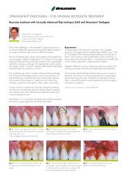
Dissolution Of Discrete Calcium Phosphate Crystals From CandidateTi-based Implant SurfacesPadina Pezeshki, Stanley Lugowski, John E. Davies, Faculty of Dentistry, Institute of Biomaterials andBiomedical Engineering, Faculty of Engineering, University of TorontoCanadian Biomaterials Society25th Annual Meeting (May 26–28, 2006, Calgary, Alberta,Canada)IntroductionPlasma-spraying of dental and orthopedic implants withcalcium phosphate (CaP) was introduced in the mid-1980sas a means of accelerating early peri-implant bone healingand rendering the implant surface bonebonding [1]. While some success has been,and continues to be, achieved clinically;problems have also been identified due todisintegration and delamination of suchrelatively thick (approx. 50 micron) coatings.The latter, due to the high temperature plasmaspraymethod, results in a heterogeneous CaPcoating composition comprising severalpossible CaP phases of differing solubilities.Thus, the solubility of these and other coatingsachieved through biomimetic strategies or solgeltechnology has been a subject ofconsiderable study and different fabricationmethods have aimed to modify the propertiesthat influence the CaP dissolution rate such asphase, particle size, surface area, and porosity[2,3]. Recently, a new means of modifyingmetallic surfaces with CaP has been developedthat comprises the deposition of discretecrystals of CaP from solution onto the metaloxide surface. In suspension the CaP crystalsare of the order of 20-100nms, which can bedeposited on the surface as a function ofimmersion time. Our current study wasdesigned to quantify the amount anddissolution kinetics of these discrete CaPcrystals from Ti6Al4V alloy dental implantsurfaces and compare their behavior withcommercially-available plasma sprayed CaPcoated implants of a similar size. Sinceactivated macrophages and osteoclasts canlocally reduce the extracellular pH to four, wechose an investigational pH range of 4-7 forour experiments.Materials And MethodsTwo Ti6Al4V implant types were employed. TheDCD-treated implants were IOSS615 (6mmdiameter x 15mm length) and plasma sprayedCaP implants were HT 613 (6mm diameter x1.501.000.500.0035.0030.0025.0020.0015.0010.005.000.0013mm length): both supplied by BIOMET 3i in Palm BeachGardens, Florida. Experiments were carried out to (i) measurethe total amount of Ca on the surface of the implants and (ii)the amount of Ca released in pH-specific solutions. In orderto measure the traces of Ca, all experimental equipment wasinitially cleaned to clear Ca contamination. For determinationof total amount of Ca on the surfaces, each DCD-treatedimplant was immersed in 10ml of 1 M Ultrex Grade HCl (pHFigure 1: Comparison of total [Ca] on plasma sprayed and DCD-treated implants4.003.503.002.502.00Ca = -1.9205 pH 3 + 32.805 pH 2 - 188.26 pH + 383.04Ca = 0.0826 pH 3 - 0.9697pH 2 + 1.8537pH + 6.27783 3.5 4 4.5 5 5.5 6 6.5 7 7.5pH3 3.5 4 4.5 5 5.5 6 6.5 7 7.5pHFigure 2: Comparison of different implants’ dissolution response3