- Page 1 and 2:
March 7-11F inalFinal ProgrammeThe
- Page 3:
Bienvenidosa Viena!!
- Page 6:
25 th European Congress of Radiolog
- Page 10 and 11:
WelcomeIt is a great pleasure and a
- Page 13 and 14:
Committees12 ESR Executive Council1
- Page 15 and 16:
Programme Planning CommitteePostgra
- Page 17 and 18:
Scientific SubcommitteesMolecular I
- Page 19:
Topic CoordinatorsCategorical Cours
- Page 22 and 23:
George Simpson Bisset IIIHouston, T
- Page 24 and 25:
Tarek A. El-DiastyMansoura/EGHonora
- Page 26 and 27:
Gary Glazer †Stanford, CA/USHonor
- Page 28 and 29:
José CáceresBarcelona/ESGold Meda
- Page 30 and 31:
Johannes LammerVienna/ATGold Medall
- Page 32 and 33:
Maximilian F. ReiserMunich/DEGold M
- Page 34 and 35:
Jesús PrietoPamplona/ESOpening Lec
- Page 36 and 37:
Carlo CatalanoRome/ITHonorary Lectu
- Page 38 and 39:
Jean-François ‘Jeff’GeschwindB
- Page 40 and 41:
Luis Martí-BonmatíValencia/ESHono
- Page 42 and 43:
Customiseyour congress!Plan and per
- Page 44 and 45:
General InformationInformation from
- Page 46 and 47:
General InformationInformation from
- Page 48 and 49:
General InformationInformation from
- Page 50 and 51:
BOOSTYOURCAREER.TAKE THE EUROPEANDI
- Page 52 and 53:
General InformationCME at ECR 2013G
- Page 54 and 55:
General InformationESR Meets Sessio
- Page 56 and 57:
myESRWhen you‘ve checked in to EC
- Page 58 and 59:
General InformationInformation from
- Page 60 and 61:
Activities 2013Visiting SchoolsVisi
- Page 62 and 63:
EIBIR SUMMER SCHOOLon Neurology Ima
- Page 64 and 65:
General InformationEPOS - Scientifi
- Page 66 and 67:
Visit the EPOS TM Area on the secon
- Page 68 and 69:
General InformationECR 2013SPECIALE
- Page 70 and 71:
EnjoyVienna‘sculturalhighlightsVi
- Page 72 and 73:
Visit the Technical Exhibition!And
- Page 74 and 75:
Top radiologists read more than jus
- Page 76 and 77:
Insights into ImagingEducation and
- Page 78 and 79:
Johannes Krisch in Der Alpenkönig
- Page 80 and 81:
Floor PlansU2 - Lower LevelU2 - LOW
- Page 82 and 83:
Floor PlansOE - Entrance LevelOE -
- Page 84 and 85:
Floor PlansO1 - First LevelO1 - FIR
- Page 86 and 87:
Floor PlansO3 - Third LevelO3 - THI
- Page 89 and 90:
ProgrammeOverviews88 Thursday, Marc
- Page 91 and 92:
Programme OverviewThursday, March 7
- Page 93 and 94:
Programme OverviewFriday, March 8I/
- Page 95 and 96:
Programme OverviewSaturday, March 9
- Page 97 and 98:
Programme OverviewSunday, March 10I
- Page 99:
Programme OverviewMonday, March 11I
- Page 102 and 103:
New Horizons SessionsFriday, March
- Page 104 and 105:
Special Focus SessionsFriday, March
- Page 106 and 107:
Special Focus SessionsSunday, March
- Page 108 and 109:
Special Focus SessionsMonday, March
- Page 110 and 111:
Multidisciplinary SessionsManaging
- Page 112 and 113:
Categorical CoursesOncologic Imagin
- Page 114 and 115:
Mini CoursesOrgans from A to Z: Hea
- Page 116 and 117:
Mini CoursesJoint Course of ESR and
- Page 118 and 119:
Refresher Courses / Scientific Sess
- Page 120 and 121:
Refresher Courses / Scientific Sess
- Page 122 and 123:
Refresher Courses / Scientific Sess
- Page 124 and 125:
Refresher Courses / Scientific Sess
- Page 126 and 127:
Refresher Courses / Scientific Sess
- Page 128 and 129:
Refresher Courses / Scientific Sess
- Page 130 and 131:
Refresher Courses / Scientific Sess
- Page 132 and 133:
Refresher Courses / Scientific Sess
- Page 134 and 135:
Refresher Courses / Scientific Sess
- Page 136 and 137:
Refresher Courses / Scientific Sess
- Page 138 and 139:
E 3 - European Excellence in Educat
- Page 140 and 141:
Gustav Klimt, Design Drawing Tree,
- Page 142 and 143:
Accompanying SessionsSaturday, Marc
- Page 144 and 145:
EIBIR presents IMAGINEThursday Marc
- Page 146 and 147:
Rising Stars ProgrammeBasic Session
- Page 148 and 149:
Update Your Skills(Practical Course
- Page 150 and 151:
Florian Boesch in Radamisto by Geor
- Page 152 and 153:
Satellite SymposiaFriday, March 8,
- Page 154 and 155:
Satellite SymposiaSaturday, March 9
- Page 156 and 157:
Zubin Mehta at the Musikverein© ww
- Page 158 and 159:
Postgraduate Educational ProgrammeA
- Page 160 and 161:
Postgraduate Educational ProgrammeA
- Page 162 and 163: Postgraduate Educational ProgrammeA
- Page 164 and 165: Postgraduate Educational ProgrammeA
- Page 166 and 167: Postgraduate Educational ProgrammeA
- Page 168 and 169: Postgraduate Educational ProgrammeA
- Page 170 and 171: Postgraduate Educational ProgrammeA
- Page 172 and 173: Postgraduate Educational ProgrammeA
- Page 174 and 175: Postgraduate Educational ProgrammeA
- Page 176 and 177: Postgraduate Educational Programme1
- Page 178 and 179: Postgraduate Educational ProgrammeA
- Page 180 and 181: Postgraduate Educational ProgrammeA
- Page 182 and 183: Postgraduate Educational ProgrammeA
- Page 184 and 185: Postgraduate Educational ProgrammeA
- Page 186 and 187: Postgraduate Educational ProgrammeA
- Page 188 and 189: Postgraduate Educational ProgrammeA
- Page 190 and 191: Postgraduate Educational ProgrammeA
- Page 192 and 193: Postgraduate Educational ProgrammeA
- Page 194 and 195: Postgraduate Educational ProgrammeA
- Page 196 and 197: Postgraduate Educational ProgrammeA
- Page 198 and 199: Postgraduate Educational ProgrammeA
- Page 200 and 201: Postgraduate Educational ProgrammeA
- Page 202 and 203: Postgraduate Educational ProgrammeA
- Page 204 and 205: Postgraduate Educational ProgrammeA
- Page 206 and 207: Postgraduate Educational ProgrammeA
- Page 208 and 209: Postgraduate Educational ProgrammeA
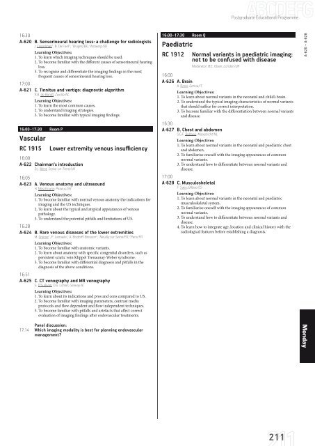
- Page 210 and 211: Postgraduate Educational ProgrammeA
- Page 215 and 216: ScientificSessionsSession numbers a
- Page 217 and 218: Scientific Sessions11:51B-0020 Rela
- Page 219 and 220: Scientific Sessions11:42B-0060 The
- Page 221 and 222: Scientific Sessions11:33B-0098 Eval
- Page 223 and 224: Scientific Sessions11:51B-0140 Radi
- Page 225 and 226: Scientific Sessions14:00-15:30 Room
- Page 227 and 228: Scientific Sessions15:12B-0218 Imag
- Page 229 and 230: Scientific Sessions15:03B-0257 CT a
- Page 231 and 232: Scientific Sessions10:30-12:00 Room
- Page 233 and 234: Scientific Sessions10:30-12:00 Room
- Page 235 and 236: Scientific Sessions10:30-12:00 Room
- Page 237 and 238: Scientific Sessions14:00-15:30 Room
- Page 239 and 240: Scientific Sessions15:03B-0447 Brow
- Page 241 and 242: Scientific Sessions15:03B-0487 Vali
- Page 243 and 244: Scientific Sessions10:30-12:00 Room
- Page 245 and 246: Scientific Sessions10:30-12:00 Room
- Page 247 and 248: Scientific Sessions11:42B-0588 Prev
- Page 249 and 250: Scientific Sessions10:30-12:00 Room
- Page 251 and 252: Scientific Sessions10:30-12:00 Room
- Page 253 and 254: Scientific Sessions11:42B-0697 MRI
- Page 255 and 256: Scientific Sessions11:33B-0736 Whol
- Page 257 and 258: Scientific Sessions10:30-12:00 Room
- Page 259 and 260: Scientific Sessions11:51B-0788 Meta
- Page 261 and 262: Scientific Sessions11:51B-0828 Non-
- Page 263 and 264:
Scientific Sessions11:33B-0866 Repe
- Page 265 and 266:
Scientific Sessions15:21B-0908 Diff
- Page 267 and 268:
Scientific Sessions15:03B-0946 Asse
- Page 269 and 270:
Scientific Sessions14:54B-0985 Esti
- Page 271:
Scientific Sessions14:00-15:30 Room
- Page 274 and 275:
List of Authors and Co-AuthorsAA. R
- Page 276 and 277:
List of Authors and Co-AuthorsBiffa
- Page 278 and 279:
List of Authors and Co-AuthorsConlo
- Page 280 and 281:
List of Authors and Co-AuthorsEurin
- Page 282 and 283:
List of Authors and Co-AuthorsGüm
- Page 284 and 285:
List of Authors and Co-AuthorsJerec
- Page 286 and 287:
List of Authors and Co-AuthorsLamal
- Page 288 and 289:
List of Authors and Co-AuthorsMasui
- Page 290 and 291:
List of Authors and Co-AuthorsOoste
- Page 292 and 293:
List of Authors and Co-AuthorsReise
- Page 294 and 295:
List of Authors and Co-AuthorsSeraf
- Page 296 and 297:
List of Authors and Co-AuthorsTomš
- Page 298 and 299:
List of Authors and Co-AuthorsWille
- Page 300 and 301:
List of ModeratorsAAbolmaali N.: SS
- Page 302:
CreditsCoordination:ESR Office | Ne


