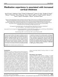
P ERSPECTIVES ON DISEASEU. <strong>Bellugi</strong> <strong>et</strong> <strong>al</strong>. – Linking cognition <strong>and</strong> the brainABCWMS (173) WMS (117)WMSWMSLNdTHLNvNorm<strong>al</strong>Norm<strong>al</strong>spared, while spati<strong>al</strong>-integrative functions aremarkedly impaired). Perhaps cortic<strong>al</strong> systems that subserv<strong>et</strong>he slower, but higher resolution, processes associatedwith the parvocellular pathway are selectivelyspared in WMS, while in DNS the two pathways couldboth be affected 54 .<strong>Brain</strong> cytoarchitectonic findings in WMSLNdLNvFig. 7. <strong>Brain</strong> cytoarchitectonic in Williams syndrome (WMS). (A) Thearrows indicate a marked indentation of the temporo–pari<strong>et</strong><strong>al</strong> regionsin the area of the sulcus. The whole posterior-pari<strong>et</strong><strong>al</strong> regions <strong>and</strong>occipit<strong>al</strong> regions are sm<strong>al</strong>l. (B) Amygd<strong>al</strong>ar nuclei in WMS <strong>and</strong> norm<strong>al</strong>brains, showing that in WMS the dors<strong>al</strong> portion of the later<strong>al</strong> nucleus(LNd) appears to be reduced <strong>and</strong> has an unusu<strong>al</strong> shape. The arrowindicates a curtailment in the later<strong>al</strong> nucleus of the amygd<strong>al</strong>a. In thisspecimen, the nucleus was estimated to be about h<strong>al</strong>f the size of theaverage amygd<strong>al</strong>a in norm<strong>al</strong> subjects. Also, note that the tempor<strong>al</strong>horn (TH) is placed more dors<strong>al</strong>ly in WMS individu<strong>al</strong>s than in norm<strong>al</strong>subjects. (C) Unlike in the norm<strong>al</strong> brain, where the centr<strong>al</strong> sulcusreaches to the interhemispheric fissure <strong>and</strong> proceeds a variable distance<strong>al</strong>ong the medi<strong>al</strong> surface of the hemisphere (arrows), the centr<strong>al</strong> sulcusin the WMS brain ends substanti<strong>al</strong>ly before it reaches the midline(arrows). Abbreviation: LNv, ventr<strong>al</strong> later<strong>al</strong> nucleus.An opportunity to link brain findings with cognitivedeficits can <strong>al</strong>so be found in the study of foc<strong>al</strong>cognitive deficits of individu<strong>al</strong>s with WMS at the levelof brain cytoarchitectonics. Four autopsy brains ofindividu<strong>al</strong>s with WMS have been studied byG<strong>al</strong>aburda <strong>and</strong> colleagues 55–57 (see Fig. 7). Microenceph<strong>al</strong>y<strong>and</strong> the relative curtailment of the occipit<strong>al</strong><strong>and</strong> posterior-pari<strong>et</strong><strong>al</strong> areas were evident in three ofthe brains (Fig. 7A). One of the four brains showed amarked reduction in the size of the pari<strong>et</strong><strong>al</strong>, posteriortempor<strong>al</strong><strong>and</strong> occipit<strong>al</strong> regions in comparison with themore rostr<strong>al</strong> portions of the hemispheres. Theseabrupt <strong>and</strong> dramatic reductions led to the brainappearing as though a b<strong>and</strong> had constricted its posteriorportions 55 . Another brain showed dramaticreduction in the size of the amygd<strong>al</strong>a (Fig. 7B), whichTHcould be associated with abnorm<strong>al</strong> soci<strong>al</strong> behaviorthat occurs in subjects with WMS. The MRI data <strong>al</strong>socorroborated the gener<strong>al</strong> finding of a reduction in thesize of posterior areas. Curtailment of the dors<strong>al</strong>-pari<strong>et</strong><strong>al</strong>regions <strong>and</strong> posterior-tempor<strong>al</strong> areas mightindeed be relevant to the extreme visuo–spati<strong>al</strong>deficits seen in individu<strong>al</strong>s with WMS. The four brainsshow largely norm<strong>al</strong> over<strong>al</strong>l sulc<strong>al</strong> patterns, except forsome simplification of tertiary sulcation <strong>and</strong> a consistentlynon-opercularized dors<strong>al</strong> centr<strong>al</strong> sulcus. Thecentr<strong>al</strong> sulci in norm<strong>al</strong> brains reach <strong>al</strong>l the way to theinterhemispheric fissure <strong>and</strong> then a short distance furtheronto the medi<strong>al</strong> surfaces of the hemispheres, butin <strong>al</strong>l the available WMS cases the centr<strong>al</strong> sulcus ends noless than a centim<strong>et</strong>er later<strong>al</strong> to the interhemisphericfissure (Fig. 7C). This finding could indicate abnorm<strong>al</strong>development of the medio–dors<strong>al</strong> cortices, whichhave been associated with visuo–spati<strong>al</strong> functions.The blocks of the cerebr<strong>al</strong> cortex of WMS individu<strong>al</strong>sthat have been examined show well-developedcytoarchitectonic areas with <strong>al</strong>l main divisions identifiable.However, there are subtle distortions in thearchitecture. Morphom<strong>et</strong>ric studies suggest that neuron<strong>al</strong>-cell-packingdensity is diminished with a concurrentincrease in gli<strong>al</strong> numbers, which possibly indicatesa substanti<strong>al</strong> decrease in tot<strong>al</strong> number of neurons.The observed cell numbers <strong>and</strong> cell-packing densitiessuggest early development<strong>al</strong> arrest (for example, prenat<strong>al</strong>lyor before the second year of age), or regressiveevents that occur postnat<strong>al</strong>ly into the middle of thefirst decade of life 55 . Research that involves establishinglinks b<strong>et</strong>ween the genomic changes <strong>and</strong> the changes inproduction of mRNA <strong>and</strong> protein that lead to theunusu<strong>al</strong> development of the WMS brain, might shedlight on norm<strong>al</strong> brain <strong>and</strong> behavior<strong>al</strong> development 56,57 .In gener<strong>al</strong>, these findings provide unusu<strong>al</strong> opportunitiesfor linking brain findings to cognitive deficits<strong>and</strong> their neur<strong>al</strong> underpinnings.A molecular-gen<strong>et</strong>ic profile for WMSWilliams syndrome is caused by a microdel<strong>et</strong>ion onchromosome 7 that involves the gene encodingelastin (ELN) 58 <strong>and</strong> perhaps 20 other genes, includingthe human homolog of the Drosophila gene, frizzled(FZD3) 59 , LIM-kinase 1 (LIMK1) 60 , syntaxin 1A (STX1A) 61 ,replication factor C2 (RFC2) 62 , CLYNZ (Ref. 63), FKPB6(Ref. 64), WSTF (Ref. 65), WS-bTRP, WS-bHLH, BCL7B(Ref. 66), <strong>and</strong> a duplicated gene, GTF2I (gener<strong>al</strong> transcriptionfactor 2I) 67 . Korenberg <strong>and</strong> colleagues areconstructing a physic<strong>al</strong> map of the del<strong>et</strong>ed region ofchromosome 7 b<strong>and</strong> 7q11.23 by using multi-colorfluorescence in situ hybridization (FISH) of bacteri<strong>al</strong>artifici<strong>al</strong> chromosomes (BACs) on m<strong>et</strong>aphase <strong>and</strong> interphasechromosomes, large-fragment library screening,genomic Southern blot <strong>and</strong> pulsed-field-gel an<strong>al</strong>yses,STS (sequence tagged site) <strong>and</strong> polymorphic-markeran<strong>al</strong>yses. Bacteri<strong>al</strong> artifici<strong>al</strong> chromosomes were chosento construct the physic<strong>al</strong> map because they are clonedin a stable vector <strong>and</strong> contain large genomic fragmentsof up to 300 kb that are stable <strong>and</strong> readilymanipulated. Therefore, they are suitable for geneisolation <strong>and</strong> DNA sequencing. These map reagentswere used to investigate the size <strong>and</strong> extent of thedel<strong>et</strong>ions in individu<strong>al</strong>s with WMS in whom subs<strong>et</strong>s offeatures, including neurocognitive profiles, brainstructures <strong>and</strong> brain functions, were d<strong>et</strong>erminedsimultaneously 68–70 .204 TINS Vol. 22, No. 5, <strong>1999</strong>
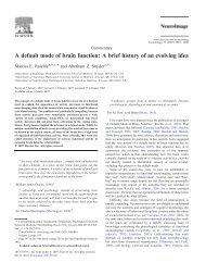
Fig. 8. The molecular-gen<strong>et</strong>ic basis ofWilliams syndrome (WMS). (A) The region ofchromosome 7, b<strong>and</strong> 7qll.23, that is commonlydel<strong>et</strong>ed in WMS is represented by thedark-blue box in the ideogram. This region isexp<strong>and</strong>ed to the right to illustrate its genomicorganization, a region of mainly single copygenes – the homolog of the Drosophila gene,frizzled (FZD3), syntaxin 1A (STX1A), elastin(ELN), LIM-kinase 1 (LIMK1), WSCR1, replicationfactor C2 (RFC2) – flanked by a seriesof genomic duplications (green, p<strong>al</strong>e blue)containing genes (for example, GTF2I),pseudogenes (for example, GTF2IP, PMS2P),<strong>and</strong> duplicate markers (for example,D7S489L). The regions used in the commonbreakpoints are indicated by dark-blue bars.The map positions of independent bacteri<strong>al</strong>artifici<strong>al</strong> chromosomes (BACs) used in part forthis an<strong>al</strong>ysis are shown as green dots to theleft of the ideogram. (B) Vertic<strong>al</strong> lines indicat<strong>et</strong>he regions del<strong>et</strong>ed <strong>and</strong> the number of subjectscarrying the common WMS del<strong>et</strong>ion,which are associated with some of the typic<strong>al</strong>faci<strong>al</strong> features, ment<strong>al</strong> r<strong>et</strong>ardation <strong>and</strong> heartdisease, or carrying sm<strong>al</strong>ler del<strong>et</strong>ions, includingsubregions of STX1A through to RFC2,which are associated with only the typic<strong>al</strong>heart disease, SVAS. Subject VI <strong>al</strong>so has asubtle defect in visu<strong>al</strong>–spati<strong>al</strong> processing.Sm<strong>al</strong>l square brack<strong>et</strong>s indicate del<strong>et</strong>ed regionsthat differ among subjects <strong>and</strong>, therefore,provide the potenti<strong>al</strong> del<strong>et</strong>ion to assign specificWMS features to single regions or genes.The large square brack<strong>et</strong>s indicate regionsthat, from the current data, are likely to containa gene or genes that when del<strong>et</strong>ed contributein some measure to the characteristicfeatures of WMS. The significance of thesedata is that del<strong>et</strong>ion of STX1A, ELN, LIMK1,WSCR1 <strong>and</strong> RFC2 do not appear to be associatedwith the characteristic faci<strong>al</strong> features orment<strong>al</strong> r<strong>et</strong>ardation seen in WMS, <strong>al</strong>thoughthey could contribute. This is the first step indefining single genes whose del<strong>et</strong>ion is ultimatelyresponsible for the distinctive cognitivefeatures of WMS. Subject I is a typic<strong>al</strong> WMSindividu<strong>al</strong> 67,68,71,74,75 , subject II has a largerdel<strong>et</strong>ion than a typic<strong>al</strong> WMS individu<strong>al</strong> 71,76 ,subject III can be found in the It<strong>al</strong>ian casesd<strong>et</strong>ailed in Ref. 69, d<strong>et</strong>ails of subjects IV <strong>and</strong>V can be found in Ref. 77, <strong>and</strong> d<strong>et</strong>ails of subjectsVI <strong>and</strong> VII can be found in Refs 78 <strong>and</strong>60, respectively.A working model of the genomeorganization that characterizeschromosome b<strong>and</strong> 7q11.2 <strong>and</strong>incorporates other maps 71,72 was developed 73 <strong>and</strong> itsuggests that this region includes highly homologouschromosom<strong>al</strong> duplications that are <strong>al</strong>so characterizedby a number of repeat-sequence families, genes <strong>and</strong>pseudogenes. The tot<strong>al</strong>ity is organized as a nestedrepeated structure that surrounds the largely uniqueregion occupied by elastin <strong>and</strong> the other del<strong>et</strong>ed genes(Fig. 8A). This suggests that the region of DNA del<strong>et</strong>edin WMS individu<strong>al</strong>s is located within an apparentlysingle copy region of chromosome 7 that appears to besurrounded by a series of genomic duplications, someof which must be recent <strong>and</strong> others of which mighthave been duplicated earlier in primate evolution.ABChromosome7222115.315.215.114131211.211.111.111.2111.2211.2321.121.221.32231.131.231.33233343536GenesBACsD7S653D7S489UFZD3STX1AELNLIMK1WSCR1RFC2U. <strong>Bellugi</strong> <strong>et</strong> <strong>al</strong>. – Linking cognition <strong>and</strong> the brain P ERSPECTIVES ON DISEASEChromosome7222115.315.114131211.111.111.2211.2321.121.221.32231.131.231.3GTF2I146 2D7S489L 2D7S6753233343536WMS1GTF2IPPMS2PD7489UFZD3STX1AELNLIMK1WSCR1RFC2GTF2ID7S489LPMS2PSVASI II III IV V VI VIISubject2DuplicatedregionsMeiotic mispairing of subs<strong>et</strong>s of the numerousrepeated sequences might ultimately contribute to thedel<strong>et</strong>ion 71 . Therefore, it is not unexpected that thedel<strong>et</strong>ion breakpoints in WMS occur largely in commonregions <strong>and</strong> most, though not <strong>al</strong>l, individu<strong>al</strong>swith WMS have the same genes del<strong>et</strong>ed 68,71,74 .However, it is studies of the uncommon individu<strong>al</strong>swith sm<strong>al</strong>ler del<strong>et</strong>ions that are beginning to provideclues to the genes responsible for the subs<strong>et</strong>s of WMS features.For example, from studies of individu<strong>al</strong>s with isolateddel<strong>et</strong>ions <strong>and</strong> mutations of elastin, it appears thatthe absence of one copy of the gene is probably responsiblefor the heart defect, SVAS, that is typic<strong>al</strong>ly found in11PhenotypesBreakpointWMSdel<strong>et</strong>ionBreakpointSome WMS MRI <strong>and</strong>cognitive featuresSome WMS faci<strong>al</strong>featuresHeart diseaseSome visuo–spati<strong>al</strong>featuresNorm<strong>al</strong> IQSome WMS MRI <strong>and</strong>cognitive featuresSome WMS faci<strong>al</strong>featuresTINS Vol. 22, No. 5, <strong>1999</strong> 205