

Laryngocoele, laryngocele - Vula - University of Cape Town
Laryngocoele, laryngocele - Vula - University of Cape Town
Laryngocoele, laryngocele - Vula - University of Cape Town

Create successful ePaper yourself
Turn your PDF publications into a flip-book with our unique Google optimized e-Paper software.
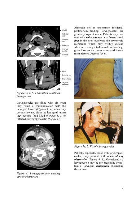
abHyoidExternalsacInternalsacEpiglottisInternaljugularCarotidarteryAlthough not an uncommon incidentalpostmortem finding, laryngocoeles aregenerally asymptomatic. Patients may presentwith voice change or a lateral swellingin the neck overlying the thyrohyoidmembrane which may visibly distendwhen increasing intraluminal pressure e.g.glass blowers and trumpet or reed instrumentplayers (Figures 7a, b).aHyoidExternal sacInternal sacThyroidcartilageFigures 5 a, b: Fluid-filled combinedlaryngocoele<strong>Laryngocoele</strong>s are filled with air whenthey retain a communication with thelaryngeal lumen (Figures 1, 4); when theybecome isolated from the laryngeal lumenthey become fluid-filled (Figures 3, 5) orinfected (laryngopyocoele) (Figure 6).bFigure 7a, b: Visible laryngocoelesFigure 6: Laryngopyocoele causingairway obstructionPatients, especially those with laryngopyocoeles,may present with acute airwayobstruction (Figure 6, 8). Occasionally alaryngocoele may be the presenting symptom<strong>of</strong> laryngeal malignancy obstructingthe saccule.2


