

Laryngocoele, laryngocele - Vula - University of Cape Town
Laryngocoele, laryngocele - Vula - University of Cape Town
Laryngocoele, laryngocele - Vula - University of Cape Town

You also want an ePaper? Increase the reach of your titles
YUMPU automatically turns print PDFs into web optimized ePapers that Google loves.
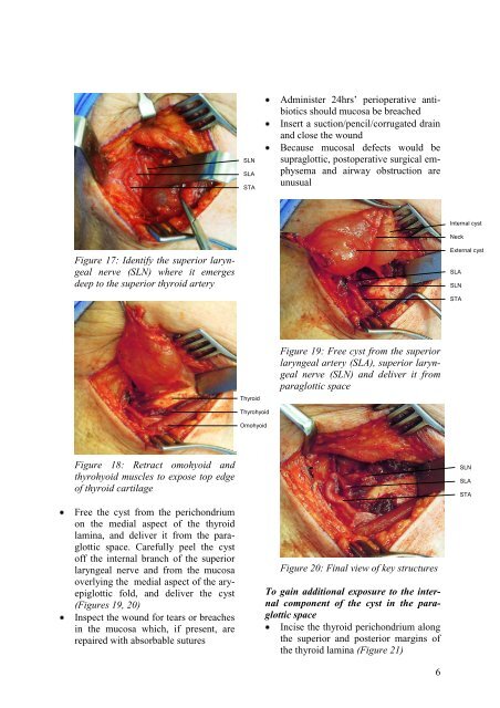
SLNSLASTAAdminister 24hrs’ perioperative antibioticsshould mucosa be breachedInsert a suction/pencil/corrugated drainand close the woundBecause mucosal defects would besupraglottic, postoperative surgical emphysemaand airway obstruction areunusualInternal cystNeckFigure 17: Identify the superior laryngealnerve (SLN) where it emergesdeep to the superior thyroid arteryExternal cystSLASLNSTAThyroidFigure 19: Free cyst from the superiorlaryngeal artery (SLA), superior laryngealnerve (SLN) and deliver it fromparaglottic spaceThyrohyoidOmohyoidFigure 18: Retract omohyoid andthyrohyoid muscles to expose top edge<strong>of</strong> thyroid cartilageSLNSLASTAFree the cyst from the perichondriumon the medial aspect <strong>of</strong> the thyroidlamina, and deliver it from the paraglotticspace. Carefully peel the cyst<strong>of</strong>f the internal branch <strong>of</strong> the superiorlaryngeal nerve and from the mucosaoverlying the medial aspect <strong>of</strong> the aryepiglotticfold, and deliver the cyst(Figures 19, 20)Inspect the wound for tears or breachesin the mucosa which, if present, arerepaired with absorbable suturesFigure 20: Final view <strong>of</strong> key structuresTo gain additional exposure to the internalcomponent <strong>of</strong> the cyst in the paraglotticspaceIncise the thyroid perichondrium alongthe superior and posterior margins <strong>of</strong>the thyroid lamina (Figure 21)6


