

Laryngocoele, laryngocele - Vula - University of Cape Town
Laryngocoele, laryngocele - Vula - University of Cape Town
Laryngocoele, laryngocele - Vula - University of Cape Town

You also want an ePaper? Increase the reach of your titles
YUMPU automatically turns print PDFs into web optimized ePapers that Google loves.
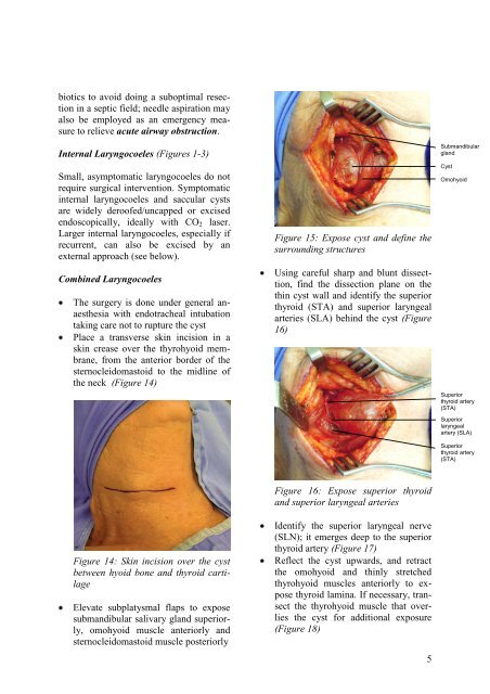
iotics to avoid doing a suboptimal resectionin a septic field; needle aspiration mayalso be employed as an emergency measureto relieve acute airway obstruction.Internal <strong>Laryngocoele</strong>s (Figures 1-3)Small, asymptomatic laryngocoeles do notrequire surgical intervention. Symptomaticinternal laryngocoeles and saccular cystsare widely dero<strong>of</strong>ed/uncapped or excisedendoscopically, ideally with CO 2 laser.Larger internal laryngocoeles, especially ifrecurrent, can also be excised by anexternal approach (see below).Combined <strong>Laryngocoele</strong>sThe surgery is done under general anaesthesiawith endotracheal intubationtaking care not to rupture the cystPlace a transverse skin incision in askin crease over the thyrohyoid membrane,from the anterior border <strong>of</strong> thesternocleidomastoid to the midline <strong>of</strong>the neck (Figure 14)Figure 15: Expose cyst and define thesurrounding structuresUsing careful sharp and blunt dissecttion,find the dissection plane on thethin cyst wall and identify the superiorthyroid (STA) and superior laryngealarteries (SLA) behind the cyst (Figure16)SubmandibularglandCystOmohyoidSuperiorthyroid artery(STA)Superiorlaryngealartery (SLA)Superiorthyroid artery(STA)Figure 14: Skin incision over the cystbetween hyoid bone and thyroid cartilageElevate subplatysmal flaps to exposesubmandibular salivary gland superiorly,omohyoid muscle anteriorly andsternocleidomastoid muscle posteriorlyFigure 16: Expose superior thyroidand superior laryngeal arteriesIdentify the superior laryngeal nerve(SLN); it emerges deep to the superiorthyroid artery (Figure 17)Reflect the cyst upwards, and retractthe omohyoid and thinly stretchedthyrohyoid muscles anteriorly to exposethyroid lamina. If necessary, transectthe thyrohyoid muscle that overliesthe cyst for additional exposure(Figure 18)5


