Wireless capsule endoscopy: from diagnostic devices to ...
Wireless capsule endoscopy: from diagnostic devices to ...
Wireless capsule endoscopy: from diagnostic devices to ...

Create successful ePaper yourself
Turn your PDF publications into a flip-book with our unique Google optimized e-Paper software.
236 Biomed Micro<strong>devices</strong> (2007) 9:235–243<br />
WCE may integrate mechanisms for active locomotion inside<br />
the GI tract, cutting edge microsensors for diagnosis<br />
and therapy and micro<strong>to</strong>ols for minimally invasive surgery<br />
(MIS) (Fireman et al., 2004; Polla et al., 2000; Bashir, 2004;<br />
Zahn et al., 2000).<br />
In this regard, several privately held companies and research<br />
centers throughout the world are trying <strong>to</strong> leverage<br />
their technological background in order <strong>to</strong> enhance the capabilities<br />
of WCE <strong>from</strong> a mere video acquisition <strong>to</strong>ol for<br />
diagnosis <strong>to</strong> a complete and au<strong>to</strong>nomous medical platform<br />
also for therapy and MIS, thanks <strong>to</strong> the advancements and<br />
harmonization of various engineering disciplines, including:<br />
microelectromechanical systems (MEMS), user friendly<br />
control interfaces and high precision mechanics.<br />
Present of wireless <strong>capsule</strong> <strong>endoscopy</strong><br />
Given Imaging<br />
The idea of a <strong>capsule</strong> able <strong>to</strong> take pictures inside the small<br />
bowel traces back <strong>to</strong> the 1980s, although at that time it<br />
seemed mere figment because of the technological limitations<br />
hampering its feasibility, among which the high power<br />
consumption of charged couple device (CCD) image sensors.<br />
However, in the early 1990s complementary metal oxide<br />
semiconduc<strong>to</strong>r (CMOS) technology, along with progresses<br />
in application specific integrated circuit (ASIC) for control<br />
systems and light emitting diode (LED) for illumination,<br />
paved the way for the advent of WCE.<br />
In 2000, during the Digestive Disease Week Conference<br />
in San Diego, California (USA), P. Swain, an endoscopist<br />
<strong>from</strong> the London Royal Hospital, and Given Imaging Ltd.,<br />
a Yoqneam (Israel) based company, announced the M2A TM<br />
pill, the first swallowable <strong>capsule</strong> able <strong>to</strong> capture images<br />
inside the small bowel (Iddan et al., 2000; Pennisi and Kerr,<br />
2004; Moayyedi and Ford, 2002). This has been possible<br />
thanks <strong>to</strong> the collaboration between P. Swain, working on<br />
wireless <strong>endoscopy</strong>, and G. Iddan, an engineer dealing with<br />
imaging <strong>devices</strong> for defense missiles at Rafael Ltd., the R&D<br />
group of the Israeli Ministry of Defense (Meron, 2000; Iddan<br />
et al., 2004).<br />
M2A TM and PillCam TM SB<br />
The first ingestible <strong>capsule</strong> by Given Imaging for diagnosis<br />
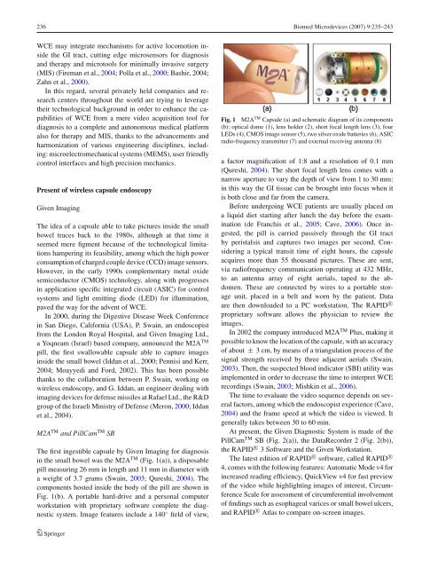
in the small bowel was the M2A TM (Fig. 1(a)), a disposable<br />
pill measuring 26 mm in length and 11 mm in diameter with<br />
a weight of 3.7 grams (Swain, 2003; Qureshi, 2004). The<br />
components hosted inside the body of the pill are shown in<br />
Fig. 1(b). A portable hard-drive and a personal computer<br />
workstation with proprietary software complete the <strong>diagnostic</strong><br />
system. Image features include a 140 ◦ field of view,<br />
Springer<br />
Fig. 1 M2A TM Capsule (a) and schematic diagram of its components<br />
(b): optical dome (1), lens holder (2), short focal length lens (3), four<br />
LEDs (4), CMOS image sensor (5), two silver oxide batteries (6), ASIC<br />
radio-frequency transmitter (7) and external receiving antenna (8)<br />
a fac<strong>to</strong>r magnification of 1:8 and a resolution of 0.1 mm<br />
(Qureshi, 2004). The short focal length lens comes with a<br />
narrow aperture <strong>to</strong> vary the depth of view <strong>from</strong> 1 <strong>to</strong> 30 mm:<br />
in this way the GI tissue can be brought in<strong>to</strong> focus when it<br />
is both close and far <strong>from</strong> the camera.<br />
Before undergoing WCE patients are usually placed on<br />
a liquid diet starting after lunch the day before the examination<br />
(de Franchis et al., 2005; Cave,2006). Once ingested,<br />
the pill is carried passively through the GI tract<br />
by peristalsis and captures two images per second. Considering<br />
a typical transit time of eight hours, the <strong>capsule</strong><br />
acquires more than 55 thousand pictures. These are sent,<br />
via radiofrequency communication operating at 432 MHz,<br />
<strong>to</strong> an antenna array of eight aerials, taped <strong>to</strong> the abdomen.<br />
These are connected by wires <strong>to</strong> a portable s<strong>to</strong>rage<br />
unit, placed in a belt and worn by the patient. Data<br />
are then downloaded <strong>to</strong> a PC workstation. The RAPID R○<br />
proprietary software allows the physician <strong>to</strong> review the<br />
images.<br />
In 2002 the company introduced M2A TM Plus, making it<br />
possible <strong>to</strong> know the location of the <strong>capsule</strong>, with an accuracy<br />
of about ± 3 cm, by means of a triangulation process of the<br />
signal strength received by three adjacent aerials (Swain,<br />
2003). Then, the suspected blood indica<strong>to</strong>r (SBI) utility was<br />
implemented in order <strong>to</strong> decrease the time <strong>to</strong> interpret WCE<br />
recordings (Swain, 2003; Mishkin et al., 2006).<br />
The time <strong>to</strong> evaluate the video sequence depends on several<br />
fac<strong>to</strong>rs, among which the endoscopist experience (Cave,<br />
2004) and the frame speed at which the video is viewed. It<br />
generally takes between 30 <strong>to</strong> 60 min.<br />
At present, the Given Diagnostic System is made of the<br />
PillCam TM SB (Fig. 2(a)), the DataRecorder 2 (Fig. 2(b)),<br />
the RAPID R○ 3 Software and the Given Workstation.<br />
The latest edition of RAPID R○ software, called RAPID R○<br />
4, comes with the following features: Au<strong>to</strong>matic Mode v4 for<br />
increased reading efficiency, QuickView v4 for fast preview<br />
of the video while highlighting images of interest, Circumference<br />
Scale for assessment of circumferential involvement<br />
of findings such as esophageal varices or small bowel ulcers,<br />
and RAPID R○ Atlas <strong>to</strong> compare on-screen images.