Wireless capsule endoscopy: from diagnostic devices to ...
Wireless capsule endoscopy: from diagnostic devices to ...
Wireless capsule endoscopy: from diagnostic devices to ...

Create successful ePaper yourself
Turn your PDF publications into a flip-book with our unique Google optimized e-Paper software.
Biomed Micro<strong>devices</strong> (2007) 9:235–243<br />
DOI 10.1007/s10544-006-9025-3<br />
<strong>Wireless</strong> <strong>capsule</strong> <strong>endoscopy</strong>: <strong>from</strong> <strong>diagnostic</strong> <strong>devices</strong> <strong>to</strong><br />
multipurpose robotic systems<br />
Andrea Moglia · Arianna Menciassi ·<br />
Marc Oliver Schurr · Paolo Dario<br />
Published online: 12 December 2006<br />
C○ Springer Science + Business Media, LLC 2007<br />
Abstract In the recent past, the introduction of miniaturised<br />
image sensors with low power consumption, based on complementary<br />
metal oxide semiconduc<strong>to</strong>r (CMOS) technology,<br />
has allowed the realisation of an ingestible wireless <strong>capsule</strong><br />
for the visualisation of the small intestine mucosa. The device<br />
has received approval <strong>from</strong> Food and Drug Administration<br />
and has gained momentum since it has been more successful<br />
than traditional techniques in the diagnosis of small intestine<br />
disorders. In 2004 an esophagus specific <strong>capsule</strong> was<br />
launched, while a solution for colon is still under development.<br />
However, present solutions suffer <strong>from</strong> several limitations:<br />
they move passively by exploiting peristalsis, are<br />
not able <strong>to</strong> s<strong>to</strong>p intentionally for a prolonged diagnosis, they<br />
receive power <strong>from</strong> an internal battery with short length, and<br />
their usage is restricted <strong>to</strong> one organ, either small bowel or<br />
esophagus. However the steady progresses in many branches<br />
of engineering, including microelectromechanical systems<br />
(MEMS), are envisaged <strong>to</strong> affect the performances of capsular<br />
<strong>endoscopy</strong>. The near future foreshadows <strong>capsule</strong>s able<br />
<strong>to</strong> pass actively through the whole gastrointestinal tract, <strong>to</strong><br />
retrieve views <strong>from</strong> all organs and <strong>to</strong> perform drug delivery<br />
and tissue sampling. In the long term, the advent of robotics<br />
could lead <strong>to</strong> au<strong>to</strong>nomous medical platforms, equipped with<br />
the most advanced solutions in terms of MEMS for therapy<br />
A. Moglia (�) · A. Menciassi · P. Dario<br />
Center for Applied Research in Micro Engineering (CRIM Lab),<br />
Scuola Superiore Sant’Anna, Pisa, Italy<br />
e-mail: andrea.moglia@crim.sssup.it<br />
M. O. Schurr<br />
novineon Healthcare Technology Partners GmbH,<br />
Tuebingen, Germany<br />
M. O. Schurr<br />
Institute of Healthcare Industries at Steinbeis University Berlin,<br />
Berlin, Germany<br />
and diagnosis of the digestive tract. In this review, we discuss<br />
the state of the art of wireless <strong>capsule</strong> <strong>endoscopy</strong> (WCE): after<br />
a description on the current status, we present the most<br />
promising solutions.<br />
Keywords <strong>Wireless</strong> <strong>capsule</strong> . Capsule <strong>endoscopy</strong> . Active<br />
<strong>capsule</strong>s . Medical robots . Robotic <strong>capsule</strong><br />
Introduction<br />
Endoscopy, intended as a methodology <strong>to</strong> assess the mucosa<br />
of the gastrointestinal (GI) tract, was born in 1868 when<br />
G. and R. Schindler pioneered methods of performing gastroscopy<br />
with a semiflexible gastroscope (Salmore, 1998).<br />
Nowadays flexible endoscopes enable diagnosis inside<br />
esophagus, s<strong>to</strong>mach, colon and in part <strong>to</strong> small bowel, thus<br />
helping <strong>to</strong> understand its structure, functions and pathologies.<br />
Flexible <strong>endoscopy</strong> induces little trauma <strong>to</strong> the patient<br />
and is generally well accepted.<br />
However, some areas of the GI tract are still difficult <strong>to</strong><br />
reach with flexible endoscopes, such as the largest part of<br />
the small bowel. At least a fraction of the patients fears flexible<br />
<strong>endoscopy</strong> due <strong>to</strong> pain and. This limits the willingness<br />
of patients <strong>to</strong> undergo <strong>endoscopy</strong>, especially in the field of<br />
cancer prevention and screening.<br />
In this sense wireless <strong>capsule</strong> <strong>endoscopy</strong> (WCE) has been<br />
playing an important role since its introduction and promises<br />
<strong>to</strong> revolutionize <strong>endoscopy</strong> because it can greatly reduce the<br />
level of discomfort <strong>to</strong> be <strong>to</strong>lerated by patients (Bradbury,<br />
2000). While this has initially been limited <strong>to</strong> small bowel<br />
<strong>endoscopy</strong>, WCE is now branching off in<strong>to</strong> other indications,<br />
such as esophagoscopy or colonoscopy, although this<br />
expansion of indications will not happen <strong>to</strong> a larger extent<br />
without significant technology advancements. In the future<br />
Springer
236 Biomed Micro<strong>devices</strong> (2007) 9:235–243<br />
WCE may integrate mechanisms for active locomotion inside<br />
the GI tract, cutting edge microsensors for diagnosis<br />
and therapy and micro<strong>to</strong>ols for minimally invasive surgery<br />
(MIS) (Fireman et al., 2004; Polla et al., 2000; Bashir, 2004;<br />
Zahn et al., 2000).<br />
In this regard, several privately held companies and research<br />
centers throughout the world are trying <strong>to</strong> leverage<br />
their technological background in order <strong>to</strong> enhance the capabilities<br />
of WCE <strong>from</strong> a mere video acquisition <strong>to</strong>ol for<br />
diagnosis <strong>to</strong> a complete and au<strong>to</strong>nomous medical platform<br />
also for therapy and MIS, thanks <strong>to</strong> the advancements and<br />
harmonization of various engineering disciplines, including:<br />
microelectromechanical systems (MEMS), user friendly<br />
control interfaces and high precision mechanics.<br />
Present of wireless <strong>capsule</strong> <strong>endoscopy</strong><br />
Given Imaging<br />
The idea of a <strong>capsule</strong> able <strong>to</strong> take pictures inside the small<br />
bowel traces back <strong>to</strong> the 1980s, although at that time it<br />
seemed mere figment because of the technological limitations<br />
hampering its feasibility, among which the high power<br />
consumption of charged couple device (CCD) image sensors.<br />
However, in the early 1990s complementary metal oxide<br />
semiconduc<strong>to</strong>r (CMOS) technology, along with progresses<br />
in application specific integrated circuit (ASIC) for control<br />
systems and light emitting diode (LED) for illumination,<br />
paved the way for the advent of WCE.<br />
In 2000, during the Digestive Disease Week Conference<br />
in San Diego, California (USA), P. Swain, an endoscopist<br />
<strong>from</strong> the London Royal Hospital, and Given Imaging Ltd.,<br />
a Yoqneam (Israel) based company, announced the M2A TM<br />
pill, the first swallowable <strong>capsule</strong> able <strong>to</strong> capture images<br />
inside the small bowel (Iddan et al., 2000; Pennisi and Kerr,<br />
2004; Moayyedi and Ford, 2002). This has been possible<br />
thanks <strong>to</strong> the collaboration between P. Swain, working on<br />
wireless <strong>endoscopy</strong>, and G. Iddan, an engineer dealing with<br />
imaging <strong>devices</strong> for defense missiles at Rafael Ltd., the R&D<br />
group of the Israeli Ministry of Defense (Meron, 2000; Iddan<br />
et al., 2004).<br />
M2A TM and PillCam TM SB<br />
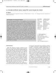
The first ingestible <strong>capsule</strong> by Given Imaging for diagnosis<br />
in the small bowel was the M2A TM (Fig. 1(a)), a disposable<br />
pill measuring 26 mm in length and 11 mm in diameter with<br />
a weight of 3.7 grams (Swain, 2003; Qureshi, 2004). The<br />
components hosted inside the body of the pill are shown in<br />
Fig. 1(b). A portable hard-drive and a personal computer<br />
workstation with proprietary software complete the <strong>diagnostic</strong><br />
system. Image features include a 140 ◦ field of view,<br />
Springer<br />
Fig. 1 M2A TM Capsule (a) and schematic diagram of its components<br />
(b): optical dome (1), lens holder (2), short focal length lens (3), four<br />
LEDs (4), CMOS image sensor (5), two silver oxide batteries (6), ASIC<br />
radio-frequency transmitter (7) and external receiving antenna (8)<br />
a fac<strong>to</strong>r magnification of 1:8 and a resolution of 0.1 mm<br />
(Qureshi, 2004). The short focal length lens comes with a<br />
narrow aperture <strong>to</strong> vary the depth of view <strong>from</strong> 1 <strong>to</strong> 30 mm:<br />
in this way the GI tissue can be brought in<strong>to</strong> focus when it<br />
is both close and far <strong>from</strong> the camera.<br />
Before undergoing WCE patients are usually placed on<br />
a liquid diet starting after lunch the day before the examination<br />
(de Franchis et al., 2005; Cave,2006). Once ingested,<br />
the pill is carried passively through the GI tract<br />
by peristalsis and captures two images per second. Considering<br />
a typical transit time of eight hours, the <strong>capsule</strong><br />
acquires more than 55 thousand pictures. These are sent,<br />
via radiofrequency communication operating at 432 MHz,<br />
<strong>to</strong> an antenna array of eight aerials, taped <strong>to</strong> the abdomen.<br />
These are connected by wires <strong>to</strong> a portable s<strong>to</strong>rage<br />
unit, placed in a belt and worn by the patient. Data<br />
are then downloaded <strong>to</strong> a PC workstation. The RAPID R○<br />
proprietary software allows the physician <strong>to</strong> review the<br />
images.<br />
In 2002 the company introduced M2A TM Plus, making it<br />
possible <strong>to</strong> know the location of the <strong>capsule</strong>, with an accuracy<br />
of about ± 3 cm, by means of a triangulation process of the<br />
signal strength received by three adjacent aerials (Swain,<br />
2003). Then, the suspected blood indica<strong>to</strong>r (SBI) utility was<br />
implemented in order <strong>to</strong> decrease the time <strong>to</strong> interpret WCE<br />
recordings (Swain, 2003; Mishkin et al., 2006).<br />
The time <strong>to</strong> evaluate the video sequence depends on several<br />
fac<strong>to</strong>rs, among which the endoscopist experience (Cave,<br />
2004) and the frame speed at which the video is viewed. It<br />
generally takes between 30 <strong>to</strong> 60 min.<br />
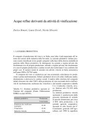
At present, the Given Diagnostic System is made of the<br />
PillCam TM SB (Fig. 2(a)), the DataRecorder 2 (Fig. 2(b)),<br />
the RAPID R○ 3 Software and the Given Workstation.<br />
The latest edition of RAPID R○ software, called RAPID R○<br />
4, comes with the following features: Au<strong>to</strong>matic Mode v4 for<br />
increased reading efficiency, QuickView v4 for fast preview<br />
of the video while highlighting images of interest, Circumference<br />
Scale for assessment of circumferential involvement<br />
of findings such as esophageal varices or small bowel ulcers,<br />
and RAPID R○ Atlas <strong>to</strong> compare on-screen images.
Biomed Micro<strong>devices</strong> (2007) 9:235–243 237<br />
Fig. 2 PillCam TM SB (a) and DataRecorder 2 (b)<br />
Remote <strong>capsule</strong> <strong>endoscopy</strong> is a recent modality <strong>to</strong> transfer<br />
the acquired data by WCE <strong>from</strong> Satellite Sites, equipped<br />
with a DataRecorder, <strong>to</strong> a Central PillCam Site with a Given<br />
Workstation. The usage of this technology should be encouraged<br />
in those geographical areas, such as the Scandinavian<br />
mainland, with scattered population, high distances between<br />
hospitals and where travelling is difficult, especially during<br />
winter.<br />
The device obtained approval <strong>from</strong> Food and Drug Administration<br />
(FDA) in 2003.<br />
In Oc<strong>to</strong>ber, 2003 Given Imaging got clearance <strong>to</strong> market<br />
<strong>capsule</strong> <strong>endoscopy</strong> for use in pediatrics patients with an age<br />
<strong>from</strong> 10 <strong>to</strong> 18 years.<br />
Clinical applications<br />
To date over 340,000 patients worldwide have experienced<br />
WCE without the invasiveness and discomfort of more conventional<br />
procedures. In fact WCE does not require sedation,<br />
nor entails radiation absorption for patients.<br />
Thanks <strong>to</strong> the ability <strong>to</strong> acquire images, while passing<br />
through the entire small intestine, WCE has been shown<br />
successful, in many cases providing better results than traditional<br />
techniques, in the diagnosis of occult gastrointestinal<br />
bleeding (OGIB). This is defined as bleeding of unknown<br />
origin which persists or recurs after a negative initial or primary<br />
<strong>endoscopy</strong> result, e.g. esophagogastroduodenoscopy<br />
(EGD) or colonoscopy (Zuckerman et al., 2000). In particular,<br />
each year in the United States 1 new case of GI bleeding<br />
per 1000 persons arises, with obscure bleeding representing<br />
5% of all new recurrences of GI bleeding (Pennazio et al.,<br />
2005; Triester et al., 2005).<br />
Crohn’s disease is another clinical application which can<br />
benefit <strong>from</strong> WCE (Herfarth et al., 2005). It is an inflamma<strong>to</strong>ry<br />
bowel disease (IBD) of unknown origin which can recur<br />
in any site of the digestive tract, in 30–40% of cases in the<br />
small intestine (Kornbluth et al., 2005; Hara et al., 2006).<br />
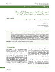
In Fig. 3 the jejunum of patient with suspected Crohn’s<br />
disease, showing thickened infiltrated folds, is illustrated.<br />
The possibility <strong>to</strong> view the intestinal mucosa has allowed<br />
<strong>to</strong> detect the villous atrophy, which is a common symp<strong>to</strong>m<br />
Fig. 3 Jejunum of patient with suspected Crohn’s disease showing<br />
thickened infiltrated folds<br />
of celiac disease (Kesari et al., 2005). This is an inflamma<strong>to</strong>ry<br />
intestinal pathology affecting people with in<strong>to</strong>lerance <strong>to</strong><br />
gluten, a wheat protein, with a prevalence of almost 1 in 100<br />
persons in western countries (Green et al., 2006; Dube et al.,<br />
2005; Maki et al., 2003). Interesting findings have been reported<br />
also in the diagnosis of small bowel tumours (Urbain<br />
et al., 2006).<br />
Then, WCE has found applications in pediatrics. It detected<br />
the presence of OGIB even in 2.5 years old child, so<br />
far the youngest patient who has received WCE treatment<br />
(Kavin et al., 2006).<br />
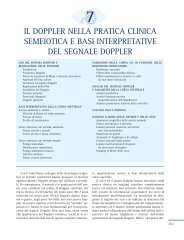
PillCam TM ESO<br />
Besides PillCam TM SB, Given Imaging produces PillCam TM<br />
ESO (Fig. 4), marketed in the U.S. by the InScope Division<br />
of Ethicon Endo-Surgery Inc., Johnson & Johnson Group,<br />
Cincinnati, Ohio (USA), which may represent an alternative<br />
Fig. 4 PillCam TM ESO<br />
Springer
238 Biomed Micro<strong>devices</strong> (2007) 9:235–243<br />
Fig. 5 Image of an esophagitis taken by PillCam TM ESO<br />
<strong>to</strong> traditional upper GI <strong>endoscopy</strong> techniques for the investigation<br />
of pathologies in the esophagus.<br />
The body of PillCam TM ESO harbors two cameras, one<br />
on each side and, while it proceeds along the esophagus, it<br />
captures 14 images per second. Other features, concerning<br />
dimensions, field of view and resolution, are the same as the<br />
PillCam TM SB.<br />
In this case the <strong>diagnostic</strong> procedure begins when, after<br />
a two-hour fast, the patient swallows the pill lying down.<br />
Then he is raised in a series of inclinations over a <strong>to</strong>tal of<br />
five min. During this stage the PillCam TM ESO acquires<br />
images which are directly transmitted <strong>to</strong> the sensor arrays<br />
and then <strong>to</strong> the s<strong>to</strong>rage unit. At this point, the patient is<br />
allowed <strong>to</strong> get up and can either walk or remain seated<br />
for additional 15 min <strong>to</strong> ensure the <strong>capsule</strong> has entered the<br />
s<strong>to</strong>mach.<br />
The first clinical trial on PillCam TM ESO concerned gastroesophageal<br />
reflux disease (GERD) (Eliakim et al., 2004).<br />
GERD is a frequent and/or severe form of heartburn, when<br />
the contents of the s<strong>to</strong>mach backflow in<strong>to</strong> the esophagus. Persons<br />
with chronic GERD may develop Barrett’s esophagus, a<br />
premalignant condition, whose incidence has been increasing<br />
steadily since 1970s. Barrett’s esophagus may in turn<br />
lead <strong>to</strong> esophageal adenocarcinoma, one of the fastest growing<br />
cancers in western countries (Ronkainen et al., 2005;<br />
Bollschweiler et al., 2001).<br />
Controindications<br />
Both PillCam TM SB and PillCam TM ESO are contraindicated<br />
in those patients with known or suspected GI obstruction,<br />
Springer<br />
Fig. 6 Patency <strong>capsule</strong><br />
strictures, or fistulas, swallowing disorders and cardiac<br />
pacemakers or other implanted electromedical <strong>devices</strong>. In<br />
particular, Leigh<strong>to</strong>n has proven that patients with portable<br />
electric cardiac <strong>devices</strong>, such as pacemakers, can undergo<br />
WCE examination (Leigh<strong>to</strong>n et al., 2005).<br />
Furthermore, a serious concern is represented by <strong>capsule</strong><br />
retention, which may lead <strong>to</strong> acute small bowel obstruction<br />
and in some cases surgery may be required <strong>to</strong> remove it. In<br />
the case of OGIB the most quoted retention rate is 0.75%<br />
(Barkin et al., 2002). For patients with Crohn’s disease, retention<br />
may happen in 6.7% (Buchman et al., 2004). It represents<br />
a concrete risk also for those undergoing esophageal <strong>capsule</strong><br />
<strong>endoscopy</strong>, if they have a his<strong>to</strong>ry of dysphagia, i.e. a difficulty<br />
<strong>to</strong> swallow. In order <strong>to</strong> avoid this inconvenience, Given<br />
Imaging has developed the Agile TM Patency System (Spada<br />
et al., 2005), a self-disintegrating pill, shown in Fig. 6. This<br />
has the same dimensions as the PillCam TM SB (11 mm ×<br />
26 mm), is made of a lac<strong>to</strong>se body, has a paraffin plug, contains<br />
a microchip for radiofrequency identification (RFID),<br />
a timer and 100 ml of barium sulfate <strong>to</strong> enable detection by<br />
fluoroscopy. A parylene layer covers the whole body, with<br />
the exception of the area in contact <strong>to</strong> the plug. The dissolution<br />
of the patency <strong>capsule</strong> starts approximately 40 hours<br />
after ingestion and is due <strong>to</strong> lactase, an enzyme produced by<br />
intestinal mucosa cells in the jejunum (Gay et al., 2005). The<br />
RFID plaque, measuring 2 mm × 12 mm and placed inside<br />
the body, can be detected by a RFID scanner which stays<br />
outside (Boivin et al., 2005). The Agile TM Patency <strong>capsule</strong><br />
received approval <strong>from</strong> FDA in May 2006.<br />
Olympus<br />
In Oc<strong>to</strong>ber 2005 Olympus Corporation, Tokyo (Japan),<br />
launched in Europe EndoCapsule, a disposable and passive<br />
pill for small bowel <strong>endoscopy</strong>, 26 mm in length and 11 mm<br />
in diameter, with a CCD sensor camera (Fig. 7(a)). This<br />
boasts a depth of field varying <strong>from</strong> zero <strong>to</strong> 20 mm, au<strong>to</strong>matic<br />
brightness control <strong>to</strong> adjust the illumination, ensured<br />
by 6 LEDs, and an operational time of 8 h. An antenna sends<br />
two images per sec <strong>to</strong> the recorder. Moreover, thanks <strong>to</strong> the<br />
Real Time Viewer (Fig. 7(b)), physicians can view real-time<br />
images and estimate the <strong>capsule</strong> position.
Biomed Micro<strong>devices</strong> (2007) 9:235–243 239<br />
Fig. 7 Olympus WCE system: EndoCapsule (a) and Real Time<br />
Viewer (b)<br />
Fig. 8 Image of normal villi structure, taken by EndoCapsule: without<br />
(a) and with structure enhancement (b)<br />
Then, the EndoCapsule Software allows the management<br />
of the acquired data. Its main features are: a multi display<br />
function for optimal observation, structure enhancement and<br />
enlargement display function <strong>to</strong> highlight the tiniest details<br />
(Fig. 8), red colour detection function <strong>to</strong> highlight suspected<br />
bleeding symp<strong>to</strong>ms, and au<strong>to</strong> speed adjustment function <strong>to</strong><br />
optimise the review speed Clinical testing is still under way<br />
in the United States and Japan, and the company needs <strong>to</strong><br />
receive full approval before selling its device in these markets<br />
(Fuyuno, 2005).<br />
The efficacy of EndoCapsule was demonstrated During<br />
Digestive Disease Week 2006, in Los Angeles, California<br />
(USA).<br />
Technical limitations<br />
The usage of an internal power source, such as a battery, is<br />
a critical issue as <strong>to</strong> <strong>to</strong>xicity and length. In particular, the<br />
Table 1 Comparison of<br />
existing WCE solutions of<br />
Given Imaging and Olympus<br />
latter sets a limit on the amount of available energy devoted<br />
<strong>to</strong> the acquisition of pictures. This affects the choice of the<br />
image sensor: CMOS technology may be preferable since it<br />
ensures a lower power consumption than CCD, but at a price<br />
of a poorer image quality (Litwiller, 2005).<br />
Moreover, the limited energy provided by a battery makes<br />
it impossible <strong>to</strong> add other functional modules <strong>to</strong> endoscopic<br />
<strong>capsule</strong>s, especially those for the locomotion and control of<br />
movement. If a <strong>capsule</strong> had an active locomotion it could<br />
vary its speed, thus reducing the transit time, and with s<strong>to</strong>p<br />
mechanisms it might stay at a specific point of <strong>diagnostic</strong><br />
interest for a prolonged time. This would enable the physician<br />
<strong>to</strong> conduct a more thorough analysis, whenever he spots<br />
interesting sites. The integration of sensors could also make<br />
other <strong>diagnostic</strong> and therapeutic tasks feasible, e.g. biopsy<br />
and drug delivery.<br />
Table 1 reports a comparison of the WCE which have<br />
received approval for usage in clinical environments.<br />
Future prospects of WCE: better images, therapy and<br />
active locomotion<br />
The importance of image sensors with finer resolution than<br />
current systems is motivated by the need <strong>to</strong> enable the detection<br />
of pathologies at an earlier stage, which are smaller and<br />
for this reason invisible at present. As a consequence, they<br />
may overcome images dis<strong>to</strong>rtion, which recurs during the<br />
zoom of a picture every time an enlarged view is required.<br />
However the higher the resolution, the higher the power consumption<br />
of the RF transmitter. In this sense Lin et al. (2006)<br />
have developed the GICam pill with a low power image compression<br />
processor. On the other hand, if a <strong>capsule</strong> were able<br />
<strong>to</strong> move actively it would decrease the length of its journey.<br />
Furthermore, with the capability <strong>to</strong> s<strong>to</strong>p at specific sites, it<br />
would help the physician <strong>to</strong> perform a thorough diagnosis<br />
in areas of major interest and <strong>to</strong> enable drug release (Raju<br />
et al., 2002). Then, steady advancements in the BioMEMS<br />
field foreshadow <strong>capsule</strong>s with sensors enabling new tasks<br />
(Gourley, 2005).<br />
In this regard, a wireless ingestible <strong>capsule</strong> endowed with<br />
three sensors for the moni<strong>to</strong>ring of pH, temperature and<br />
Given Imaging Olympus<br />
WCE Name PillCam TM SB PillCam TM ESO EndoCapsule<br />
Dimensions (D × L) 11 mm× 26 mm<br />
Operative environment Small intestine Esophagus Small intestine<br />
Image sensor type CMOS CCD<br />
Frame rate 2 images/second 14 images/second 2 images/second<br />
Image acquisition Off line Real time<br />
Power source Internal battery<br />
Source of motion Peristalsis<br />
Springer
240 Biomed Micro<strong>devices</strong> (2007) 9:235–243<br />
Fig. 9 Working principle of Olympus WCE with active locomotion<br />
pressure inside the GI tract was developed by SmartPill Corporation<br />
(Dickman et al., 2006). In July 2006, this device<br />
received approval by FDA for the sale and use in the United<br />
States.<br />
Swallowable <strong>capsule</strong>s with active locomotion, based on<br />
magnetic actuation and electrical stimulation, have been recently<br />
demonstrated (Sendoh et al., 2003; Mosse et al., 2001;<br />
Woo et al., 2005; Swain et al., 2005).<br />
In a not <strong>to</strong>o distant future, robotics will significantly contribute<br />
<strong>to</strong> make WCE au<strong>to</strong>nomous <strong>devices</strong> capable <strong>to</strong> bring a<br />
set of MEMS in targeted areas of the GI tract, independently<br />
<strong>from</strong> an external opera<strong>to</strong>r.<br />
Magnetic actuation and therapy<br />
Olympus<br />
Since November 2004, Olympus has been developing also a<br />
self propelled <strong>capsule</strong> for all parts of the GI tract and with the<br />
possibility <strong>to</strong> control its movement (Kusuda, 2005). In this<br />
case, an external magnetic field genera<strong>to</strong>r using three pairs<br />
of opposing electromagnets creates a uniform magnetic field<br />
in any direction (Fig. 9). By varying this magnetic field,<br />
Fig. 10 Norika WCE<br />
Springer<br />
position, orientation and posture of the pill can be controlled,<br />
thanks <strong>to</strong> a built in permanent magnet. For example, the<br />
magnetic field is used <strong>to</strong> generate a second and rotating<br />
magnetic field which rotates the <strong>capsule</strong>, generating thrust<br />
through the spiral on the outer surface of the pill. In this way,<br />
control of forward and reverse motion could be achieved.<br />
Additionally, the pill is provided with an external power<br />
source: coils outside the body transmit energy <strong>to</strong> those inside<br />
the <strong>capsule</strong>, by exploiting the principle of electromagnetic<br />
induction, hence eliminating the need of an internal battery.<br />
This has two advantages: increasing the pictures acquisition<br />
rate up <strong>to</strong> 5 images per second and the possibility <strong>to</strong> remain<br />
inside the GI tract for more than eight hours. Other features<br />
include a deflatable balloon and a negatively-pressurized<br />
space, both controlled <strong>from</strong> outside the body, for respectively<br />
drug delivery and body fluid sampling. Moreover,<br />
the pill will also integrate an ultrasound scanning module,<br />
promising <strong>to</strong> deliver high resolution images.<br />
Norika<br />
Since 1998 at RF SYSTEM Lab, Nagano (Japan), the Norika<br />
Project Team has been dealing with WCE, in particular on<br />
an externally powered <strong>capsule</strong> (Fig. 10), which integrates a<br />
CCD image sensor (Uehara et al., 2003). The <strong>diagnostic</strong> system<br />
includes a pill, 23 mm in length and 9 mm in diameter,<br />
a camera, 4 LEDs for illumination, an external controller, a<br />
transmitter vest and a workstation. Data processing is performed<br />
by a digital signal processor (DSP) which consumes<br />
94% of the <strong>to</strong>tal power and for this reason is placed outside<br />
the body. The camera is able <strong>to</strong> capture images at distances<br />
ranging <strong>from</strong> a few millimeters <strong>to</strong> some centimeters <strong>from</strong> its<br />
lens and features also focus adjustment.<br />
In order <strong>to</strong> enrich diagnosis, the <strong>capsule</strong> possesses some<br />
degree of rotation enabling images acquisition at different<br />
positions during its transit. A schema of the working principle<br />
is depicted in Fig. 11.
Biomed Micro<strong>devices</strong> (2007) 9:235–243 241<br />
Fig. 11 Working principle of Norika WCE<br />
Three coils are placed inside the <strong>capsule</strong>, acting as ro<strong>to</strong>r<br />
coils. Three coils, embedded in a vest-like jacket, act as<br />
sta<strong>to</strong>rs. The direction of sta<strong>to</strong>rs magnetic field sets the<br />
rotative direction. Localization and tilt can be detected with<br />
respectively 5 mm and 15 ◦ of deviation. Moreover Norika<br />
Project Team has been evaluating the use of near-infrared<br />
light in the acquisition of translucent images of the s<strong>to</strong>mach<br />
in order <strong>to</strong> see mucosal pathology behind gastric folds. In<br />
fact near-infrared light, with a wavelength ranging between<br />
800 and 1200 nm, is able <strong>to</strong> pass through human body 10<br />
times better than other types of radiation whose wavelength<br />
does not belong <strong>to</strong> this range.<br />
In the body of the pill about 40% of the volume is free and<br />
might host reservoirs for drug delivery or miniaturized knives<br />
and scissors for MIS or sensors for advanced <strong>diagnostic</strong><br />
tasks.<br />
According <strong>to</strong> industry insiders, the Norika pill needs<br />
further years of development, before entering the market<br />
(Fuyuno, 2005).<br />
Au<strong>to</strong>nomous robotic <strong>capsule</strong><br />
Intelligent Microsystem Center (IMC), Seoul (South<br />
Korea) has launched the EMILOC project (Endoscopic<br />
MIcro<strong>capsule</strong> LOcomotion and Control) and since 2004<br />
has been supporting a research team of Scuola Superiore<br />
Sant’Anna (SSSA), Pisa (Italy), <strong>to</strong> develop a robotic WCE<br />
able <strong>to</strong> move au<strong>to</strong>nomously in the GI tract.<br />
In this regard, the advent of robotics paves the way for<br />
a swallowable and disposable complete medical platform<br />
integrating actua<strong>to</strong>rs for active locomotion, sensors for diagnosis<br />
and therapy, and <strong>to</strong>ols for MIS inside the digestive<br />
tract (Menciassi et al., 2005). In the recent past robotics has<br />
Fig. 12 EMILOC robotic <strong>capsule</strong> developed at Scuola Superiore<br />
Sant’Anna<br />
been successfully applied <strong>to</strong> colonoscopy (Dario et al., 2004;<br />
Swain, 2005).<br />
The medical rationale of using micro-robotics <strong>to</strong> enable<br />
active <strong>capsule</strong> locomotion and control lies in the need <strong>to</strong><br />
actively steer the <strong>capsule</strong> inside the digestive tract for improved<br />
<strong>diagnostic</strong> and especially therapeutic means. This<br />
medical need is unmet by current capsular technologies.<br />
In particular the SSSA robotic <strong>capsule</strong> has an active locomotion<br />
based on a set of bioinspired legs. This solution<br />
offers various advantages: an accurate control on the trajec<strong>to</strong>ry<br />
allowing the <strong>capsule</strong> <strong>to</strong> pass over critical areas without<br />
<strong>to</strong>uching them; adaptability <strong>to</strong> organs with different shape,<br />
such as the large and the small intestine; fine position control,<br />
by varying the extension and orientation of the legs;<br />
high velocity <strong>to</strong> reduce the length of the journey inside the<br />
GI tract.<br />
The dimensions of the <strong>capsule</strong> shown in Fig. 12 are a diameter<br />
of 12 mm and a length of 30 mm. It has six legs, made<br />
of superelastic shape memory alloy (SMA), a well known<br />
material in the biomedical industry for its biocompatibility.<br />
Each leg has a round tip, ending with 200 µm high hooks <strong>to</strong><br />
provide friction for locomotion. Legs can fit <strong>to</strong> organs with<br />
different geometry thanks <strong>to</strong> a flexible knee. Despite their<br />
sharpness, the hooks did not damaged <strong>to</strong>o heavily the tissue<br />
during both in vitro and in vivo tests on animals, confirming<br />
what Atuma et al. had previously found (2001). Each leg is<br />
independently actuated by a SMA actua<strong>to</strong>r, whereas a human<br />
machine interface (HMI) is responsible for the control of the<br />
movement.<br />
The camera, based on CMOS technology, has a resolution<br />
of 320 × 320 pixels.<br />
Springer
242 Biomed Micro<strong>devices</strong> (2007) 9:235–243<br />
Table 2 Comparison of the<br />
future WCE of Olympus,<br />
Norika and SSSA<br />
Another feature of this <strong>capsule</strong> is an inflatable balloon<br />
useful <strong>to</strong> open the lumen, thus enabling the camera<br />
<strong>to</strong> view the largest area as possible, and <strong>to</strong> foster the<br />
locomotion.<br />
During both in vitro tests in phan<strong>to</strong>ms and in vivo tests on<br />
animals, conducted in collaboration with novineon Healthcare<br />
Technology Partners GmbH, Tuebingen, (Germany), the<br />
pill has proven <strong>to</strong> proceed successfully and s<strong>to</strong>p by spreading<br />
the legs against the wall of the small bowel. In this case<br />
force exertion of the legs is only local and over a relatively<br />
small surface of tissue, making this type of locomotion very<br />
atraumatic.<br />
When the pill will be ready <strong>to</strong> undergo clinical tests on<br />
humans, the body will be covered by a biocompatible and<br />
biodegradable layer <strong>to</strong> avoid the accidental deployment in<br />
the mouth and esophagus. Once in the s<strong>to</strong>mach, the layer<br />
will be dissolved by the gastric juices and the legs will be<br />
free <strong>to</strong> move, enabling the <strong>capsule</strong> <strong>to</strong> move au<strong>to</strong>nomously<br />
until the end of its journey.<br />
Table 2 compares the most promising WCE solutions<br />
combining active locomotion with therapeutic tasks.<br />
Concluding remarks<br />
In the past few years wireless ingestible <strong>capsule</strong>s opened<br />
the chance <strong>to</strong> conduct in a non invasive way diagnosis of a<br />
wide range of pathologies of the digestive tract, including<br />
OGIB, Crohn’s disease, celiac disease and GERD. Today,<br />
cutting-edge technologies make diagnosis feasible, while<br />
in the future high efficiency power sources and micro actua<strong>to</strong>rs<br />
promise <strong>to</strong> increase the potentialities of WCE. In<br />
this sense robotics and MEMS will play a significant role<br />
<strong>to</strong> develop a complete medical platform also for therapy<br />
and MIS. An additional input may come <strong>from</strong> the increasingly<br />
wide number of research centers and private companies<br />
engaged <strong>to</strong> achieve the supremacy in this fast growing<br />
field.<br />
Springer<br />
Olympus Norika SSSA<br />
Dimensions (D × L) 11 mm× 26 mm 9 mm × 23 mm 12 mm × 30 mm<br />
Operative environment Whole gastrointestinal tract<br />
Image sensor type CCD CMOS<br />
Frame rate 5 images/second 30 images/second 2 images/second<br />
Power source <strong>Wireless</strong> power transmission Battery<br />
Source of motion External magnetic field Bioinspired legs<br />
Motion control Yes<br />
S<strong>to</strong>p capability Yes<br />
Other features Availability of space <strong>to</strong> lodge sensors for therapy and micro <strong>to</strong>ols for MIS<br />
Advanced image acquisition<br />
by ultrasound<br />
Inflatable balloon<br />
References<br />
C. Atuma, V. Strugala, A. Allen, and L. Holm, American Journal<br />
of Physiology-Gastrointestinal and Liver Physiology 280, 922<br />
(2001).<br />
J.S. Barkin and S. Friedman, Amer. J. Gastroenter. 97, A83 (2002).<br />
R. Bashir, Adv. Drug Delivery Rev. 56, 1656 (2004).<br />
M.L. Boivin, H. Lochs, and W.A. Voderholzer, Endoscopy 37, 808<br />
(2005).<br />
E. Bollschweiler, E. Wolfgarten, C. Gutschow, and A.H. Holscher,<br />
Cancer 923, 549 (2001).<br />
J. Bradbury, The Lancet 356, 2074 (2000).<br />
L. Buchman, F.H. Miller, A. Wallin, A.A. Chowdhry, and C. Ahn,<br />
Amer. J. Gastroenter. 99, 2171 (2004).<br />
D.R. Cave, Gastrointest. Endos. Clin. North Amer. 14, 17 (2004).<br />
D.R. Cave, Nat. Clin. Pract. Gastroenterol. Hepa<strong>to</strong>l. 3, 158 (2006).<br />
P. Dario, P. Ciarletta, A. Menciassi, and B. Kim, Intern. J. Robot. Rese.<br />
23, 549 (2004).<br />
R. de Franchis, A. Avgerinos, J. Barkin, D. Cave, and B. Filoche,<br />
Endoscopy 37, 1040 (2005).<br />
R. Dickman and R. Fass, Digest. Dis. 24, 313 (2006).<br />
C. Dube, A. Ros<strong>to</strong>m, R. Sy, A. Cranney, N. Saloojee, C. Garritty, M.<br />
Sampson, L. Zhang, F. Yazdi, V. Mamaladze, I. Pan, J. Macneil,<br />
D. Mack, D. Patel, and D. Moher, Gastroenterology 128, S57<br />
(2005).<br />
R. Eliakim, K. Yassin, I. Shlomi, A. Suissa, and G.M. Eisen,<br />
Alimentary Pharmacololy & Therapeutics 20, 1083 (2004).<br />
Z. Fireman, A. Glukhovsky, and E. Scapa, Gastrointest. Endosc. Clin.<br />
North Amer. 14, 219 (2004).<br />
I. Fuyuno, Nature 438, 913 (2005).<br />
G. Gay, M. Delvaux, V. Laurent, N. Reibel, D. Regent, G. Grosdidier,<br />
and J.F. Roche, Endoscopy 37, 174 (2005).<br />
P.L. Gourley, Biotechnology Progress 21, 2 (2005).<br />
P.H.R. Green and B. Jabri B, Ann. Rev. Med. 57, 207 (2006).<br />
A.K. Hara, J.A. Leigh<strong>to</strong>n, R.I. Heigh, V.K. Sharma, A.C. Silva, G. De<br />
Petris, J.G. Hentz, and D.E. Fleischer, Radiology 238, 128 (2006).<br />
H. Herfarth and G. Rogler, Endoscopy 37, 42 (2005).<br />
G. Iddan, G. Meron, A. Glukhovsky, and P. Swain, Nature 405, 17<br />
(2000).<br />
G.J. Iddan and C.P. Swain, His<strong>to</strong>ry and development of <strong>capsule</strong><br />
<strong>endoscopy</strong>, Gastrointest. Endosc. Clin. North Amer. 14, 1 (2004).<br />
H. Kavin, J. Berman, T.L. Martin, A. Feldman, and K. Forsey-Koukol,<br />
Pediatrics 117, 539 (2006).<br />
Kesari, R.K. Bobba, and E.L. Arsura, Gastrointest. Endosc. 62, 796<br />
(2005).<br />
Kornbluth, J.F. Colombel, J.A. Leigh<strong>to</strong>n, and E. Loftus, Endoscopy<br />
37, 1051 (2005).
Biomed Micro<strong>devices</strong> (2007) 9:235–243 243<br />
Y. Kusuda, Sens. Rev. 25, 259 (2005).<br />
J.A. Leigh<strong>to</strong>n, K. Srivathsan, E.J. Carey, V.K. Sharma, R.I. Heigh,<br />
J.K. Post, P.J. Erickson, S.R. Robinson, J.L. Bazzell, and D.E.<br />
Fleischer, Amer. J. Gastroenterol. 100, 1728 (2005).<br />
M.C. Lin, L.R. Dung, and P.K. Weng, Biomed. Engin. Online 5, 14<br />
(2006).<br />
S. Liangpunsakul, L. Mays, and D.K. Rex, Amer. J. Gastroenterol. 98,<br />
2676 (2003).<br />
D. Litwiller, Pho<strong>to</strong>n. Spectra 8, 54 (2005).<br />
M. Maki, K. Mustalahti, J. Kokkonen, P. Kulmala, M. Haapalahti,<br />
T. Karttunen, J. Ilonen, K. Laurila, I. Dahlbom, T. Hansson,<br />
P. Hopfl, and M. Knip, New Engl. J. Med. 348, 2517<br />
(2003).<br />
A. Menciassi, A. Moglia, S. Gorini, G. Pernorio, C. Stefanini, and P.<br />
Dario, J. Micromech. Microeng. 15, 2045 (2005).<br />
G. Meron, Gastrointest. Endosc. 52, 817 (2000).<br />
D.S. Mishkin, R. Chuttani, J. Croffie, J. Disario, J. Liu, R. Shah, L.<br />
Somogyi, W. Tierney, L.M. Song, and B.T. Petersen, Gastrointest.<br />
Endosc. 63, 539 (2006).<br />
P. Moayyedi and A. Ford, Brit. Med. J. 325, 1399 (2002).<br />
C.A. Mosse, T.N. Mills, M.N. Appleyard, S.S. Kadirkamanathan, and<br />
C.P. Swain, Gastrointest. Endosc. 54, 79 (2001).<br />
M. Pennazio, G. Eisen, and N. Goldfarb, Endoscopy 37, 1046<br />
(2005).<br />
E. Pennisi and R.A. Kerr, Science 306, 2001 (2004).<br />
D.L. Polla, A.G. Erdman, W.P. Robbins, D.T. Markus, J. Diaz-Diaz, R.<br />
Rizq, Y. Nam, H.T. Brickner, A. Wang, and P. Krulevitch, Ann.<br />
Rev. Biomed. Engin. 2, 551 (2000).<br />
W.A. Qureshi, Nat. Rev. Drug Disc. 3, 447 (2004).<br />
G.S. Raju and T. Yusuf, Gastroenterology 122, S28 (2002).<br />
J. Ronkainen, P. Aro, T. S<strong>to</strong>rskrubb, S.E. Johansson T. Lind, E.<br />
Bolling-Sternevald, M. Vieth, M. S<strong>to</strong>lte, N.J. Talley, and L.<br />
Agreus, Gastroenterology 129, 1825 (2005).<br />
R. Salmore, Gastroenterol. Nurs. 21, 40, (1998).<br />
J. T. Santini, M.J. Cima, and R.A. Langer, Nature 397, 335 (1999).<br />
M. Sendoh, K. Ishiyama, and K.I. Arai, IEEE Trans. Magnet. 39, 3232<br />
(2003).<br />
Spada, G. Spera, M. Riccioni, L. Biancone, L. Petruzziello, A. Tringali,<br />
P. Familiari, M. Marchese, G. Onder, M. Mutignani, V. Perri, C.<br />
Petruzziello, F. Pallone, and G. Costamagna, Endoscopy 37, 793<br />
(2005).<br />
P. Swain, Gastrointest. Endosc. Clin. North Amer. 15, 839 (2005).<br />
P. Swain, Gut 52, iv48 (2003).<br />
P. Swain, T. Mills, B. Kelleher, L. Schmitz, S. Mosse, P. Burke, K. Ikeda,<br />
and A. Fritscher-Ravens, Gastrointest. Endosc. 61, AB101 (2005).<br />
S.L. Triester, J.A. Leigh<strong>to</strong>n, G.I. Leontiadis, D.E. Fleischer, A.K Hara,<br />
R.I. Heigh, A.D. Shiff, and V.K. Sharma, Amer. J. Gastroenterol.<br />
100, 2407 (2005).<br />
A. Uehara, and K. Hoshina, Minimally Invasive Therapy & Allied<br />
Technol. 12, 227 (2003).<br />
D. Urbain, D. De Looze, I. Demedts, E. Louis, O. Dewit, E. Macken,<br />
and A. Van Gossum, Endoscopy 38, 408 (2006).<br />
S.H. Woo, J.Y. Yang, E.S. Jung, J.H. Lee, Y.K. Moon, T.W. Kim, C.H.<br />
Won, H.C. Choi, and J.H. Cho, New Method of Moving Control<br />
for <strong>Wireless</strong> Endoscopic Capsule Using Electrical Stimuli. IEICE<br />
Trans. Fundament. Electr. Commun. Comp. Sci. 6, 1476 (2005).<br />
J.D. Zahn, N.H. Talbot, D. Liepmann, and A.P. Pisano, Biomed.<br />
Microdev. 2, 295 (2000).<br />
G.R. Zuckerman, C. Prakash, M.P. Askin, and B.S. Lewis, Gastroenterology<br />
118, 201 (2000).<br />
http://www.givenimaging.com. Accessed in 2006.<br />
http://www.olympus.co.jp/en/news/2005b/nr051013capsle.cfm.<br />
Accessed in 2006.<br />
http://www.smartpillcorp.com/ Accessed in 2006.<br />
http://www.rfnorika.com/. Accessed in 2006.<br />
http://www.sssup.it/sssup/jsp/section.jsp?sec id1 = 528&sec id2 =<br />
65918&lang = it. Accessed in 2006.<br />
http://www.novineon.com. Accessed in 2006.<br />
Springer