Biomedical Imaging Equipment Technician (BIET) - ETA International
Biomedical Imaging Equipment Technician (BIET) - ETA International
Biomedical Imaging Equipment Technician (BIET) - ETA International

You also want an ePaper? Increase the reach of your titles
YUMPU automatically turns print PDFs into web optimized ePapers that Google loves.
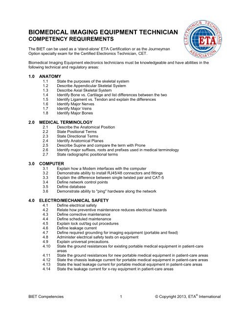
BIOMEDICAL IMAGING EQUIPMENT TECHNICIANCOMPETENCY REQUIREMENTSThe <strong>BIET</strong> can be used as a ‘stand-alone’ <strong>ETA</strong> Certification or as the JourneymanOption specialty exam for the Certified Electronics <strong>Technician</strong>, CET.<strong>Biomedical</strong> <strong>Imaging</strong> <strong>Equipment</strong> electronics technicians must be knowledgeable and have abilities in thefollowing technical and regulatory areas:1.0 ANATOMY1.1 State the purposes of the skeletal system1.2 Describe Appendicular Skeletal System1.3 Describe Axial Skeletal System1.4 Identify Bone vs. Cartilage and list differences between the two1.5 Identify Ligament vs. Tendon and explain the differences1.6 Identify Major Nerves1.7 Identify Major Veins1.8 Identify Major Bones2.0 MEDICAL TERMINOLOGY2.1 Describe the Anatomical Position2.2 State Positional Terms2.3 State Directional Terms2.4 Identify Anatomical Planes2.5 Describe Supine and compare the term with Prone2.6 Identify major suffixes, roots and prefixes used in medical terminology2.7 State radiographic positional terms3.0 COMPUTER3.1 Explain how a Modem interfaces with the computer3.2 Demonstrate ability to install RJ45/48 connectors and fittings3.3 Explain the difference between single twisted pair and CAT-53.4 Define network control points3.5 Define database3.6 Demonstrate ability to "ping" hardware along the network4.0 ELECTRO/MECHANICAL SAFETY4.1 Define electrical safety4.2 Relate how preventive maintenance reduces electrical hazards4.3 Define corrective maintenance4.4 Define scheduled maintenance4.5 Explain lock out/tag out procedures4.6 Define leakage current4.7 Define required grounding for imaging equipment (portable and fixed)4.8 Administer electrical safety tests on equipment4.9 Explain universal precautions.4.10 State the ground resistances for existing portable medical equipment in patient-careareas4.11 State the ground resistances for new portable medical equipment in patient-care areas4.12 State the chassis leakage current for portable medical equipment in patient-care areas4.13 State the lead leakage current for portable medical equipment in patient-care areas4.14 State the leakage current for x-ray equipment in patient-care areas<strong>BIET</strong> Competencies 1 © Copyright 2013, <strong>ETA</strong> ® <strong>International</strong>
<strong>ETA</strong> ® <strong>International</strong> – <strong>Biomedical</strong> <strong>Imaging</strong> <strong>Equipment</strong> <strong>Technician</strong> Competencies5.0 PICTURE ARCHIVE COMMUNICATION SYSTEM5.1 Explain electrical surge potentials5.2 List ways of preventing damage from electrical surges5.3 Describe the Internet and its application to imaging modalities5.4 Explain TCP/IP duties and protocols5.5 Describe security problems with the Internet5.6 Describe Tele-radiology5.7 Describe Picture Archive Communication system5.8 List major components of Picture Archive Communication system5.9 Describe the intranet and its application to imaging modalities5.10 Explain basic computer/ network maintenance procedures6.0 DIAGNOSTIC ULTRASOUND EQUIPMENT6.1. List the functions of the five basic components of a diagnostic medical ultrasoundmachine6.2. Identify the unique characteristics for each of the types of transducer scan heads used inreal-time ultrasound6.3. Describe current ultrasound image display formats (pie-shaped, rectangular, trapezoidal,circular)6.4. Describe the different ultrasound image recording formats (Polaroid film, single emulsionfilm, thermal paper, magnetic tape, magnetic disks, and optical disks)6.5 Describe A-Mode6.6 Describe B-Mode6.7 Describe M-Mode7.0 BUILDING WIRING7.1 List standards used in the electrical wiring of medical buildings7.2 Explain methods of pre-wiring and ways to wire existing buildings7.3 Explain NEC or other safety rules pertaining to building wiring and grounding8.0 BASIC RADIOGRAPHIC EQUIPMENT8.1 List the main function of an X-ray machine8.2 State the different types of X-ray machines (Fluoroscope, cine, chest, dental)8.3 Sketch a circuit diagram of an X-ray tube8.4 Sketch a circuit diagram of an X-ray machine8.5 Describe the "heel effect"8.6 Describe the focal spot8.7 Explain the purpose of grids8.8 Explain the purpose of the ‘bucky’8.9 Identify dental X-ray machine components8.10 Identify portable X-ray machine components8.11 Identify general ‘rad-room’ components8.12 Identify ‘cath lab’ components9.0 FILM PROCESSING9.1 Describe Wet Processing9.2 Identify Chemicals and Functions9.3 Describe Dry Processing9.4 Identify and Describe Laser <strong>Imaging</strong> Process9.5 Describe function and makeup of X-ray Cassettes9.6 Describe and Identify X-ray film types9.7 State dark-room procedures9.8 Describe film duplication process9.9 Demonstrate proper cassette loading technique10.0 TEST EQUIPMENT10.1 Explain the purpose of a Dosimeter<strong>BIET</strong> Competencies 2 © Copyright 2013, <strong>ETA</strong> ® <strong>International</strong>
<strong>ETA</strong> ® <strong>International</strong> – <strong>Biomedical</strong> <strong>Imaging</strong> <strong>Equipment</strong> <strong>Technician</strong> Competencies10.2 Demonstrate proper operation of an Oscilloscope10.3 Demonstrate proper operation of a DVM10.4 Demonstrate proper operation of a milliammeter10.5 Explain the application of an Ion chamber10.6 Explain the application of the half-value layer11.0 MAGNETIC RESONANCE IMAGING11.1 Identify Magnet types11.2 Describe Fourier Process11.3 Identify Cryogens11.4 Describe T1 and T211.5 State purpose of Gradients11.6 Identify Coils11.7 State purpose of auxiliary coils11.8 Identify RF leakage11.9 Identify image produced with metal in bore12.0 COMPUTED TOMOGRAPHY12.1 Define computed tomography12.2 Identify the components of computed tomography (gantry • tube/detectors • generator •couch - computers • applications • reconstruction • display)12.3 Describe the formation of the image12.4 Describe computed tomography dose index (CTDI)12.5 Describe multiple scan average dose (MSDA)12.6 Describe beam geometry12.7 Describe measuring dose12.8 Describe Protocol selection options (i.e. kvp, mAs, slice thickness, feed, matrix,algorithm)13.0 NUCLEAR MEDICINE13.1. Identify the major components of a scintillation camera and label them correctly on adiagram13.2. List the function of scintillation camera collimators13.3 Identify the material of which scintillation camera collimators are made.13.4 Identify the chemical composition of a scintillation crystal and its physical characteristics13.5 List the environmental factors that can adversely affect a scintillation crystal13.6 Identify the purpose of a photo multiplier tube in a scintillation detector system13.7 Describe the function of a pulse height analyzer in a scintillation detector system13.8. Differentiate between planar, SPECT, and PET14.0 CODES AND REGULATIONS14.1 State pertinent NFPA 99 chapters14.2 Explain ACR regulations14.3 List the Labeling Criteria per 21CFR14.4 List the safety indicators required per 21CFR14.5 Enumerate fluoroscopic time limits14.6 State required accuracy of mA measurements14.7 State required accuracy of kVp measurements14.8 State required accuracy of timer14.9 State required accuracy of light field14.10 State the three major organizations involved in setting the safe limits of radiation dosage.<strong>BIET</strong> Competencies 3 © Copyright 2013, <strong>ETA</strong> ® <strong>International</strong>
<strong>ETA</strong> ® <strong>International</strong> – <strong>Biomedical</strong> <strong>Imaging</strong> <strong>Equipment</strong> <strong>Technician</strong> Competencies15.0 TROUBLESHOOTING15.1 Demonstrate proper usage of test equipment15.2 Describe "Last good, first Bad" method of troubleshooting15.3 Describe "divide and conquer" method of troubleshooting15.4 Demonstrate how to use static-arresting test procedures15.5 Demonstrate diagnosis and repair of defective electronic imaging equipment16.0 RADIATION SAFETY16.1 State the importance of exposure time in regard to safety16.2 State the importance of shielding in regard to safety16.3 State the importance of distance from source in regard to radiation safety16.4 Describe the safe handling of Isotopes16.5 Describe the safe handling of Cryogens16.6 Describe the reasons for non-ferrous tools in the MRI suite16.7 Describe the Thomson Effect16.8 Describe the purpose of a film badge16.9 State the inverse square law16.10 State the potential lethal dose of x-radiation for humans17.0 RADIATION PHYSICS17.1 Define Ionizing radiation17.2 State the diagnostic (measurement) function of an X-ray machine17.3 Explain how X-rays are produced17.4 Explain decay rate17.5 Describe hard and soft radiation18.0 LINEAR ACCELERATORS18.1. Describe a cyclotron18.2 Explain how a cyclotron may be utilized for treatment18.3. Discuss how a neutron beam is generated18.4. Describe the betatron18.5. Discuss the major differences between a cyclotron and betatron18.6. Name the types of isotope treatment units18.7. State the function of a linear accelerator treatment unit18.8. Name the types of beams produced by a linear accelerator and state their uses18.9. List types of linear accelerator designs utilized to accelerate electrons18.10. List the functions of the major block diagram components and auxiliary systems of amedical linear accelerator18.11 Name the common types of external beams utilized in radiotherapyNotes:End of <strong>Biomedical</strong> <strong>Imaging</strong> <strong>Equipment</strong> <strong>Technician</strong> Competencies(with 18 major Categories)The purpose in distributing the above Competencies list is to provide a detailed syllabus forelectronics educational institutions and instructors. Also to go further and explain what the studentshould be able to do with each of the items included in the Categories and Competencies listings.Find An <strong>ETA</strong> Test Site:http://www.eta-i.org/testing.htmlSuggested Study Materials:<strong>Biomedical</strong> Instrumentation Systems; Chatterjee and Miller; ISBN 978-1418018665; Delmar CengageLearning; 2010; ppg 704. Available through <strong>ETA</strong> ® <strong>International</strong> at 800-288-3824 or www.eta-i.orgIntroduction to <strong>Biomedical</strong> Instrumentation: The Technology of Patient Care; Christie; ISBN 978-0521515122; Cambridge University Press; 2009; ppg 248.Introduction to <strong>Biomedical</strong> <strong>Equipment</strong> Technology, 4E; Carr, Brown; ISBN 978-0130104922; PrenticeHall, 2000; ppg. 743.Glen Wolfe, CET, Chairman<strong>BIET</strong> Competencies 4 © Copyright 2013, <strong>ETA</strong> ® <strong>International</strong>