

Hammer and gouge mastoidectomy for acute mastoiditis - Vula ...
Hammer and gouge mastoidectomy for acute mastoiditis - Vula ...
Hammer and gouge mastoidectomy for acute mastoiditis - Vula ...

You also want an ePaper? Increase the reach of your titles
YUMPU automatically turns print PDFs into web optimized ePapers that Google loves.
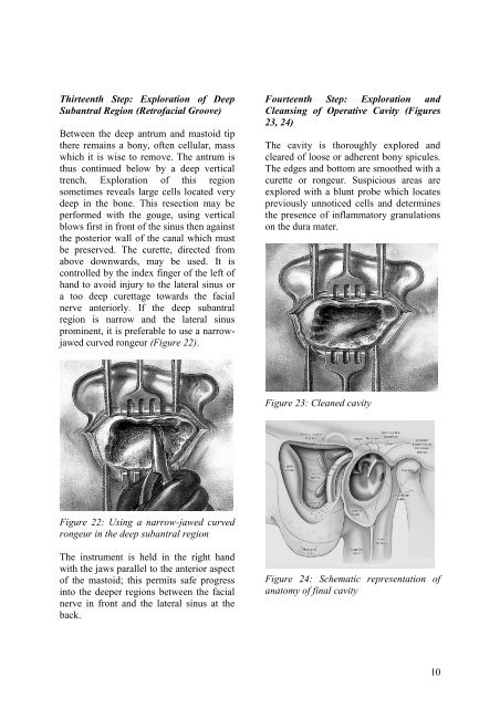
Thirteenth Step: Exploration of DeepSubantral Region (Retrofacial Groove)Between the deep antrum <strong>and</strong> mastoid tipthere remains a bony, often cellular, masswhich it is wise to remove. The antrum isthus continued below by a deep verticaltrench. Exploration of this regionsometimes reveals large cells located verydeep in the bone. This resection may beper<strong>for</strong>med with the <strong>gouge</strong>, using verticalblows first in front of the sinus then againstthe posterior wall of the canal which mustbe preserved. The curette, directed fromabove downwards, may be used. It iscontrolled by the index finger of the left ofh<strong>and</strong> to avoid injury to the lateral sinus ora too deep curettage towards the facialnerve anteriorly. If the deep subantralregion is narrow <strong>and</strong> the lateral sinusprominent, it is preferable to use a narrowjawedcurved rongeur (Figure 22).Fourteenth Step: Exploration <strong>and</strong>Cleansing of Operative Cavity (Figures23, 24)The cavity is thoroughly explored <strong>and</strong>cleared of loose or adherent bony spicules.The edges <strong>and</strong> bottom are smoothed with acurette or rongeur. Suspicious areas areexplored with a blunt probe which locatespreviously unnoticed cells <strong>and</strong> determinesthe presence of inflammatory granulationson the dura mater.Figure 23: Cleaned cavityFigure 22: Using a narrow-jawed curvedrongeur in the deep subantral regionThe instrument is held in the right h<strong>and</strong>with the jaws parallel to the anterior aspectof the mastoid; this permits safe progressinto the deeper regions between the facialnerve in front <strong>and</strong> the lateral sinus at theback.Figure 24: Schematic representation ofanatomy of final cavity10


