

Hammer and gouge mastoidectomy for acute mastoiditis - Vula ...
Hammer and gouge mastoidectomy for acute mastoiditis - Vula ...
Hammer and gouge mastoidectomy for acute mastoiditis - Vula ...

You also want an ePaper? Increase the reach of your titles
YUMPU automatically turns print PDFs into web optimized ePapers that Google loves.
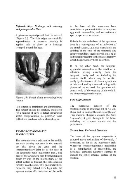
Fifteeeth Step: Drainage <strong>and</strong> suturing<strong>and</strong> postoperative CareA glove/corrugated/pencil drain is inserted(Figure 25). The skin edges are carefullyapproximated. A pressure dressing isapplied held in place by a b<strong>and</strong>agewrapped around the head.Figure 25: Pencil drain protruding fromwoundPost-operative antibiotics are administered.The patient should be carefully monitored<strong>for</strong> a number of days to detect intracranialseptic complications, as posterior fossacollections can have subtle clinical signs.TEMPOROZYGOMATICMASTOIDITISThe pneumatic cells adjacent to the middleear may develop not only in the mastoidbut also above the canal <strong>and</strong> thetemporomaxillary joint i.e. at the base ofthe squamous bone (zygomatic arch). Thebase of the squamosa may be pneumatizedeither by way of the intermediary of theantral system or through the cells openingdirectly into the attic. This pneumatization,however, may extend very high into thesquama temporalis. Infection of the cellsin the base of the squamous boneconstitutes a para<strong>mastoiditis</strong> or temporozygomatic<strong>mastoiditis</strong>, <strong>and</strong> necessitates aspecial operative technique.If the infection in the base of the squamousbone is a consequence of an infection ofthe antral system, i.e. a true <strong>mastoiditis</strong>, theopening of the cells of the tympanic <strong>and</strong>temporomaxillary segments will only be anadditional procedure to the <strong>mastoidectomy</strong>,which has previously been described.If, on the other h<strong>and</strong>, the temporozygomatic<strong>mastoiditis</strong> is the result of aninfection arising directly from thetympanic cavity <strong>and</strong> not including themastoid itself, which may be verifiedeasily by the absence of clinical symptomsat this level <strong>and</strong> by a normal radiographicpicture of the mastoid, the operation willconsist only of the opening of the cells inthe temporozygomatic region.First Step: IncisionThe cutaneous incision of the<strong>mastoidectomy</strong> is extended 3.0 or 4.0 cm.above <strong>and</strong> in front of the auricle of the ear.This incision obliquely crosses the lineatemporalis; it goes through to the bone,including the temporal muscle <strong>and</strong> theperiosteum.Second Step: Periosteal ElevationThe bone of the squama temporalis isexposed above <strong>and</strong> in front of the canal, ifnecessary, as far as the zygomatic arch.Whenever temporozygomatic <strong>mastoiditis</strong>is a complication of <strong>mastoiditis</strong>, theperiosteal elevation naturally extends toinclude the entire external surface of themastoid.11


