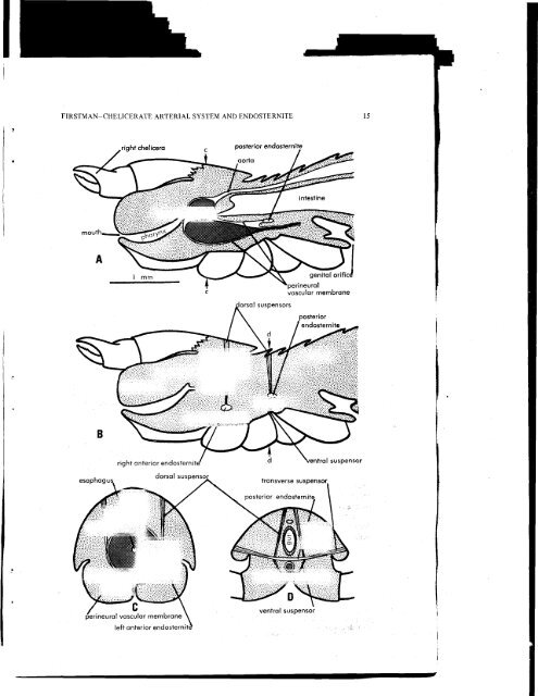
14 <strong>THE</strong> JOURNAL <strong>OF</strong> ARACHNOLOGYchambered pulsating vessel with a pair of ostia and an aorta running forward to a periganglionicsinus."The endosternite of A. persicus is divided into anterior and posterior parts: the anteriorendosternite consists of a pair of thickened portions of the perineural vascular membraneon the right and left sides of the brain. This condition resembles that of thecyphophthalmid endosternite (described above). Each side receives the insertions of twopairs of dorsoventral muscles (Fig. 9B and D). Extending posteriad from the anteriorendosternite, on each side of the central nervous System, is a skeletal ridge of thickenedperineural vascular membrane which receives the origins of many appendicular muscles,mainly coxal rotators. Morphologically, this ridge must be regarded äs part of the anteriorendosternite; it is labeled in Fig. 9B. The posterior endosternite is formed principallyby the tendonified medial portion of a single transverse muscle (Fig. 9A, B, C). It iscontinuous with a posterior extension of the perineural vascular membrane. Immediatelyposterior to the transverse band of the posterior endosternite, a single pair of dorsoventralmuscles is bisected by the perineural vascular membrane, which forms a horizontal membranousseptum for a short distance behind the transverse band (Fig. 9B). Hence, theendosternite of this tick (anterior plus posterior portions) involves three pairs of dorsoventralmuscles and one pair of transverse muscles. In the past, certain authors (e.g.,Pagenstecher, 1862) have confused the true dorsoventral muscles with coxal elevators.The latter are powerful muscles, originating on the carapace, which extend ventradto insert on coxal apodemes, whereas the true dorsoventral muscles originale on thecarapace and sternum and insert on the endosternite.I have studied wholemount slides (prepared by I. M. Newell) of Caloglyphus sp., anastigmatic mite (suborder Sarcoptiformes), which show an endosternite that is moreextensively developed than in ticks. It is continuous with a perineural vascular membranewhich receives a dorsal vessel, though a functional heart is said not to be present in theSarcoptiformes. The endosternite of this mite is more similar to that of a harvestmanthan is the tick endosternite.IIOrder Pseudoscorpionida—Morphological treatments of the pseudoscorpion date backäs far äs the Vermischte Schriften, by Treviranus (1816), who examined Chelifer sp. Theearliest investigation of the internal anatomy is that of Menge (1855), who examinedvarious genera, though description of the circulatory structures was not attempted until1880, by Daday, in Chernes hahnii. A general treatment of internal morphology wasprepared in 1888, by Croneberg, who based his report upon earlier findings, and upon hisown observations of C. hahnii.Croneberg (1888), in describing the brain of C. hahnii, distinguished an "inner neurilemma"from an "outer neurilemma," and I infer that the latter is a vestige of theperineural vascular membrane. In his Fig. 17, he showed that the "outer neurilemma" isFig. lO.-Generalized diagram of the endosternite and perineural vascular membrane in pseudoscorpions,based upon my observations of Microcreagris sp. and Garypus californicus, and upon thedescriptions of Vachon (1949) in Chelifer cancroides, and of Croneberg (1888) in Chernes hahnii.A: midsagittal view of the cephalothoracic region, seen from the left; B: lateral view of the endosternites,showing the dorsal and ventral suspensor muscles, äs seen from the left; C: transverse sectionthrough the anterior endosternite, seen from the anterior; the plane of the section is indicated in Fig.A by the arrows, cc. D: transverse section through the posterior endosternite, seen from the anterior;the plane of the section is indicated in Fig. B by the arrows, dd. The central nervous System is shownwith dark shading.
FIRSTMAN-CHEEICERATE ARTERIAE <strong>SYSTEM</strong> AND ENDOSTERNITEperineuralvascular membranetransverse suspensorposterior endosternitperineural vascular membraneleft anterior endosterni