32 <strong>THE</strong> JOURNAL <strong>OF</strong> ARACHNOLOGYmental in the formation of the cephalothoracic endosternite. In accordance to the comparativeevidence put forth in this paper, I believe the transverse suspensors of thecephalothoracic endosternite are derivatives of serial transverse muscles which lay primitivelyover the nervous System. In this connection, Snodgrass (1935, 1952) pointed outthat in insects there are serial transverse muscles which lie over the nervous System (Fig.28).Class PycnogonidaThe Pycnogonid Arterial System-Early observations of circulation in pycnogonidswere made Johnston (1837), Henri Milne-Edwards (1840), Quatrefages (1845), and VanBeneden (1846). None of these authors described a heart, although Van Beneden sawsome of the movements of blood beneath the dorsal integument of a living specimen ofNymphon. Cole (1910) similarly described circulatory movements which he observed inliving specimens of Endeis. A heart was described in Nymphon by Zenker (1852), inEndeis by Krohn (1885), and in Colossendeis and certain other genera by Hoek(1881). A detailed description of the pycnogonid circulatory apparatus, based principallyupon Endeis and Nymphon, was given by Dohrn in 1881. He agreed with Hoek indescribing the heart äs a tube which attaches dorsally to the integument and ventrally tothe gut. It was Dohrn who first pointed out that the pycnogonid hemocoel is dividedlongitudinally by a double-walled, horizontal, vascular septum which separates dorsal andventral blood cavities; blood in the dorsal cavity is directed anteriad, whereas blood in theventral cavity flows posteriad. The ventral surface of the heart is continuous with thisseptum along its midline; rhythmic undulations of the septum coincide with the cardiacsystole and diastole, and these undulatory movements create the pressures which aspirateblood in and out of the paired appendages. The concept of Dohrn's horizontal vascularseptum has been reviewed and diagrammed by Cole (1910).My own dissections of pycnogonids include the following species: Pycnogonum littorale,P. rhinoceros, Endeis sp., Colossendeis scotti, Decolopoda australis, Nymphoncharcoti, Pentanymphon antarcticum, undAmmothea striata. These dissections show thepresence of a perivisceral arterial membrane, continuous with the aorta, which envelopsthe intestine and the central nervous System (Figs. 24, 25). As in Limulus and thearachnids, this membrane encloses a perivisceral arterial blood sinus. The membrane iscontinuous with the double-walled horizontal septum (described above) that extendslaterad to the body wall, separating the venous hemocoel into dorsal and ventralcavities. The horizontal septum is partially muscularized by means of transverse musclefiber bands that originale on the exoskeleton; it extends horizontally through the coxaeinto the walking legs (the legs protrude laterally from the trunk), separating their luminainto dorsal and ventral venous channels.The horizontal vascular septum (of Dohrn) is present in all the pycnogonids I haveexamined, although the exact vertical position of its horizontal plane, with respect to thegut, differs from family to family. Whereas in Colossendeis it is situated immediatelybeneath the heart, in Endeis it extends laterad from the sides of the gut, and in Pycnogonumit extends from the base of the gut; in all casses it is a continuation of theperivisceral arterial membrane (Fig. 27A, B, C). Between the two layers of the horizontalseptum there lies a thin arterial blood sinus which is continuous with the rest of theperivisceral blood sinus.Loman (1917) studied the blood circulation in Nymphon. He described the aorta äsbifurcating to go around the optic nerve ("läuft ringförmig um den Augennerv") and
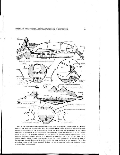
FIRSTMAN-<strong>CHELICERATE</strong> <strong>ARTERIAL</strong> <strong>SYSTEM</strong> AND ENDOSTERNITE 33optictuberclyestiges of fhe neurointestina! omentumrineural vascular membranerineural vascular membrane'vestigial hearthorizontal vascular septum (of Dohrn)perineural vascular membrane'PycnogonumFig. 24.—A: midsagittal view of Colossendeis scoiti (class Pycnogonida), seen from the left. The fülllength of the proboscis is not shown. The oval Windows above the nerve cord are perforations in theneurointestinal omentum; the ovate Windows below the nerve cord are perforations in the ventralmesentery. B: transverse section through the plane indicated by the arrows in Fig. A. C: an enlargeddetail of Fig. A. The structure indicated by "see legend" is the uppermost membrane (the top) ofDohrn's horizontal vascular septum; it is thickened at this point and it receives the insertions ofcardiac muscles which extend dorsoventrally frorn the dorsal integument. D: midsagittal view ofPycnogonum littorale. E: transverse section through the plane indicated by the arrows in Fig. D. Thecentral nervous System is shown with dark shading. The venous hemocoel is Stippled; the heart, arterialSystem and gut are unshaded.