16 <strong>THE</strong> JOURNAL <strong>OF</strong> ARACHNOLOGYcontinuous with a dorsal vessel which I take to be the anterior aorta. Although Cronebergmade no comment about this relationship, he did say that the aorta extends forwarduntil it reaches the posterior face of the supraesophageal ganglionic mass.The pseudoscorpion endosternite has been described by Vachon (1949), based mainlyupon Chelifer cancroides. The endosternite of this arachnid, like that of Argas persicus(the fowl tick), is divided completely into separate anterior and posterior portions. Theanterior endosternite is paired, lying on the right and left sides of the supraesophagealganglionic mass (Fig. l OB, C). A single pair of dorsal suspensor muscles inserts into theanterior endosternite, and several pairs of appendicular and pharyngeal muscles originalefrom it. The posterior endosternite has been described by Vachon äs "... a simple transverse,tendinous band." He illustrated it in both lateral and dorsal views, and Croneberg(1888) illustrated it in transverse view. A single pair of dorsoventral suspensor musclesinserts onto the posterior endosternite, and at least three pairs of appendicular musclesoriginale from it. According to Vachon (1949), the anterior and posterior endosternitesare derived each from three segments: he said that the anterior endosternite is derivedfrom the pedipalpal and the first and second walking-leg segments, whereas the posteriorendosternite is derived from the third and fourth walking-leg and the first abdominalsegments.I have examined the arterial System and endosternite of Microcreagris sp. and ofGarypus californicus. In both of these genera, I found a perineural vascular membrane(Fig. 10A, C) which is similar to that already described for other apulmonate arachnids.It is somewhat fragmentary, however, and it exists apparently äs a vestige whichmay no longer have a vascular function. It is most plainly developed in those regionswhere it is continuous with the endosternites. Weygoldt (l 969) in his midsagittal view ofthe anterior end of an embryonic Neobisium sp. (his Fig. 92) illustrated a membranewhich is continuous with the posterior endosternite, and I believe this is the same membrane(the perineural vascular membrane) which I have observed in Microcreagris sp. andG. californicus.The endosternites of these two pseudoscorpions correspond exactly to the earlierdescriptions of Croneberg (1888) and Vachon (1949). The anterior endosternite, lyingon each side of the brain, is continuous with the perineural vascular membrane; I interpretit äs the morphological equivalent of the lateral horns (the anterior cornua) of themore completely developed endosternites of other arachnids. The posterior endosterniteresembles the posterior portion of the tick endosternite because, morphologically, it isthe tendinous, medial axis of a transverse muscle which originales, on both sides, fromthe carapace (Fig. l OA, D).The pseudoscorpion endosternite, despite its morphological similarity to the argasidendosternite, is more reduced (i.e., more vestigial) than the latter: the anterior endosterniteof the pseudoscorpion receives the insertion of only a single pair of dorsoventralmuscles, whereas that of the argasid tick has two such insertions on each side. Moreover,the endosternal ridge, of the anterior endosternite of the tick, is not developed in thepseudoscorpion. The apodemal endoskeleton is more highly developed in pseudoscorpionsthan it is in argasic ticks. I believe this Supports my hypothesis that there is ageneral correlation in all apulmonate arachnids between the extent of apodemal developmentand the degree of reduction of the mesodermal endosternite.Order Ricinuleida-The first morphological treatment of the Ricinuleida was that ofHansen and Sorensen (1904), who dealt primarily with the external anatomy of various
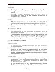
FIRSTMAN-<strong>CHELICERATE</strong> <strong>ARTERIAL</strong> <strong>SYSTEM</strong> AND ENDOSTERNITE 17rightcheliceraesophagusaortacuculusperineural vascular membrane*abdominal nerve trunk0.5 mmerineural vascular membranestomodeal muscle^estigial dorsal suspensor musclemidgutperineuralvascular membranvestigial endosternal area of theperineural vascular membraneFig. 11.-A: midsagittal view of the cephalothoracic region of Cryptocellus boneti (orderRidnuleida), seen from the left; B: lateral view of same, showing the vestigial endosternal area of theperineural vascular membrane; the central nervous System is shown with dark shading.