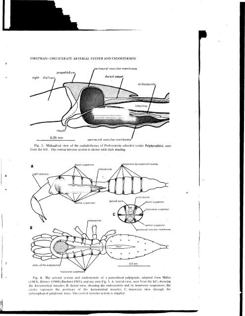
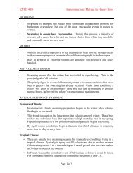
6 <strong>THE</strong> JOURNAL <strong>OF</strong> ARACHNOLOGYside. A few specimens were cut transversely and dissected from the anterior or posteriorsurface. Illumination was reflected from two sides, and in order to enchance detail, thetissues were stained, äs required, with Shaeffer's washable blue Skrip ink., OBSERVATIONS AND FINDINGS>The Apulmonate Arachnid Orders:PalpigradidaOpilionidaAcaridaPseudoscorpionidaRicinuleidaSolpugidaOrder Palpigradida— Most arachnologists regard palpigrades äs the most generalized ofthe living arachnids, i.e., with the greatest number of primitive features, and with fewestspecializations (Roewer, 1934). The carapace is metamerized externally, and there arefive cephalothoracic sternites, the anteriormost of which belongs to the cheliceral segment.This is the only living order in which there is a distinct cheliceral sternite (Snodgrass,1952). Sternarthron zitteli, stated to be a fossil palpigrade of Upper Jurassic age,possesses six cephalothoracic sternites (Petrunkevitch, 1955). Palpigrades bear close resembalanceto the superorder Uropygida (schizomids and thelyphonids), and most arachnologistsagree that modern palpigrades have emerged from the ancestral stock that gaverise to the non-scorpion pulmonate Orders. However, palpigrades are not pulmonates, forthey do not possess book lungs. Some palpigrades possess three pairs of abdominal"lung-sacs" which some investigators have interpreted äs respiratory organs. Rucker(1901) believed that lung-sacs were the phyletic antecedants of both book lungs 'andtracheal spiracles.The earliest published description of the internal anatomy of a palpigrade is that ofRucker (1901), who said of the circulatory System of Prokoenenia wheeleri only that". . . the simplest condition possible exists." She said that a heart is lacking, althoughBörner (1904) described a heart with four pairs of ostia in Eukoenenia mirabilis. Theendosternite of E. mirabilis has been described by Börner (1904), but the most detailedstudies have been those of Millot (1942b, 1943, 1949b). Millot described six pairs ofventral suspensors of the endosternite; this number is regarded äs primitive, since presumablythere was one pair of dorsoventral muscles in each of the six appendage-bearingsegments of the cephalothorax of ancestral arachnids. Only four pairs of suspensorspersist on the dorsal side of the endosternite. In addition to the dorsal and ventralsuspensors, Millot described five pairs of transverse suspensors (he called them "lateralsuspensors") which originate from the sides of the carapace and extend horizontally to• their insertions on the lateral margins of the endosternite (Fig. 4B and C).I have examined Prokoenenia wheeleri, of central Texas, and I have found that thecentral nervous System is invested by a perineural vascular membrane which encloses aperiganglionic arterial sinus (Fig. 3), äs in the other apulmonate chelicerates. This membraneis continuous with the borders of the endosternite. The same membrane is continuousalso with a dorsal vessal, probably an aorta, in the cephalorthorax. I did not tracethis vessel into the abdomen to confirm the presence of a heart, but my diagram of ageneralized palpigrade (Fig. 4) shows a heart because Börner (1904) described one in E.
FIRSTMAN-<strong>CHELICERATE</strong> <strong>ARTERIAL</strong> <strong>SYSTEM</strong> AND ENDOSTERNITErightchelicerapropeltidiumperineural vascular membranedorsal vesselperineural vascular membraneFig. 3.—Midsagittal view of the caphalothorax of Prokoenenia wheeleri (order Palpigradida), seenfrom the left. The central nervous System is shown with dark shading.dorsal suspensorsibdominal dorsoventral musclescornu of the endosternitetransverse suspensorsFig. 4. The arterial system and endosternite of a generalized palpigrade, adapted from Millot(1943), Börner (1904),Rucker(1901), and my own Fig. 3. A: lateral view, seen from the left, showingthe dorsoventral muscles; B: dorsal view, showing the endosternite and its transverse suspensors; thecircles represent the positions of the dorsoventral muscles; C: transverse view through thesubesophageal ganglionic mass. The eentral nervous system is Stippled.