18 <strong>THE</strong> JOURNAL <strong>OF</strong> ARACHNOLOGYi'species ofRicinoides. The internal anatomy was not studied in detail until Millot (1945a,b, c) dissected R. feae. He described a reduced heart and an anterior aorta which becomeslost at the posterior surface of the supraesophageal ganglionic mass. He noted athin "fibro-muscular sheet" associated with the brain and commented that this mayrepresent the vestige of an endosternite.I have examined two species of Cryptocellus (Fig. 11): C. boneti, from Morelos,Mexico, and C. osorioi, from caves in San Luis Potosf, Mexico. I find in both of thesespecies of Cryptcellus the typical apulmonate condition of the arterial system: the centralnervous System is invested with a perineural vascular membrane which is continuous withthe aorta. Associated with this membrane, there is a certain region which I call theendosternal area (Fig. 11 B). For the following two reasons, I Interpret this area äs avestigial endosernite: (l)arising from the endosternal area is a pair of Strands of connectivetissue; these Strands are probably non-contractile, but they attach to the carapaceand appear to be vestiges of dorsal suspensor muscles; (2) a pair of stomedeal musclesoriginales from the anterior portion of the endosternal area.The apodemal endoskeleton in ricinuleids is more strongly developed than in pseudoscorpions,but less so than in solpugids; the development of the mesodermal endosterniteseems to be inversely proportional to the development of the apodemal endoskeleton, äsis also the case in the other apulmonate arachnids.;'•!'Order Solpugida-In 1896, Bernard observed in Galeodes that ". . . the anterior end ofthe heart is produced into an aorta, which . . . appears to discharge the blood direct onthe central nerve-mass." My own dissections of Eremobates sp. (Fig. 12) confirm thisreport; the central nervous System is enveloped by a perineural vascular membrane whichencloses a periganglionic arterial sinus. The order Solpugida is unique in that all itsmembers lack a mesodermal endosternite (Bernard, 1896; Giltay, 1925; Millot, 1949a). Inits place, solpugids possess a highly developed apodemal endoskeleton. An apodemal archarises from the floor of the tritosternal segment (Bernard, 1896; Millot and Vachon,1949b). This arch passes over the dorsal surface of the subesophageal ganglionic mass,where it serves äs a functional analogue of the mesodermal endosternite.The solpugid endoskeleton was studied first by Kittary (1848) in Galeodes, and laterby Dufour (1862) in Galeodes, Bernard (1896) in Galeodes, Sorensen (1914) in Daesia,Solpuga, Galeodes, Rhagodes, and especially by Roewer (1934) in various genera. Unfortunately,Bernard's erroneous interpretation is this structure was adopted äs valid bycertain influential arachnologists, such äs Comstock (1948). Bernard attempted to homologjzeall arachnid endosternites with the apodemal endoskeleton of the solpugid,which he regarded äs a primitive arachnid. Apparently, he was motivated by a determinationto demonstrate unequivocally that arachnids cannot at all be closely related toLimulus, which he regarded äs a crustacean (Bernard, 1892a, b). Bernard's interpretationof the arachnid endosternite is not in agreement with that of Pocock (1902), nor orMillot (1949a), nor of my own. Millot said that the interpretation of the solpugid tritosternalapodeme (incontestably an ectodermal structure) äs a homologue of the scorpionendosternite, is an indefensible conception. Moreover, he pointed out that embryologistsuniversally recognize the mesodermal origin of the endosternite (p. 287):L'apodeme tritosternal des Solifuges a ete parfois homologue ä l'endosternite des Scorpions et,pai son intermediaire, ä celui des autres Arachnides. Cette conception ne paraft pas defendable.L'apodeme tritosternal, incontestablement ectodermique, ne peut etre comaree ä l'endosternitedont l'origine mesodermique est reconnue-par tous les embryologistes.
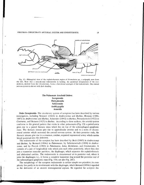
FIRSTMAN-<strong>CHELICERATE</strong> <strong>ARTERIAL</strong> <strong>SYSTEM</strong> AND ENDOSTERNITE 19^apodemal endoskeletondorsal vesse!pharynx'perineural vascular membrane'l mm coxa of leg 2abdominal nerve trunkFig. 12,-Midsagittal view of the cephalothoracic tegion of Eremobates sp., a solpugid, seen fromthe left. Note that a mesodermal endosternite is lacking. An apodemal invagination of the exoskeleton,derived from the tritosternum, forms a functional analogue of the endosternite. The centralnervous System is shown with dark shading.The Pulmonate Arachnid Orders:ScorpionidaThelyphonidaSchizomidaAmblypygidaAraneidaOrder Scorpionida—The circulatory System of scorpions has been described by variousinvestigators, including Newport (1843) in Androctonus and Buthus, Houssay (1886,1887) in Androctonus and Buthus, Schneider (1892) inButhus, Petrunkevitch (1922) inCentrurus, and Buisson (1925) in Buthus. According to these authors, the arterial Systemconforms to the general pattern that exists in other pulmonates (Fig. 13): a paired aortagives rise to a paired thoracic sinus which lies on top of the subesophageal ganglionicmass. The thoracic sinuses give rise to appendicular arteries and to a series of circumneuralarteries which surround the central nervous System. At their posterior ends, thethoracic sinuses give rise to a common, median, unpaired supraneural artery which carriesblood posteriad into the abdomen.The endosternite of the scorpion has been described by Beck (1885) in Androctonusand Buthus, by Bernard (1894c) in Palamnaeus, by Schimkewitsch (1894) in Androctonus,and by Pocock (1902) in Palamnaeus, lurus, Bothriurus, and Centruroides. 1tconsists of a pair of longitudinal rods which join each other posteriorly, where they alsojoin a transverse muscular partition, the diaphragm, which separates the cephalothoracicand abdominal cavities. The endosternite is circumneural at its posterior end, where itjoins the diaphragm; i.e., it forms a complete transverse ring around the posterior end ofthe subesophageal ganglionic mass (Fig. 14A; see also Fig. 26C).The morphology of the scorpion endosternite is neither simple nor primitive; its complexitylies partly in its involvement with the diaphragm, which Bernard (1894c) regardedäs the derivative of an ancient intersegmental septum. He regarded the scorpion dia-