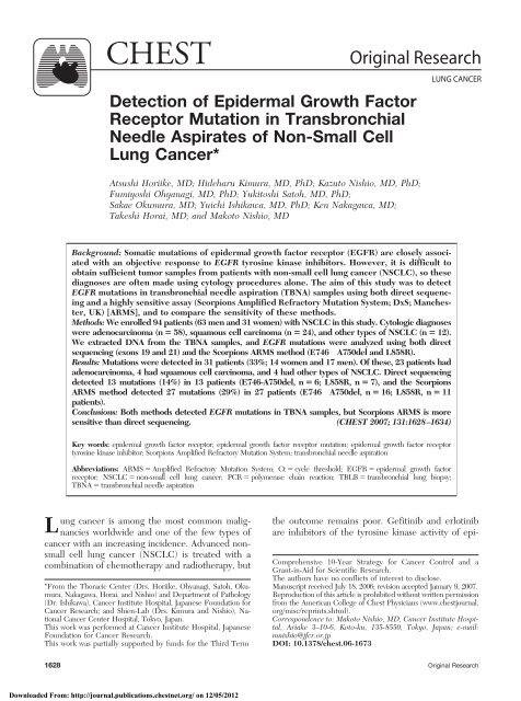
Detection of Epidermal Growth Factor Receptor Mutation in ...
Detection of Epidermal Growth Factor Receptor Mutation in ...
Detection of Epidermal Growth Factor Receptor Mutation in ...

You also want an ePaper? Increase the reach of your titles
YUMPU automatically turns print PDFs into web optimized ePapers that Google loves.
CHEST Orig<strong>in</strong>al Research<br />
<strong>Detection</strong> <strong>of</strong> <strong>Epidermal</strong> <strong>Growth</strong> <strong>Factor</strong><br />
<strong>Receptor</strong> <strong>Mutation</strong> <strong>in</strong> Transbronchial<br />
Needle Aspirates <strong>of</strong> Non-Small Cell<br />
Lung Cancer*<br />
Atsushi Horiike, MD; Hideharu Kimura, MD, PhD; Kazuto Nishio, MD, PhD;<br />
Fumiyoshi Ohyanagi, MD, PhD; Yukitoshi Satoh, MD, PhD;<br />
Sakae Okumura, MD; Yuichi Ishikawa, MD, PhD; Ken Nakagawa, MD;<br />
Takeshi Horai, MD; and Makoto Nishio, MD<br />
Background: Somatic mutations <strong>of</strong> epidermal growth factor receptor (EGFR) are closely associated<br />
with an objective response to EGFR tyros<strong>in</strong>e k<strong>in</strong>ase <strong>in</strong>hibitors. However, it is difficult to<br />
obta<strong>in</strong> sufficient tumor samples from patients with non-small cell lung cancer (NSCLC), so these<br />
diagnoses are <strong>of</strong>ten made us<strong>in</strong>g cytology procedures alone. The aim <strong>of</strong> this study was to detect<br />
EGFR mutations <strong>in</strong> transbronchial needle aspiration (TBNA) samples us<strong>in</strong>g both direct sequenc<strong>in</strong>g<br />
and a highly sensitive assay (Scorpions Amplified Refractory <strong>Mutation</strong> System; DxS; Manchester,<br />
UK) [ARMS], and to compare the sensitivity <strong>of</strong> these methods.<br />
Methods: We enrolled 94 patients (63 men and 31 women) with NSCLC <strong>in</strong> this study. Cytologic diagnoses<br />
were adenocarc<strong>in</strong>oma (n � 58), squamous cell carc<strong>in</strong>oma (n � 24), and other types <strong>of</strong> NSCLC (n � 12).<br />
We extracted DNA from the TBNA samples, and EGFR mutations were analyzed us<strong>in</strong>g both direct<br />
sequenc<strong>in</strong>g (exons 19 and 21) and the Scorpions ARMS method (E746 A750del and L858R).<br />
Results: <strong>Mutation</strong>s were detected <strong>in</strong> 31 patients (33%; 14 women and 17 men). Of these, 23 patients had<br />
adenocarc<strong>in</strong>oma, 4 had squamous cell carc<strong>in</strong>oma, and 4 had other types <strong>of</strong> NSCLC. Direct sequenc<strong>in</strong>g<br />
detected 13 mutations (14%) <strong>in</strong> 13 patients (E746-A750del, n � 6; L858R, n � 7), and the Scorpions<br />
ARMS method detected 27 mutations (29%) <strong>in</strong> 27 patients (E746 A750del, n � 16; L858R, n � 11<br />
patients).<br />
Conclusions: Both methods detected EGFR mutations <strong>in</strong> TBNA samples, but Scorpions ARMS is more<br />
sensitive than direct sequenc<strong>in</strong>g. (CHEST 2007; 131:1628–1634)<br />
Key words: epidermal growth factor receptor; epidermal growth factor receptor mutation; epidermal growth factor receptor<br />
tyros<strong>in</strong>e k<strong>in</strong>ase <strong>in</strong>hibitor; Scorpions Amplified Refractory <strong>Mutation</strong> System; transbronchial needle aspiration<br />
Abbreviations: ARMS � Amplified Refractory <strong>Mutation</strong> System; Ct � cycle threshold; EGFR � epidermal growth factor<br />
receptor; NSCLC � non-small cell lung cancer; PCR � polymerase cha<strong>in</strong> reaction; TBLB � transbronchial lung biopsy;<br />
TBNA � transbronchial needle aspiration<br />
Lung cancer is among the most common malignancies<br />
worldwide and one <strong>of</strong> the few types <strong>of</strong><br />
cancer with an <strong>in</strong>creas<strong>in</strong>g <strong>in</strong>cidence. Advanced nonsmall<br />
cell lung cancer (NSCLC) is treated with a<br />
comb<strong>in</strong>ation <strong>of</strong> chemotherapy and radiotherapy, but<br />
*From the Thoracic Center (Drs. Horiike, Ohyanagi, Satoh, Okumura,<br />
Nakagawa, Horai, and Nishio) and Department <strong>of</strong> Pathology<br />
(Dr. Ishikawa), Cancer Institute Hospital, Japanese Foundation for<br />
Cancer Research; and Shien-Lab (Drs. Kimura and Nishio), National<br />
Cancer Center Hospital, Tokyo, Japan.<br />
This work was performed at Cancer Institute Hospital, Japanese<br />
Foundation for Cancer Research.<br />
This work was partially supported by funds for the Third Term<br />
LUNG CANCER<br />
the outcome rema<strong>in</strong>s poor. Gefit<strong>in</strong>ib and erlot<strong>in</strong>ib<br />
are <strong>in</strong>hibitors <strong>of</strong> the tyros<strong>in</strong>e k<strong>in</strong>ase activity <strong>of</strong> epi-<br />
Comprehensive 10-Year Strategy for Cancer Control and a<br />
Grant-<strong>in</strong>-Aid for Scientific Research.<br />
The authors have no conflicts <strong>of</strong> <strong>in</strong>terest to disclose.<br />
Manuscript received July 18, 2006; revision accepted January 9, 2007.<br />
Reproduction <strong>of</strong> this article is prohibited without written permission<br />
from the American College <strong>of</strong> Chest Physicians (www.chestjournal.<br />
org/misc/repr<strong>in</strong>ts.shtml).<br />
Correspondence to: Makoto Nishio, MD, Cancer Institute Hospital,<br />
Ariake 3–10-6, Koto-ku, 135-8550, Tokyo, Japan; e-mail:<br />
mnishio@jfcr.or.jp<br />
DOI: 10.1378/chest.06-1673<br />
1628 Orig<strong>in</strong>al Research<br />
Downloaded From: http://journal.publications.chestnet.org/ on 12/05/2012
dermal growth factor receptor (EGFR), and have<br />
recently been used to treat advanced NSCLC. 1<br />
These agents are dramatically effective <strong>in</strong> some<br />
For editorial comment see page 1619<br />
patients yet completely <strong>in</strong>effective <strong>in</strong> others. The<br />
response rate to gefit<strong>in</strong>ib is high among <strong>in</strong>dividuals<br />
with an Asian background. 2<br />
In May and June <strong>of</strong> 2004, two <strong>in</strong>dependent groups<br />
reported an association between somatic EGFR mutations<br />
and a dramatic cl<strong>in</strong>ical response to gefit<strong>in</strong>ib,<br />
respectively. 3,4 Thereafter, EGFR mutations were<br />
extensively <strong>in</strong>vestigated. 5–17 The mutations consist <strong>of</strong><br />
small, <strong>in</strong>-frame deletions or substitutions clustered<br />
around the adenos<strong>in</strong>e triphosphate-b<strong>in</strong>d<strong>in</strong>g site <strong>in</strong><br />
exons 18, 19, and 21 <strong>of</strong> the EGFR gene, and<br />
approximately 90% <strong>of</strong> patients with EGFR mutations<br />
have one <strong>of</strong> two major mutations. One is a 15-base<br />
pair nucleotide <strong>in</strong>-frame deletion (E746 A750del)<br />
<strong>in</strong> exon 19, and the other is a po<strong>in</strong>t mutation<br />
<strong>in</strong>volv<strong>in</strong>g the replacement <strong>of</strong> leuc<strong>in</strong>e with arg<strong>in</strong><strong>in</strong>e at<br />
codon 858 (L858R) <strong>in</strong> exon 21. 18 The above studies<br />
<strong>in</strong>cluded genetic analyses <strong>of</strong> surgical tissues or biopsy<br />
specimens. However, to obta<strong>in</strong> sufficient amounts <strong>of</strong><br />
tumor samples from <strong>in</strong>operable NSCLC patients is<br />
<strong>of</strong>ten difficult. Some studies 19,20 <strong>of</strong> patients with<br />
advanced NSCLC have found a correlation between<br />
cl<strong>in</strong>ical manifestations and EGFR mutation status<br />
obta<strong>in</strong>ed from small tumor samples, such as those<br />
obta<strong>in</strong>ed us<strong>in</strong>g standard transbronchial lung biopsy<br />
(TBLB). All <strong>of</strong> the above studies are limited by the<br />
fact that the rate <strong>of</strong> usable samples obta<strong>in</strong>ed from<br />
enrolled patients is very low. Therefore, a method is<br />
required to detect mutant EGFR, especially the two<br />
major mutations, us<strong>in</strong>g samples other than surgical<br />
tissues from NSCLC patients. We addressed this<br />
problem us<strong>in</strong>g a sensitive technique for actual tumor<br />
sampl<strong>in</strong>g, and a highly sensitive assay for detect<strong>in</strong>g<br />
EGFR mutations.<br />
Pulmonary lesions are most <strong>of</strong>ten cl<strong>in</strong>ically diagnosed<br />
us<strong>in</strong>g flexible bronchoscopy. Common bronchoscopic<br />
sampl<strong>in</strong>g techniques used for pulmonary<br />
lesions are transbronchial needle aspiration (TBNA)<br />
and TBLB. One report has <strong>in</strong>dicated that TBNA is<br />
superior to TBLB <strong>in</strong> diagnos<strong>in</strong>g pulmonary lesions:<br />
Gaspar<strong>in</strong>i et al 21 found that the diagnostic sensitivity<br />
<strong>of</strong> these techniques is 50.0% for TBLB, 70.1% for<br />
TBNA, and 76.0% for TBLB and TBNA together.<br />
We thus presumed that TBNA is a highly sensitive<br />
means <strong>of</strong> tumor sampl<strong>in</strong>g, and that DNA obta<strong>in</strong>ed<br />
from such specimens might provide useful <strong>in</strong>formation<br />
about the mutation status <strong>of</strong> the EGFR gene.<br />
We postulated that Scorpions Amplified Refractory<br />
<strong>Mutation</strong> System (ARMS) [DxS; Manchester,<br />
UK] technology would enhance the sensitivity <strong>of</strong><br />
detect<strong>in</strong>g EGFR mutations. Scorpion primers are<br />
used with a fluorescence-based method that specifically<br />
detects polymerase cha<strong>in</strong> reaction (PCR) products.<br />
22 A “scorpion” consists <strong>of</strong> a specific probe<br />
sequence held <strong>in</strong> a hairp<strong>in</strong> loop configuration by<br />
complementary stem sequences on the 5� and 3�<br />
ends <strong>of</strong> the probe. A scorpion can be comb<strong>in</strong>ed with<br />
ARMS to enable the detection <strong>of</strong> s<strong>in</strong>gle-base mutations.<br />
22,23 The ARMS method is used for allele<br />
discrim<strong>in</strong>ation, and additional mismatches have been<br />
<strong>in</strong>troduced near the 3� term<strong>in</strong>i <strong>of</strong> the primers to<br />
enhance specificity. The ARMS method is superior<br />
to both direct sequenc<strong>in</strong>g and the WAVE method<br />
(Transgenomic; Omaha, NE) for detect<strong>in</strong>g EGFR<br />
mutations. 24 Here, we aimed to detect major EGFR<br />
mutations <strong>in</strong> TBNA specimens and to verify the<br />
sensitivity <strong>of</strong> these methods for detect<strong>in</strong>g EGFR<br />
mutations.<br />
Patients<br />
Materials and Methods<br />
We studied patients with NSCLC diagnosed us<strong>in</strong>g specimens<br />
obta<strong>in</strong>ed by TBLB and/or TBNA. Tumors <strong>in</strong> sal<strong>in</strong>e solution were<br />
not collected from enlarged lymph nodes only. After obta<strong>in</strong><strong>in</strong>g<br />
written <strong>in</strong>formed consent from the patients to participate <strong>in</strong> all<br />
study protocols approved by the Institutional Review Board <strong>of</strong><br />
the Cancer Institute Hospital, tumor tissues, tumors <strong>in</strong> sal<strong>in</strong>e<br />
solution obta<strong>in</strong>ed us<strong>in</strong>g TBNA, and cl<strong>in</strong>ical data were collected.<br />
We recorded age at diagnosis, gender, cytologic diagnosis <strong>of</strong><br />
NSCLC, cl<strong>in</strong>ical stage, and smok<strong>in</strong>g status. Cytologic diagnoses<br />
were based on the World Health Organization pathology classification.<br />
Cl<strong>in</strong>icopathologic stag<strong>in</strong>g was determ<strong>in</strong>ed accord<strong>in</strong>g to<br />
the International Union Aga<strong>in</strong>st Cancer TNM classification <strong>of</strong><br />
malignant tumors. Nonsmokers were def<strong>in</strong>ed as those who had<br />
smoked � 100 cigarettes <strong>in</strong> their lifetime. We obta<strong>in</strong>ed detailed<br />
<strong>in</strong>formation about smok<strong>in</strong>g history, <strong>in</strong>clud<strong>in</strong>g age at first cigarette,<br />
packs per day, and number <strong>of</strong> smok<strong>in</strong>g and smoke-free<br />
years (after quitt<strong>in</strong>g). Patients were categorized as follows: never<br />
smoked (� 100 lifetime cigarettes), former smokers (quit � 1<br />
year ago), or current smokers (quit � 1 year ago).<br />
TBNA Sampl<strong>in</strong>g<br />
Four experienced operators performed standard flexible bronchoscopy<br />
(Olympus P260F; Olympus; Tokyo, Japan) us<strong>in</strong>g 21gauge<br />
cytology needles and aspirated for 10 s <strong>in</strong> the standard<br />
fashion. 25 Paired samples consisted <strong>of</strong> two aspirates that were<br />
obta<strong>in</strong>ed <strong>in</strong> immediate succession <strong>in</strong> an identical manner, with<br />
the needle <strong>in</strong>sertion po<strong>in</strong>ts ideally 1 mm apart. At least four<br />
aspirates (two pairs) were obta<strong>in</strong>ed from each site. For cytologic<br />
analysis, the aspirate was immediately placed onto a glass slide,<br />
covered with a second slide, and the slides were drawn apart<br />
under cont<strong>in</strong>uous gentle pressure. The smear was spray-fixed<br />
us<strong>in</strong>g ethanol, processed rout<strong>in</strong>ely and visualized by Papanicolaou<br />
sta<strong>in</strong><strong>in</strong>g. The second aspirate was mixed <strong>in</strong>to 2 mL <strong>of</strong> sal<strong>in</strong>e<br />
solution and stored at – 80°C until DNA extraction.<br />
DNA Extraction<br />
Samples obta<strong>in</strong>ed by TBNA <strong>in</strong> sal<strong>in</strong>e solution were digested<br />
with prote<strong>in</strong>ase K, and then DNA was extracted with phenol-<br />
www.chestjournal.org CHEST / 131 /6/JUNE, 2007 1629<br />
Downloaded From: http://journal.publications.chestnet.org/ on 12/05/2012
chlor<strong>of</strong>orm and precipitated with ethanol. Precipitated DNA was<br />
eluted <strong>in</strong> 50 �L <strong>of</strong> sterile, double-distilled water. The concentration<br />
and purity <strong>of</strong> the extracted DNA were determ<strong>in</strong>ed by<br />
spectrophotometry and then the DNA was stored at – 20°C.<br />
PCR Amplification and Direct Sequenc<strong>in</strong>g<br />
Genomic PCR was performed <strong>in</strong> 25-�L volumes us<strong>in</strong>g 50 ng <strong>of</strong><br />
template DNA, 0.75 U <strong>of</strong> AmpliTaq Gold DNA polymerase<br />
(Perk<strong>in</strong>-Elmer; Roche Molecular Systems; Branchburg, NJ), 2.5<br />
�L <strong>of</strong> PCR buffer (Perk<strong>in</strong>-Elmer), 0.8 �mol/L deoxynucleotide<br />
triphosphate (Perk<strong>in</strong>-Elmer), 0.5 �mol/L <strong>of</strong> each primer, and<br />
various concentrations <strong>of</strong> MgCl 2, depend<strong>in</strong>g on the polymorphic<br />
marker. Exons 19 and 21 were amplified by nested PCR. Primer<br />
sequences were obta<strong>in</strong>ed as described by Lynch et al. 3 Initial<br />
PCR analyses proceeded <strong>in</strong> a volume <strong>of</strong> 25 �L as follows: 35<br />
cycles <strong>of</strong> denaturation at 94°C for 45 s, primer anneal<strong>in</strong>g at 58°C<br />
for 30 s, and elongation at 72°C for 30 s. A f<strong>in</strong>al extension<br />
proceeded at 72°C for 10 m<strong>in</strong>. Nested PCR was performed us<strong>in</strong>g<br />
20 cycles under the same conditions as the <strong>in</strong>itial PCR. The bands<br />
<strong>of</strong> PCR products were visualized us<strong>in</strong>g a 2100 bioanalyzer and the<br />
DNA 500 Labchip kit (Agilent Technologies; Palo Alto, CA).<br />
Each sample was sequenced <strong>in</strong> duplicate <strong>in</strong> both forward and<br />
reverse directions us<strong>in</strong>g the BigDye Term<strong>in</strong>ator kit (Applied<br />
Biosystems; Foster City, CA) and an ABI prism 310 (Applied<br />
Biosystems) accord<strong>in</strong>g to manufacturer <strong>in</strong>structions. The sequences<br />
were then compared with the GenBank-archived human<br />
sequence for EGFR (accession number AY588246).<br />
Scorpions ARMS for the <strong>Detection</strong> <strong>of</strong> E746 A750del and<br />
L858R<br />
We used the EGFR Scorpions kit, which comb<strong>in</strong>es two<br />
technologies, namely ARMS and Scorpions, to detect mutations<br />
<strong>in</strong> real-time PCR reactions. All reactions proceeded <strong>in</strong> 25-�L<br />
volumes us<strong>in</strong>g 1 �L <strong>of</strong> template DNA, 7.5 �L <strong>of</strong> reaction buffer<br />
mix, 0.6 mL <strong>of</strong> primer mix, and 0.1 mL <strong>of</strong> Taq polymerase.<br />
Real-time PCR was performed us<strong>in</strong>g a SmartCycler II (Cepheid;<br />
Sunnyvale, CA) under the follow<strong>in</strong>g conditions: <strong>in</strong>itial denaturation<br />
at 95°C for 10 m<strong>in</strong>, 50 cycles <strong>of</strong> 95°C for 30 s, and 62°C for<br />
60 s with fluorescence read<strong>in</strong>g (set to FAM, which allows optical<br />
excitation at 480 nm and measurement at 520 nm) at the end <strong>of</strong><br />
each cycle. Data were analyzed us<strong>in</strong>g Cepheid SmartCycler<br />
s<strong>of</strong>tware (Version 1.2b). The cycle threshold (Ct) was def<strong>in</strong>ed as<br />
the cycle at the highest peak <strong>of</strong> the second derivative curve that<br />
represented the po<strong>in</strong>t <strong>of</strong> maximum curvature <strong>of</strong> the growth<br />
curve. Both Ct and maximum fluorescence were used for <strong>in</strong>terpretation<br />
<strong>of</strong> the results. Positive results were def<strong>in</strong>ed as Ct � 45<br />
and maximum fluorescence <strong>in</strong>tensity � 30. When only the curve<br />
that <strong>in</strong>dicated the wild-type <strong>in</strong>creased, the sample was considered<br />
wild-type with respect to EGFR. When both wild- and mutanttype<br />
curves <strong>in</strong>creased, the sample was considered mutant-type<br />
with respect to EGFR. These analyses were performed <strong>in</strong><br />
duplicate for each sample.<br />
Statistical Analysis<br />
The rates <strong>of</strong> EGFR mutation between the two groups were<br />
compared us<strong>in</strong>g � 2 or Fisher exact tests. The latter test was<br />
applied to five or fewer observations <strong>in</strong> a group. We used logistic<br />
regression models to further explore observed differences and to<br />
identify basel<strong>in</strong>e factors that might <strong>in</strong>dependently predict an<br />
EGFR mutation. Probability values <strong>of</strong> � 0.05 were def<strong>in</strong>ed as<br />
be<strong>in</strong>g statistically significant. All statistical tests were two sided.<br />
Results<br />
Patient Characteristics<br />
N<strong>in</strong>ety-four patients were enrolled <strong>in</strong> this study<br />
(63 men and 31 women; median age, 66 years)<br />
[Table 1]. Among these, 58 patients had adenocarc<strong>in</strong>oma,<br />
24 patients had squamous cell carc<strong>in</strong>oma, 5<br />
patients had large cell carc<strong>in</strong>oma, 2 patients had<br />
other classifications <strong>of</strong> NSCLC, and 5 patients had<br />
unclassified NSCLC. Disease <strong>in</strong> 70 patients was<br />
diagnosed from both TBNA and TBLB samples,<br />
disease <strong>in</strong> 23 patients was diagnosed us<strong>in</strong>g only<br />
TBNA samples, and disease <strong>in</strong> 1 patient was diagnosed<br />
us<strong>in</strong>g TBLB samples alone (Table 2). The<br />
DNA from TBNA samples <strong>in</strong> all 93 patients was<br />
extracted at a median concentration <strong>of</strong> 8.7 ng/�L<br />
(range, 0.1 to 39.0 ng/�L).<br />
<strong>Detection</strong> <strong>of</strong> EGFR <strong>Mutation</strong>s Us<strong>in</strong>g Direct<br />
Sequenc<strong>in</strong>g<br />
We performed direct sequenc<strong>in</strong>g <strong>in</strong> all patients.<br />
We could determ<strong>in</strong>e EGFR mutation status us<strong>in</strong>g<br />
direct sequenc<strong>in</strong>g <strong>in</strong> samples from 83 patients. We<br />
could not evaluate the mutation status <strong>of</strong> the other<br />
10 patients because we did not obta<strong>in</strong> sufficient PCR<br />
products; bands were undetectable for these 10<br />
patients. In 13 <strong>of</strong> the 83 patients (15.7%), EGFR<br />
mutations were detected us<strong>in</strong>g direct sequenc<strong>in</strong>g. All<br />
13 were heterozygous. E746 A750del was detected<br />
<strong>in</strong> five patients, E746 A752del <strong>in</strong>sA was detected <strong>in</strong><br />
Table 1—Patient Characteristics<br />
Characteristics No.<br />
EGFR <strong>Mutation</strong>,<br />
No. (%)<br />
Patients 94 31 (33.0)<br />
Gender<br />
Male 63 17 (27.0)<br />
Female 31 14 (45.1)<br />
Age, yr<br />
Mean 67<br />
Range 26–86<br />
Stage<br />
I 44 11 (25.0)<br />
II 3 0 (0)<br />
III 28 13 (46.4)<br />
IV 15 6 (40.0)<br />
Recurrence after surgery 4 1 (25.0)<br />
Cytologic diagnosis<br />
Adenocarc<strong>in</strong>oma 58 23 (39.7)<br />
Squamous cell carc<strong>in</strong>oma 24 4 (16.7)<br />
Large cell carc<strong>in</strong>oma 5 0 (0)<br />
Other 2 1 (50.0)<br />
Unclassified 5 3 (60.0)<br />
Smok<strong>in</strong>g history<br />
Current 26 7 (26.9)<br />
Former 34 10 (29.4)<br />
Never 34 14 (41.2)<br />
1630 Orig<strong>in</strong>al Research<br />
Downloaded From: http://journal.publications.chestnet.org/ on 12/05/2012
Table 2—Diagnostic Yield <strong>of</strong> Different Bronchoscopic<br />
Sampl<strong>in</strong>g Techniques*<br />
TBLB<br />
TBNA<br />
Positive Negative<br />
Positive 70 (74.4) 1 (1.1)<br />
Negative 23 (24.5)<br />
*Data are presented as No. (%).<br />
one patient, and L858R was detected <strong>in</strong> seven<br />
patients. E746 A750 deletion and L858R substitution<br />
mutations were frequent (12 <strong>of</strong> 13 patients with<br />
detectable EGFR mutations; 92.3%). Figure 1 shows<br />
the results <strong>of</strong> direct sequenc<strong>in</strong>g <strong>in</strong> a patient with<br />
E746 A750del (patient 50; Fig 1, top, A), and a<br />
patient with L858R (patient 70; Fig 1, bottom, B).<br />
None <strong>of</strong> the patients had more than one mutation.<br />
<strong>Mutation</strong> Analysis Us<strong>in</strong>g the Scorpions ARMS<br />
Method<br />
We performed Scorpions ARMS <strong>in</strong> all patients.<br />
We could analyze EGFR mutation status <strong>of</strong> 91<br />
patients us<strong>in</strong>g the EGFR Scorpions kit. Because<br />
curves correspond<strong>in</strong>g to neither the wild-type nor<br />
the mutant-type were detectable <strong>in</strong> two patients, we<br />
could not determ<strong>in</strong>e their EGFR mutation status.<br />
NSCLC was diagnosed <strong>in</strong> another patient with<br />
TBLB alone. Curves corresponded to EGFR mutations<br />
<strong>in</strong> 27 patients, <strong>in</strong>dicated the E746 A750del <strong>in</strong><br />
exon 19 <strong>in</strong> 16 patients, and <strong>in</strong>dicated L858R <strong>in</strong> exon<br />
21 <strong>in</strong> 11 patients (Fig 2).<br />
Comparison <strong>of</strong> the Two Methods for Detect<strong>in</strong>g the<br />
Two Major <strong>Mutation</strong>s<br />
EGFR mutations were detected <strong>in</strong> 31 patients<br />
(Table 3). Both methods together could determ<strong>in</strong>e<br />
mutation status <strong>in</strong> 9 patients, whereas either Scorpions<br />
ARMS or direct sequenc<strong>in</strong>g could do so <strong>in</strong> 18<br />
patients and 4 patients, respectively. The EGFR<br />
mutations were more frequently detected by the<br />
Scorpions ARMS method than by direct sequenc<strong>in</strong>g<br />
(Table 4).<br />
EGFR <strong>Mutation</strong> Status and Cl<strong>in</strong>ical Manifestations<br />
The frequency <strong>of</strong> EGFR mutations was higher <strong>in</strong><br />
patients with adenocarc<strong>in</strong>omas (23 <strong>of</strong> 58 patients,<br />
39.7%; vs 8 <strong>of</strong> 36 patients, 22.2% <strong>in</strong> nonadenocarc<strong>in</strong>omas),<br />
women (14 <strong>of</strong> 31 patients, 45.2%; vs 17 <strong>of</strong> 62<br />
patients, 27.4% <strong>in</strong> males), and nonsmokers (14 <strong>of</strong> 34<br />
patients, 41.2%; vs 17 <strong>of</strong> 59 patients, 28.8% <strong>of</strong><br />
current or former smokers), although the differences<br />
were not statistically significant. The EGFR status<br />
detected by direct sequenc<strong>in</strong>g alone was not statistically<br />
correlated with cytologic diagnosis, gender, or<br />
response to gefit<strong>in</strong>ib (data not shown).<br />
Figure 1. Wave figures generated by direct sequenc<strong>in</strong>g. Top, A: E746 A750 del <strong>in</strong> exon 19. Bottom,<br />
B: L858R <strong>in</strong> exon 21. All mutations were confirmed bidirectionally with forward and reverse<br />
sequenc<strong>in</strong>g.<br />
www.chestjournal.org CHEST / 131 /6/JUNE, 2007 1631<br />
Downloaded From: http://journal.publications.chestnet.org/ on 12/05/2012
Figure 2. Curves for exon 21 us<strong>in</strong>g the Scorpions ARMS<br />
method. Top, A: L858R. Bottom, B: wild-type. Top, A: Curves for<br />
both wild-type and mutant-type have <strong>in</strong>creased, so this sample<br />
was considered mutant-type with respect to EGFR. Bottom, B:<br />
Only one curve <strong>in</strong>dicat<strong>in</strong>g the presence <strong>of</strong> wild-type has <strong>in</strong>creased,<br />
so the sample was considered wild-type with respect to<br />
EGFR.<br />
Correlation With Responsiveness to Tyros<strong>in</strong>e<br />
K<strong>in</strong>ase Inhibitors<br />
Only two patients received gefit<strong>in</strong>ib, one <strong>of</strong> whom<br />
was a 63-year-old woman with a cytologic diagnosis<br />
<strong>of</strong> adenocarc<strong>in</strong>oma who had never smoked (patient<br />
70). She had partially responded to gefit<strong>in</strong>ib adm<strong>in</strong>istered<br />
from September 2005 to August 2006. Her<br />
mutation status accord<strong>in</strong>g to both direct sequenc<strong>in</strong>g<br />
and the Scorpions ARMS methods was L858R (Table<br />
3). The other patient was a 69-year-old woman<br />
with a cytologic diagnosis <strong>of</strong> adenocarc<strong>in</strong>oma who<br />
had also never smoked (patient 94). Her condition<br />
had stabilized <strong>in</strong> response to gefit<strong>in</strong>ib that had been<br />
adm<strong>in</strong>istered from August 2005 to October 2005.<br />
We determ<strong>in</strong>ed her mutation status as wild-type <strong>in</strong><br />
exons 19 and 21.<br />
Discussion<br />
We demonstrated the feasibility <strong>of</strong> detect<strong>in</strong>g<br />
EGFR mutations <strong>in</strong> DNA from TBNA samples from<br />
NSCLC patients. Furthermore, we showed that the<br />
diagnostic sensitivity <strong>of</strong> TBNA <strong>in</strong> our patients was<br />
higher than that <strong>of</strong> TBLB, which agreed with reported<br />
f<strong>in</strong>d<strong>in</strong>gs. The volume <strong>of</strong> DNA extracted from<br />
TBNA samples was measurable by spectrophotometry<br />
us<strong>in</strong>g our methods and was sufficient to analyze<br />
EGFR mutation status. Therefore, TBNA samples<br />
are apparently suited to such analysis. The mutation<br />
rate <strong>in</strong> this study was lower (33.3%) than that found<br />
by other studies <strong>of</strong> Japanese NSCLC patients. 11,12<br />
However, <strong>in</strong> l<strong>in</strong>e with previous results, we detected<br />
EGFR mutations at a higher frequency <strong>in</strong> women,<br />
adenocarc<strong>in</strong>oma patients, and nonsmokers. 6,9 We<br />
did not f<strong>in</strong>d a relationship between EGFR mutation<br />
status and response to EGFR tyros<strong>in</strong>e k<strong>in</strong>ase <strong>in</strong>hibitors<br />
such as gefit<strong>in</strong>ib. Only two patients had already<br />
received gefit<strong>in</strong>ib at the time the study was implemented,<br />
and the others were to receive gefit<strong>in</strong>ib as a<br />
second-l<strong>in</strong>e (or later) treatment. The relationship<br />
between EGFR mutation status and response to<br />
gefit<strong>in</strong>ib will be determ<strong>in</strong>ed <strong>in</strong> the near future.<br />
The results <strong>of</strong> this study suggest that the Scorpions<br />
ARMS method is more sensitive than direct sequenc<strong>in</strong>g<br />
for detect<strong>in</strong>g the two major EGFR mutations.<br />
Direct sequenc<strong>in</strong>g is currently the rout<strong>in</strong>e<br />
method <strong>of</strong> detect<strong>in</strong>g EGFR mutations <strong>in</strong> tumor<br />
samples, and a standard method for detect<strong>in</strong>g EGFR<br />
mutations <strong>in</strong> tumor specimens other than surgical<br />
tissues has been established. Our results <strong>in</strong>dicated<br />
that the EGFR Scorpions Kit is superior to direct<br />
sequenc<strong>in</strong>g for detect<strong>in</strong>g EGFR mutations, especially<br />
the major deletion mutations <strong>in</strong> exon 19 and<br />
L858R. We previously showed that EGFR mutation<br />
status <strong>in</strong> serum DNA detected us<strong>in</strong>g the Scorpions<br />
ARMS method is a useful predictive marker <strong>of</strong> the<br />
response to gefit<strong>in</strong>ib. That study showed that Scorpions<br />
ARMS is more sensitive than direct sequenc<strong>in</strong>g<br />
for detect<strong>in</strong>g EGFR mutations <strong>in</strong> a mixture <strong>of</strong><br />
normal and mutant DNA. 26 We <strong>in</strong>ferred from these<br />
results that the differences <strong>in</strong> the determ<strong>in</strong>ed mutation<br />
status for the 18 patients who tested positive<br />
us<strong>in</strong>g Scorpions ARMS and negative us<strong>in</strong>g direct<br />
sequenc<strong>in</strong>g are due to the density <strong>of</strong> tumor cells <strong>in</strong><br />
the sample. However, the reason for the differences<br />
<strong>in</strong> the determ<strong>in</strong>ed mutation status for those patients<br />
who tested negative us<strong>in</strong>g Scorpions ARMS and<br />
positive us<strong>in</strong>g direct sequenc<strong>in</strong>g rema<strong>in</strong>s obscure.<br />
The two methods detected different mutations <strong>in</strong> the<br />
same patient (patient 58), <strong>in</strong>dicat<strong>in</strong>g that the primer<br />
for the deletion mutation <strong>of</strong> exon 19 can detect not<br />
only E746 A750del but also E746 S752del <strong>in</strong>sA<br />
<strong>in</strong> the Scorpions ARMS method. The differences<br />
were frequent <strong>in</strong> patients with L858R <strong>in</strong> exon 21<br />
(21.4% <strong>of</strong> patients with L858R, 5.9% <strong>of</strong> patients with<br />
other mutations). The sensitivity <strong>of</strong> Scorpions ARMS<br />
for detect<strong>in</strong>g L858R was approximately equivalent to<br />
that for the detection <strong>of</strong> E746 A750del <strong>in</strong> our<br />
previous study. Some reports 19,27 have <strong>in</strong>dicated that<br />
the presence <strong>of</strong> EGFR gene amplification is more<br />
predictive <strong>of</strong> responses than EGFR mutation. However,<br />
this does not alter the fact that an EGFR<br />
mutation is one predictor <strong>of</strong> response. To detect<br />
EGFR gene amplification from cytology samples is<br />
complicated by the difficulty <strong>of</strong> def<strong>in</strong><strong>in</strong>g fluorescent<br />
<strong>in</strong> situ hybridization. Because there were few cancer<br />
cells <strong>in</strong> cytology samples, and these samples did not<br />
yield <strong>in</strong>terpretable signals (data not shown).<br />
1632 Orig<strong>in</strong>al Research<br />
Downloaded From: http://journal.publications.chestnet.org/ on 12/05/2012
Patient<br />
No.<br />
Table 3—EGFR <strong>Mutation</strong> Status and Characteristics <strong>of</strong> Patients With <strong>Mutation</strong>s*<br />
Cytologic<br />
Diagnosis Gender<br />
Age,<br />
yr<br />
Some <strong>in</strong>vestigators have tried to improve the<br />
sensitivity <strong>of</strong> detect<strong>in</strong>g EGFR mutations. The novel<br />
peptide nucleic acid-locked nucleic acid PCR clamp<br />
method 28 and the mutant-enriched PCR assay 29 are<br />
both rapid and sensitive. Although the m<strong>in</strong>imum<br />
detectable mutation volumes were not evaluated <strong>in</strong><br />
these studies, the sensitivity <strong>of</strong> these methods seems<br />
to be comparable to that <strong>of</strong> Scorpions ARMS and<br />
thus sufficient for cl<strong>in</strong>ical use. S<strong>in</strong>ce the Scorpions<br />
ARMS method is simple and very fast, it might be<br />
suitable for mutation screen<strong>in</strong>g. However, one limitation<br />
<strong>of</strong> the EGFR Scorpions kit is that it can detect<br />
Table 4—EGFR <strong>Mutation</strong> Analysis <strong>of</strong> Different<br />
Genetic Assays*<br />
Variables E746-A750del L858R Total<br />
Direct sequenc<strong>in</strong>g 6 (6.5) 7 (7.5) 13 (14.0)<br />
Scorpions ARMS 16 (17.2) 11 (11.8) 27 (29.0)<br />
Total 17 (18.3) 14 (15.0) 31 (33.3)<br />
*Data are presented as No. (%).<br />
Smok<strong>in</strong>g<br />
History<br />
<strong>Mutation</strong> Status<br />
Direct Sequenc<strong>in</strong>g Scorpions ARMS<br />
50 Ad Male 63 Former E746_A750del E746_A750del<br />
54 Ad Male 49 Former E746_A750del E746_A750del<br />
58 Ad Male 57 Former E746_S752del <strong>in</strong>sA E746_A750del<br />
87 Ad Female 75 Never E746_A750del E746_A750del<br />
91 Ad Female 69 Never E746_A750del E746_A750del<br />
47 Ad Male 74 Former E746_A750del Wild-type<br />
12 NS Male 86 Former Wild-type E746_A750del<br />
22 Ad Male 67 Current Wild-type E746_A750del<br />
28 Ad Female 56 Current Wild-type E746_A750del<br />
40 Ad Male 52 Current Wild-type E746_A750del<br />
43 Sq Male 70 Former Wild-type E746_A750del<br />
44 Ad Female 72 Never Wild-type E746_A750del<br />
49 Ad Male 73 Former Wild-type E746_A750del<br />
67 Ad Male 76 Former Wild-type E746_A750del<br />
77 NS Male 62 Current Wild-type E746_A750del<br />
79 Ad Female 66 Never Wild-type E746_A750del<br />
92 NS Male 68 Current Wild-type E746_A750del<br />
4 Ad Female 55 Never L858R L858R<br />
70 Ad Female 63 Never L858R L858R<br />
82 Ad Male 50 Current L858R L858R<br />
89 Ad Female 55 Never L858R L858R<br />
56 Sq Male 55 Former L858R Wild-type<br />
61 Ad Female 71 Never L858R Wild-type<br />
62 Sq Female 73 Never L858R Wild-type<br />
6 Ot Male 26 Never Wild-type L858R<br />
10 Ad Female 73 Never Wild-type L858R<br />
15 Ad Female 73 Never Wild-type L858R<br />
17 Ad Male 65 Current Wild-type L858R<br />
23 Sq Male 77 Former Wild-type L858R<br />
32 Ad Female 69 Never Wild-type L858R<br />
74 Ad Female 75 Never Wild-type L858R<br />
*Ad � adenocarc<strong>in</strong>oma; Sq � squamous cell carc<strong>in</strong>oma; NS � unclassified non-small cell carc<strong>in</strong>oma; Ot � other classification <strong>of</strong> non-small cell<br />
carc<strong>in</strong>oma.<br />
only mutations targeted by the designed Scorpions<br />
primers. Not all EGFR mutations are found at the<br />
two targeted sites, as some are clustered around the<br />
adenos<strong>in</strong>e triphosphate-b<strong>in</strong>d<strong>in</strong>g site <strong>in</strong> exons 18, 19,<br />
and 21. 3–6,9,10 M<strong>in</strong>or variations <strong>of</strong> deletional mutations<br />
<strong>in</strong> exon 19, such as E747 P753del <strong>in</strong>sS and<br />
L747 T751del, and po<strong>in</strong>t mutations other than<br />
L858R cannot be detected us<strong>in</strong>g Scorpions ARMS.<br />
Although approximately 90% <strong>of</strong> NSCLC-associated<br />
EGFR mutations comprise the two major EGFR<br />
mutations, 18 others might be missed us<strong>in</strong>g Scorpions<br />
ARMS. Moreover, a secondary mutation, a substitution<br />
<strong>of</strong> methion<strong>in</strong>e for threon<strong>in</strong>e at position 790,<br />
leads to gefit<strong>in</strong>ib resistance <strong>in</strong> NSCLC patients with<br />
EGFR mutations that are responsive to gefit<strong>in</strong>ib. 30,31<br />
These mutation states may also be critical factors for<br />
gefit<strong>in</strong>ib therapy. Scorpions primers need to be<br />
designed to detect these mutations, and further<br />
study us<strong>in</strong>g these primers is required. In conclusion,<br />
both direct sequenc<strong>in</strong>g and Scorpions ARMS can<br />
detect EGFR mutations <strong>in</strong> DNA extracted from<br />
www.chestjournal.org CHEST / 131 /6/JUNE, 2007 1633<br />
Downloaded From: http://journal.publications.chestnet.org/ on 12/05/2012
TBNA samples obta<strong>in</strong>ed from NSCLC patients, but<br />
the latter method is more sensitive.<br />
ACKNOWLEDGMENT: We thank Dr. Stephan Little (DxS,<br />
Manchester, UK) for provid<strong>in</strong>g the EGFR Scorpions kit and for<br />
technical support.<br />
References<br />
1 Frankl<strong>in</strong> WA, Veve R, Hirsch FR, et al. <strong>Epidermal</strong> growth<br />
factor receptor family <strong>in</strong> lung cancer and premalignancy.<br />
Sem<strong>in</strong> Oncol 2002; 29:3–14<br />
2 Fukuoka M, Yano S, Giaccone G, et al. Multi-<strong>in</strong>stitutional<br />
randomized phase II trial <strong>of</strong> gefit<strong>in</strong>ib for previously treated<br />
patients with advanced non-small-cell lung cancer (The<br />
IDEAL 1 Trial). J Cl<strong>in</strong> Oncol 2003; 21:2237–2246<br />
3 Lynch TJ, Bell DW, Sordella R, et al. Activat<strong>in</strong>g mutations <strong>in</strong><br />
the epidermal growth factor receptor underly<strong>in</strong>g responsiveness<br />
<strong>of</strong> non-small-cell lung cancer to gefit<strong>in</strong>ib. N Engl J Med<br />
2004; 350:2129–2139<br />
4 Paez JG, Janne PA, Lee JC, et al. EGFR mutations <strong>in</strong> lung<br />
cancer: correlation with cl<strong>in</strong>ical response to gefit<strong>in</strong>ib therapy.<br />
Science 2004; 304:1497–1500<br />
5 Pao W, Miller V, Zakowski M, et al. EGF receptor gene<br />
mutations are common <strong>in</strong> lung cancers from “never smokers”<br />
and are associated with sensitivity <strong>of</strong> tumors to gefit<strong>in</strong>ib and<br />
erlot<strong>in</strong>ib. Proc Natl Acad Sci USA2004; 101:13306–13311<br />
6 Kosaka T, Yatabe Y, Endoh H, et al. <strong>Mutation</strong>s <strong>of</strong> the<br />
epidermal growth factor receptor gene <strong>in</strong> lung cancer: biological<br />
and cl<strong>in</strong>ical implications. Cancer Res 2004; 64:8919–<br />
8923<br />
7 Huang M-J, Lim K-H, Tzen C-Y, et al. EGFR mutations <strong>in</strong><br />
malignant pleural effusion <strong>of</strong> non-small cell lung cancer: a<br />
case report. Lung Cancer 2005; 49:413–415<br />
8 Marchetti A, Martella C, Felicioni L, et al. EGFR mutations<br />
<strong>in</strong> non-small-cell lung cancer: analysis <strong>of</strong> a large series <strong>of</strong> cases<br />
and development <strong>of</strong> a rapid and sensitive method for diagnostic<br />
screen<strong>in</strong>g with potential implications on pharmacologic<br />
treatment. J Cl<strong>in</strong> Oncol 2005; 23:857–865<br />
9 Shigematsu H, L<strong>in</strong> L, Takahashi T, et al. Cl<strong>in</strong>ical and<br />
biological features associated with epidermal growth factor<br />
receptor gene mutations <strong>in</strong> lung cancers. J Natl Cancer Inst<br />
2005; 97:339–346<br />
10 Han SW, Kim TY, Hwang PG, et al. Predictive and prognostic<br />
impact <strong>of</strong> epidermal growth factor receptor mutation <strong>in</strong><br />
non-small-cell lung cancer patients treated with gefit<strong>in</strong>ib.<br />
J Cl<strong>in</strong> Oncol 2005; 23:2493–2501<br />
11 Mitsudomi T, Kosaka T, Endoh H, et al. <strong>Mutation</strong>s <strong>of</strong> the<br />
epidermal growth factor receptor gene predict prolonged<br />
survival after gefit<strong>in</strong>ib treatment <strong>in</strong> patients with non-smallcell<br />
lung cancer with postoperative recurrence. J Cl<strong>in</strong> Oncol<br />
2005; 23:2513–2520<br />
12 Takano T, Ohe Y, Sakamoto H, et al. <strong>Epidermal</strong> growth factor<br />
receptor gene mutations and <strong>in</strong>creased copy numbers predict<br />
gefit<strong>in</strong>ib sensitivity <strong>in</strong> patients with recurrent non-small-cell<br />
lung cancer. J Cl<strong>in</strong> Oncol 2005; 23:6829–6837<br />
13 Park<strong>in</strong> DM, Bray F, Ferlay J, et al. Global cancer statistics,<br />
2002. CA Cancer J Cl<strong>in</strong> 2005; 55:74–108<br />
14 Tomizawa Y, Iijima H, Sunaga N, et al. Cl<strong>in</strong>icopathologic<br />
significance <strong>of</strong> the mutations <strong>of</strong> the epidermal growth factor<br />
receptor gene <strong>in</strong> patients with non-small cell lung cancer.<br />
Cl<strong>in</strong> Cancer Res 2005; 11:6816–6822<br />
15 Tokumo M, Toyooka S, Kiura K, et al. The relationship<br />
between epidermal growth factor receptor mutations and<br />
cl<strong>in</strong>icopathologic features <strong>in</strong> non-small cell lung cancers. Cl<strong>in</strong><br />
Cancer Res 2005; 11:1167–1173<br />
16 Sonobe M, Manabe T, Wada H, et al. <strong>Mutation</strong>s <strong>in</strong> the<br />
epidermal growth factor receptor gene are l<strong>in</strong>ked to smok<strong>in</strong>g<strong>in</strong>dependent,<br />
lung adenocarc<strong>in</strong>oma. Br J Cancer 2005; 93:<br />
355–363<br />
17 Mu XL, Li LY, Zhang XT, et al. Gefit<strong>in</strong>ib-sensitive mutations<br />
<strong>of</strong> the epidermal growth factor receptor tyros<strong>in</strong>e k<strong>in</strong>ase<br />
doma<strong>in</strong> <strong>in</strong> Ch<strong>in</strong>ese patients with non-small cell lung cancer.<br />
Cl<strong>in</strong> Cancer Res 2005; 11:4289–4294<br />
18 Pao W, Miller VA. <strong>Epidermal</strong> growth factor receptor mutations,<br />
small-molecule k<strong>in</strong>ase <strong>in</strong>hibitors, and non-small-cell<br />
lung cancer: current knowledge and future directions. J Cl<strong>in</strong><br />
Oncol 2005; 23:2556–2568<br />
19 Bell DW, Lynch TJ, Haserlat SM, et al. <strong>Epidermal</strong> growth<br />
factor receptor mutations and gene amplification <strong>in</strong> nonsmall-cell<br />
lung cancer: molecular analysis <strong>of</strong> the IDEAL/<br />
INTACT gefit<strong>in</strong>ib trials. J Cl<strong>in</strong> Oncol 2005; 23:8081–8092<br />
20 Tsao MS, Sakurada A, Cutz JC, et al. Erlot<strong>in</strong>ib <strong>in</strong> lung cancer<br />
- molecular and cl<strong>in</strong>ical predictors <strong>of</strong> outcome. N Engl J Med<br />
2005; 353:133–144<br />
21 Gaspar<strong>in</strong>i S, Zuccatosta L, Zitti P, et al. Integration <strong>of</strong> TBNA<br />
and TCNA <strong>in</strong> the diagnosis <strong>of</strong> peripheral lung nodules:<br />
<strong>in</strong>fluence on stag<strong>in</strong>g. Ann Ital Chir 1999; 70:851–855<br />
22 Whitcombe D, Theaker J, Guy SP, et al. <strong>Detection</strong> <strong>of</strong> PCR<br />
products us<strong>in</strong>g self-prob<strong>in</strong>g amplicons and fluorescence. Nat<br />
Biotechnol 1999; 17:804–807<br />
23 Newton CR, Graham A, Hept<strong>in</strong>stall LE, et al. Analysis <strong>of</strong> any<br />
po<strong>in</strong>t mutation <strong>in</strong> DNA: the amplification refractory mutation<br />
system (ARMS). Nucleic Acids Res 1989; 17:2503–2516<br />
24 Wookey A, Ellison G, Donald E, et al. Comparison <strong>of</strong><br />
methods for the detection <strong>of</strong> mutations <strong>in</strong> the epidermal<br />
growth factor receptor gene. Presented at the 96th Annual<br />
Meet<strong>in</strong>g <strong>of</strong> the American Association for Cancer Research,<br />
April 16–20, 2005, Anaheim, CA; abstract 5287<br />
25 Dasgupta A, Ja<strong>in</strong> P, M<strong>in</strong>ai OA, et al. Utility <strong>of</strong> transbronchial<br />
needle aspiration <strong>in</strong> the diagnosis <strong>of</strong> endobronchial lesions.<br />
Chest 1999; 115:1237–1241<br />
26 Kimura H, Kasahara K, Kawaishi M, et al. <strong>Detection</strong> <strong>of</strong><br />
epidermal growth factor receptor mutations <strong>in</strong> serum as a<br />
predictor <strong>of</strong> the response to gefit<strong>in</strong>ib <strong>in</strong> patients with nonsmall-cell<br />
lung cancer. Cl<strong>in</strong> Cancer Res 2006; 12:3915–3921<br />
27 Hirsch FR, Varella-Garcia M, McCoy J, et al. Increased<br />
epidermal growth factor receptor gene copy number detected<br />
by fluorescence <strong>in</strong> situ hybridization associates with <strong>in</strong>creased<br />
sensitivity to gefit<strong>in</strong>ib <strong>in</strong> patients with bronchioloalveolar<br />
carc<strong>in</strong>oma subtypes: a Southwest Oncology Group study.<br />
J Cl<strong>in</strong> Oncol 2005; 23:6838–6845<br />
28 Nagai Y, Miyazawa H, Huqun, et al. Genetic heterogeneity <strong>of</strong><br />
the epidermal growth factor receptor <strong>in</strong> non-small cell lung<br />
cancer cell l<strong>in</strong>es revealed by a rapid and sensitive detection<br />
system: the peptide nucleic acid-locked nucleic acid PCR<br />
clamp. Cancer Res 2005; 65:7276–7282<br />
29 Asano H, Toyooka S, Tokumo M, et al. <strong>Detection</strong> <strong>of</strong> EGFR<br />
gene mutation <strong>in</strong> lung cancer by mutant-enriched polymerase<br />
cha<strong>in</strong> reaction assay. Cl<strong>in</strong> Cancer Res 2006; 12:43–48<br />
30 Kobayashi S, Boggon TJ, Dayaram T, et al. EGFR mutation<br />
and resistance <strong>of</strong> non-small-cell lung cancer to gefit<strong>in</strong>ib.<br />
N Engl J Med 2005; 352:786–792<br />
31 Kwak EL, Sordella R, Bell DW, et al. Irreversible <strong>in</strong>hibitors<br />
<strong>of</strong> the EGF receptor may circumvent acquired resistance to<br />
gefit<strong>in</strong>ib. Proc Natl Acad Sci USA2005; 102:7665–7670<br />
1634 Orig<strong>in</strong>al Research<br />
Downloaded From: http://journal.publications.chestnet.org/ on 12/05/2012