3D Anatomy Series
Human Anatomy Reproductions with an extra dimension
Human Anatomy Reproductions with an extra dimension

Create successful ePaper yourself
Turn your PDF publications into a flip-book with our unique Google optimized e-Paper software.
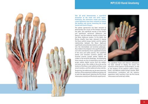
MP1530 Hand <strong>Anatomy</strong><br />
This <strong>3D</strong> print demonstrates a superficial<br />
dissection of the hand and wrist region.<br />
Anteriorly, the transverse carpal and palmar<br />
carpal ligaments have been removed to expose<br />
the tendons and nerves traversing the carpal<br />
tunnel and Canal of Guyon.<br />
The palmar aponeurosis has been removed to<br />
demonstrate the course of the tendons through<br />
the palm, the superficial muscles of the thenar<br />
and hypothenar eminences (abductors and<br />
flexors), and the lumbrical muscles arising from<br />
the flexor digitorum tendon. In the digits, the<br />
fibrous sheaths have been removed to expose<br />
the flexor pollicis longus tendon and the spatial<br />
relationships between the flexor digitorum<br />
superficialis and profundus tendons as they insert<br />
into the intermediate and terminal phalanges.<br />
Also visible in the midpalm is the superficial palmar<br />
arch with contributions from superficial branches<br />
of the ulnar and radial arteries. The superficial<br />
palmar arch branches (common palmar) and<br />
terminal arteries (proper palmar digital) are<br />
visible to the terminal phalanges. Accompanying<br />
these vessels are the corresponding common and<br />
proper palmar digital nerves from the median<br />
and ulnar nerves. Also visible in the wrist are the<br />
tendons of the flexor carpi radialis and flexor carpi<br />
ulnaris tendons, and the radial and ulnar arteries.<br />
Posteriorly, the radial artery can be seen traversing<br />
the floor of the anatomical snuffbox and giving rise<br />
to both the deep branch (piercing the first dorsal<br />
interosseous muscle) and the dorsal carpal branch.<br />
The superficial fascia and extensor retinaculum<br />
has been removed to display the course and<br />
insertions of the extensor muscle tendons, as well<br />
as the tendons of the extensor pollicis longus,<br />
brevis, and abductor pollicis longus muscles. Both<br />
intertendinous connections and the extensor<br />
expansions (with insertions from the first dorsal<br />
interosseous and lumbrical) visible.<br />
15