3D Anatomy Series
Human Anatomy Reproductions with an extra dimension
Human Anatomy Reproductions with an extra dimension

You also want an ePaper? Increase the reach of your titles
YUMPU automatically turns print PDFs into web optimized ePapers that Google loves.
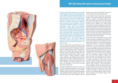
MP1765 Male left pelvis and proximal thigh<br />
This <strong>3D</strong> printed male left pelvis and proximal<br />
thigh (sectioned through the midsagittal plane<br />
in the midline and transversely through the<br />
L3/4 intervertebral disc) shows superficial and<br />
deep structures of the true and false pelves,<br />
inguinal and femoral region. In the transverse<br />
section, the epaxial musculature, abdominal<br />
wall musculature (rectus abdominis, external<br />
and internal abdominal obliques, transversus<br />
abdominis), psoas major and quadratus<br />
lumborum are visible and separated from each<br />
other and the superficial fat by fascial layers<br />
such as the rectus sheath and the thoracolumbar<br />
fascia. The psoas major muscle lies lateral to<br />
the external iliac artery, with the left testicular<br />
artery and vein lying on its superficial surface.<br />
More laterally (and moving inferiorly), the<br />
ilioinguinal nerve, the lateral cutaneous<br />
nerve of the thigh and the femoral nerve are<br />
positioned over the superficial surface of the<br />
iliacus muscle.<br />
The left common iliac artery bifurcates at the<br />
level of the sacral promontory into the external<br />
and internal iliac arteries. This specimen does not<br />
possess a clearly defined anterior and posterior<br />
division of the internal iliac artery; instead, the<br />
terminal arteries sequentially radiate from the<br />
internal iliac. The lateral sacral, inferior rectal,<br />
inferior gluteal, internal pudendal, superior<br />
vesical, obturator and umbilical arteries (which<br />
terminates in the medial umbilical ligament) are<br />
visible adjacent to the sacral ventral rami. The<br />
inferior gluteal and internal pudendal arteries<br />
have not bifurcated in this view and track<br />
inferiorly over piriformis.<br />
The deep circumflex iliac artery and vein can<br />
be seen passing deep posterior to the inguinal<br />
ligament, while the branches from the inferior<br />
epigastric artery and veins can be seen perforating<br />
rectus abdominis and the overlying rectus sheath.<br />
The left common iliac vein lies deep to the left<br />
common iliac artery; the obturator branch and<br />
the external iliac vein have been preserved.<br />
In the midline the pubic symphysis and sagittal<br />
sections of the pelvic viscera are visible: from<br />
anterior to posterior, the bladder (receiving the<br />
left ureter, which passes over the iliac vessels<br />
at the level of the pelvic brim), the left seminal<br />
vesicles and vas deferens, and rectum (with<br />
surrounding external anal sphincter muscle). The<br />
pathway of the urethra is visible from the inferior<br />
pole of the bladder through the prostate gland,<br />
pelvic diaphragm and the corpus spongiosum of<br />
the penis. Inferior to the sectioned erectile bodies<br />
(corpus cavernosa and corpus spongiosum) lies<br />
the scrotum, where the skin has been removed to<br />
reveal the parietal tunica vaginalis.<br />
On the preserved proximal thigh the fascia lata<br />
has been removed to highlight the transition of<br />
the neurovasculature and musculature from the<br />
pelvic region. Superior to the inguinal ligament<br />
a window has been cut to reveal the underlying<br />
aponeurosis of the transversus abdominis<br />
muscle. From medial to lateral, the femoral vein<br />
and artery have been removed from the femoral<br />
sheath, and the termination of the femoral nerve<br />
lies superficial to the iliopsoas muscle.. The great<br />
saphenous vein can be seen coursing medially<br />
over the pectineus, adductor longus and<br />
gracilis muscles, while branches of the femoral<br />
nerve pass over the profunda femoris artery.<br />
The thigh musculature is visible, with the cut<br />
sartorius muscle overlying the iliacus muscles<br />
and the origins of anterior thigh muscles (rectus<br />
femoris, vastus lateralis, vastus intermedius,<br />
vastus medialis). The tensor fasciae latae can<br />
be seen inserting on the anterior border of the<br />
iliotibial tract, which extends over the lateral<br />
surface of the thigh. A window has been cut to<br />
expose the underlying gluteus medius muscle,<br />
which terminates at the lateral aspect of the<br />
greater trochanter.<br />
37