3D Anatomy Series
Human Anatomy Reproductions with an extra dimension
Human Anatomy Reproductions with an extra dimension

You also want an ePaper? Increase the reach of your titles
YUMPU automatically turns print PDFs into web optimized ePapers that Google loves.
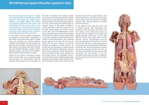
MP1400 Nervous System Dissection (posterior view)<br />
This <strong>3D</strong> printed specimen presents a unique<br />
view of axial anatomy, presenting a dorsal deep<br />
dissection of the head, neck, axillae, thorax,<br />
abdomen, and gluteal regions. The removal<br />
of the posterior portions of the cranium and<br />
laminectomy from the cervical region to the<br />
opening of the sacral canal affords a continuous<br />
view of the central nervous system structures<br />
and origin of the segmental nerves relative to<br />
other axillary and appendicular structures.<br />
In the cranium the two cerebral hemispheres<br />
are exposed in coronal section, separated by a<br />
falx cerebri that preserves the superior sagittal<br />
sinus, and supported by a partial tentorium<br />
cerebelli. The cerebellum has been removed and<br />
along the lateral margins, the sigmoid sinus has<br />
been opened. This exposes the fourth ventricle,<br />
pons and medulla oblongata, posterior inferior<br />
cerebellar arteries, and cranial nerves (CN VII –<br />
XII) arising from these brainstem structures.<br />
Inferior to the cranium, the posterior cervical<br />
portion of the spinal cord is exposed through<br />
deep dissection and laminectomies (with the<br />
exception of the posterior arch of the atlas). At<br />
this level of dissection, the vertebral arteries<br />
can be observed ascending through the vertebral<br />
foramina and curving anteriorly on the superior<br />
surface of the atlas towards the foramen magnum.<br />
The roots of the cervical and brachial plexus are<br />
exposed, resting on the scalene musculature,<br />
cervical vasculature (common carotid artery on<br />
the right, internal jugular and common carotid<br />
on the left) and sternocleidomastoid muscles<br />
and can be traced anteriorly towards the margins<br />
of the dissection. Removal of the scapulae (fully<br />
on the right, partially on the left) the more distal<br />
portions of the brachial plexus can be followed<br />
passing superior to the first ribs and into the<br />
axilla, with the cords, divisions and terminal<br />
branches surrounding the axillary arteries. On<br />
the left side, the musculature is largely removed<br />
(with parts of the deltoid, infraspinatus, and teres<br />
minor muscles preserved) and the long thoracic<br />
nerve and lateral thoracic artery descend near<br />
the serratus anterior. The subscapular artery is<br />
shown dividing into the circumflex scapular artery<br />
(passing to the triangular space) and the root of<br />
the thoracodorsal artery. On the right side, the<br />
more full removal of the scapula affords a view<br />
of the brachial plexus structures and the passage<br />
of the axillary nerve and posterior circumflex<br />
humeral artery laterally towards the surgical<br />
neck of the humerus.<br />
In the midline of the thorax the spinal cord<br />
is exposed through both laminectomy and<br />
dissection of the dura mater. The dorsal roots<br />
and rootlets of the mixed spinal nerves are<br />
exposed and pass laterally to the dorsal root<br />
ganglia (enclosed within dura). On the right<br />
side the thoracic mixed spinal nerves and the<br />
posterior thoracic musculoskeletal wall has<br />
been removed (from the 2nd rib to the level of<br />
the 11th and 12th ribs ) to expose the posterior<br />
surface of the lung and the posterior diaphragm.<br />
On the left side, most of the posterior thoracic<br />
wall has been removed, but the 3rd-5th ribs are<br />
retained to demonstrate the external intercostal<br />
musculature and the position of the 5th<br />
intercostal nerve within the space. In addition,<br />
the full sequence of intercostal nerves has been<br />
retained...*<br />
6<br />
* Complete description can be found at: www.3danatomyseries.com