3D Anatomy Series
Human Anatomy Reproductions with an extra dimension
Human Anatomy Reproductions with an extra dimension

You also want an ePaper? Increase the reach of your titles
YUMPU automatically turns print PDFs into web optimized ePapers that Google loves.
MP1500 Upper Limb<br />
Ms. Michelle Quayle<br />
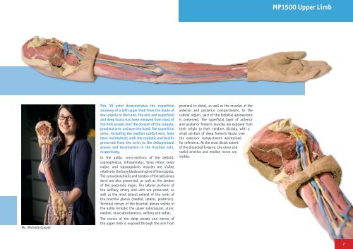
This <strong>3D</strong> print demonstrates the superficial<br />
anatomy of a left upper limb from the blade of<br />
the scapula to the hand. The skin and superficial<br />
and deep fascia has been removed from most of<br />
the limb except over the dorsum of the scapula,<br />
proximal arm, and over the hand. The superficial<br />
veins, including the median cubital vein, have<br />
been maintained; with the cephalic and basilic<br />
preserved from the wrist to the deltopectoral<br />
groove and termination in the brachial vein,<br />
respectively.<br />
In the axilla, cross-sections of the deltoid,<br />
supraspinatus, infraspinatus, teres minor, teres<br />
major, and subscapularis muscles are visible<br />
relative to the bony blade and spine of the scapula.<br />
The coracobrachialis and tendon of the latissimus<br />
dorsi are also preserved, as well as the tendon<br />
of the pectoralis major. The lateral portions of<br />
the axillary artery and vein are preserved, as<br />
well as the most lateral extend of the cords of<br />
the brachial plexus (medial, lateral, posterior).<br />
Terminal nerves of the brachial plexus visible in<br />
the axilla include: the upper subscapular, ulnar,<br />
median, musculocutaneous, axillary and radial.<br />
The course of the deep vessels and nerves of<br />
the upper limb is exposed through the arm from<br />
proximal to distal, as well as the muscles of the<br />
anterior and posterior compartments. In the<br />
cubital region, part of the bicipital aponeurosis<br />
is preserved. The superficial layer of anterior<br />
and posterior forearm muscles are exposed from<br />
their origin to their tendons distally, with a<br />
small portion of deep forearm fascia over<br />
the extensor compartment maintained<br />
for reference. At the most distal extent<br />
of the dissected forearm, the ulnar and<br />
radial arteries and median nerve are<br />
visible.<br />
7