3D Anatomy Series
Human Anatomy Reproductions with an extra dimension
Human Anatomy Reproductions with an extra dimension

You also want an ePaper? Increase the reach of your titles
YUMPU automatically turns print PDFs into web optimized ePapers that Google loves.
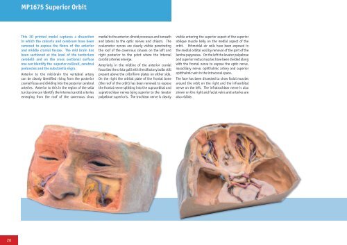
MP1675 Superior Orbit<br />
This <strong>3D</strong> printed model captures a dissection<br />
in which the calvaria and cerebrum have been<br />
removed to expose the floors of the anterior<br />
and middle cranial fossae. The mid brain has<br />
been sectioned at the level of the tentorium<br />
cerebelli and on the cross sectional surface<br />
one can identify the superior colliculi, cerebral<br />
peduncles and the substantia nigra.<br />
Anterior to the mid-brain the vertebral artery<br />
can be clearly identified rising from the posterior<br />
cranial fossa and dividing into the posterior cerebral<br />
arteries. Anterior to this in the region of the sella<br />
turcica one can identify the internal carotid arteries<br />
emerging from the roof of the cavernous sinus<br />
medial to the anterior clinoid processes and beneath<br />
and lateral to the optic nerves and chiasm. The<br />
oculomotor nerves are clearly visible penetrating<br />
the roof of the cavernous sinuses on the left and<br />
right posterior to the point where the internal<br />
carotid arteries emerge.<br />
Anteriorly in the midline of the anterior cranial<br />
fossa lies the crista galli with the olfactory bulbs still<br />
present above the cribriform plates on either side.<br />
On the right the orbital plate of the frontal bone<br />
(the roof of the orbit) has been removed to expose<br />
the frontal nerve splitting into the supraorbital and<br />
supratrochlear nerves lying superior to the levator<br />
palpebrae superioris. The trochlear nerve is clearly<br />
visible entering the superior aspect of the superior<br />
oblique muscle belly on the medial aspect of the<br />
orbit. Ethmoidal air cells have been exposed in<br />
the medial orbital wall by removal of the part of the<br />
lamina papyracea. On the left the levator palpebrae<br />
and superior rectus muscles have been divided along<br />
with the frontal nerve to expose the optic nerve,<br />
nasociliary nerve, ophthalmic artery and superior<br />
ophthalmic vein in the intraconal space.<br />
The face has been dissected to show facial muscles<br />
around the orbit on the right and the infraorbital<br />
nerve on the left. The infratrochlear nerve is also<br />
shown on the right and facial veins and arteries are<br />
also visible.<br />
26