3D Anatomy Series
Human Anatomy Reproductions with an extra dimension
Human Anatomy Reproductions with an extra dimension

You also want an ePaper? Increase the reach of your titles
YUMPU automatically turns print PDFs into web optimized ePapers that Google loves.
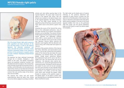
MP1783 Female right pelvis<br />
superficial and deep structures<br />
This <strong>3D</strong> printed female right pelvis preserves<br />
both superficial and deep structures of the<br />
true and false pelves, as well as the inguinal<br />
ligament, the obturator membrane and<br />
canal, and both the greater and lesser sciatic<br />
foramina. Somewhat unique is the removal of<br />
portions of the peritoneum (a grayish colour)<br />
to create ‘windows’ displaying extraperitoneal<br />
structures.<br />
The specimen has been sectioned transversely<br />
through the L4 vertebra, displaying a cross<br />
section of the colon, the epaxial musculature<br />
(psoas and quadratus lumborum muscles), and<br />
the abdominal wall musculature. The common iliac<br />
artery has been preserved from the level of the L4<br />
vertebra, and its bifurcation into the external and<br />
internal iliac arteries can be observed at the level<br />
of the sacral promontory. Deep to the arteries the<br />
common iliac vein and the origin of the inferior<br />
vena cava are visible.<br />
The external iliac artery and vein passes<br />
anteroinferiorly along the pelvic brim, giving<br />
rise to the inferior epigastric and deep circumflex<br />
arteries and veins before passing deep to the<br />
inguinal ligament. The psoas major muscle lies<br />
lateral to the external iliac artery, with the<br />
femoral nerve evident on its lateral margin close<br />
to the inguinal ligament. The lateral cutaneous<br />
nerve of the thigh travels laterally on the<br />
superficial surface of the iliacus muscle to exit the<br />
‘false’ pelvis close to the anterior superior iliac<br />
spine.<br />
Following the course of the internal iliac artery<br />
deep to the undissected peritoneum, many of<br />
the major branches of its anterior and posterior<br />
divisions can be identified. The anterior division<br />
divides (deep to the peritoneum) into the superior<br />
vesical, obturator and obliterated umbilical<br />
artery. With a course parallel to the obturator<br />
artery, the obturator nerve can be seen running<br />
over obturator internus before entering the<br />
obturator canal together with the obturator vein<br />
(nerve, artery, vein in that order from superior to<br />
inferior).<br />
Branches of the posterior division of the internal<br />
iliac artery, iliolumbar, and several lateral sacral<br />
arteries, can be seen arising from the posterior<br />
aspect of the internal iliac just below the sacral<br />
promontory. Its terminal branch, the superior<br />
gluteal, usually passes posteriorly between<br />
the lumbosacral trunk and S1 nerve, but this is<br />
hidden from view. The internal iliac vein and its<br />
tributaries - the obturator veins, uterine vein,<br />
vesical veins, etc. can be seen lying internal to the<br />
nerves and muscles. The large S1 and S2 roots and<br />
the smaller S3 nerve root can be seen emerging<br />
from the sacral foramina to pass laterally where<br />
it is joined by the lumbosacral trunk (L4 and l5<br />
roots) which is not visible, to form the sciatic<br />
nerve which exits through the greater sciatic<br />
foramen to emerge on the posterior aspect in<br />
the gluteal region. In the pelvis as these roots<br />
pass laterally they are interdigitated between the<br />
fibres of piriformis muscle.<br />
The right ureter can be clearly seen as it passes<br />
inferiorly on the posterior abdominal wall<br />
superficial to psoas muscle. It passes over the<br />
pelvic brim at the bifurcation of the common iliac<br />
artery to descend on the lateral wall of the pelvis<br />
before passing medially in the base of the broad<br />
ligament (hidden from view as the peritoneal<br />
folds that ‘drape’ over the uterine [Fallopian]<br />
tubes are still intact) to reach the lateral angles<br />
of the bladder...*<br />
40<br />
* Complete description can be found at: www.3danatomyseries.com