Ortopedická protetika Praha sro - Společnost pro pojivové tkáně
Ortopedická protetika Praha sro - Společnost pro pojivové tkáně
Ortopedická protetika Praha sro - Společnost pro pojivové tkáně

Create successful ePaper yourself
Turn your PDF publications into a flip-book with our unique Google optimized e-Paper software.
Pokroky ve výzkumu, diagnostice a terapii<br />
Vydává <strong>Společnost</strong> <strong>pro</strong> <strong>pojivové</strong> <strong>tkáně</strong> ČLS J. E. Purkyně<br />
Ambulantní centrum <strong>pro</strong> vady pohybového aparátu<br />
Katedra antropologie a genetiky člověka PřF UK v Praze<br />
Odborná společnost ortopedicko-<strong>pro</strong>tetická ČLS J. E. Purkyně<br />
ročník 14/2007 číslo 3-4<br />
EMBASE / Excerpta Medica
<strong>Ortopedická</strong> <strong><strong>pro</strong>tetika</strong> <strong>Praha</strong> s.r.o.<br />
Výrobce individuálních<br />
ortopedicko-<strong>pro</strong>tetických pomůcek<br />
zajišťuje:<br />
– Lékařské vyšetření pacienta a předpis pomůcky<br />
– Zhotovení všech individuálních ortopedických pomůcek (<strong>pro</strong>tézy HK<br />
a DK, končetinové a trupové ortézy, měkké bandáže, ortopedickou obuv,<br />
ortopedické vložky apod.<br />
<strong>pro</strong>vozní doba:<br />
po 7.30–17.00; út–čt 7.30–16.00; pá 7.30–15.00<br />
<strong>Ortopedická</strong> Protetika <strong>Praha</strong> s.r.o., Kloknerova 1/1245, 148 00 <strong>Praha</strong> 4<br />
tel.: 272 932 241–6, l. 131, tel./fax: 272 937 386, e-mail: <strong><strong>pro</strong>tetika</strong>@seznam.cz<br />
Metro C stanice Chodov, dále autobus č. 118 stanice Dědinova – budova MEDICENTRUM<br />
Partner všech zdravotních pojišťoven v ČR
POHYBOVÉ ÚSTROJÍ<br />
ročník 14, 2007, číslo 3+4<br />
datum vydání 10. 10. 2007<br />
REDAKČNÍ RADA<br />
VEDOUCÍ REDAKTOR: Doc. MUDr. Ivo Mařík, CSc.<br />
ZÁSTUPCE VEDOUCÍHO REDAKTORA: Prof. Ing. Miroslav Petrtýl, DrSc.<br />
VĚDECKÝ SEKRETÁŘ: MUDr. Miloslav Kuklík, CSc.<br />
ODPOVĚDNÝ REDAKTOR: Ing. Pavel Lorenc<br />
Prof. MUDr. Milan Adam, DrSc. Doc. MUDr. Petr Korbelář, CSc.<br />
Prof. MUDr. Jaroslav Blahoš, DrSc. Doc. MUDr. Vladimír Kříž<br />
Doc. RNDr. Pavel Bláha, CSc. Prof. Ing. František Maršík, DrSc.<br />
Prof. Ing. Jan Čulík, DrSc. Doc. RNDr. Ivan Mazura, CSc.<br />
Doc. MUDr. Ivan Hadraba, CSc. MUDr. Pavel Novosad<br />
Prof. RNDr. Karel Hajniš, CSc. Prof. MUDr. Ctibor Povýšil, DrSc.<br />
Ing. Hana Hulejová Doc. MUDr. Václav Smrčka, CSc.<br />
Prof. MUDr. Josef Hyánek, DrSc. Prof. PhDr. Jiří Straus, DrSc.<br />
Prof. MUDr. Jaromír Kolář, DrSc. MUDr. Jan Všetička<br />
EDITORIAL BOARD<br />
Prof. Dr. Ing. Romuald Bedzinski, Politechnika Doc. Dr. Med. Kazimierz S. Kozlowski,<br />
Wroclawska, Poland M.R.A.C.R., Westmead NSW 2145, Sydney<br />
Dr. Michael Bellemore, F.R.A.C.S., Prof. František Makai, MD, DSc., Bratislava, Slovakia<br />
Westmead NSW 2145, Sydney Prof. Dr. Med. Zoran Vukasinovic, Belgrade,<br />
Ass. Prof. Jacques Chêneau, MD, Saint Orens, France Yugoslavia<br />
Prof. Tomasz Karski, MD, PhD, Lublin, Poland<br />
Pohybové ústrojí. Pokroky ve výzkumu, diagnostice a terapii.<br />
ISSN 1212-4575<br />
Vydává <strong>Společnost</strong> <strong>pro</strong> <strong>pojivové</strong> <strong>tkáně</strong> ČLS J.E.Purkyně,<br />
Ambulantní centrum <strong>pro</strong> vady pohybového aparátu,<br />
Katedra antropologie a genetiky člověka, PřF UK v Praze<br />
& Odborná společnost ortopedicko – <strong>pro</strong>tetická ČLS J. E. Purkyně<br />
Excerpováno v Excerpta Medica. Tiskne PeMa, Nad Primaskou 45, <strong>Praha</strong> 10<br />
Návrh a grafická úprava obálky Rudolf Štorkán<br />
Časopis vychází 4krát ročně, nebo jako dojčíslo 2krát ročně. Každá práce je recenzována.<br />
Objednávky přijímá Ambulantní centrum <strong>pro</strong> vady pohybového aparátu,<br />
Olšanská 7, 130 00 <strong>Praha</strong> 3, tel./fax: (+420) 222 582 214,<br />
http://www.volny.cz/ambul_centrum.<br />
Rukopisy zasílejte na adresu Doc. MUDr. Ivo Mařík, CSc., Olšanská 7, 130 00 <strong>Praha</strong> 3,<br />
(ambul_centrum@volny.cz) ve formátu doc, rtf. Vydavatel<br />
upozorňuje, že za obsah inzerce odpovídá výhradně inzerent. Časopis jakožto<br />
nevýdělečný neposkytuje honoráře za otištěné příspěvky<br />
POHYBOVÉ ÚSTROJÍ, ročník 14, 2007, č. 3+4 165
166<br />
LOCOMOTOR SYSTEM<br />
Advances in Research, Diagnostics and Therapy<br />
Published by The Society for Connective Tissues, Czech Medical Association of<br />
J. E. Purkyně, Prague, Ambulant Centre for Defects of Locomotor Apparatus Prague, Dept.<br />
of Anthropology and Human Genetics, Faculty of Science Charles University in Prague &<br />
Society for Prosthetics and Orthotics, Czech Medical Association of J. E. Purkyně, Prague,<br />
Czech Republic<br />
Call for papers<br />
Support this journal by sending in your best and most interesting papers. Publication will<br />
normally be within six months of acceptance. The journal appears four times in a year.<br />
Chief editor: Ivo Mařík<br />
Associate Editor: Miroslav Petrtýl<br />
Scientific Secretary: Miloslav Kuklík<br />
Responsible Editor: Pavel Lorenc<br />
Editorial board<br />
Milan Adam Petr Korbelář<br />
Romuald Bedzinski Kazimierz Kozlowski<br />
Michael Bellemore Vladimír Kříž<br />
Jaroslav Blahoš Pavel Novosad<br />
Pavel Bláha František Makai<br />
Jacques Chêneau František Maršík<br />
Jan Čulík Ivan Mazura<br />
Ivan Hadraba Ctibor Povýšil<br />
Karel Hajniš Václav Smrčka<br />
Hana Hulejová Jiří Straus<br />
Josef Hyánek Zoran Vukasinovic<br />
Tomasz Karski Jan Všetička<br />
Jaromír Kolář<br />
Submitted papers: Locomotor System will review for publication manuscripts concerned<br />
with <strong>pro</strong>gress in research of connective tissue diagnostics, medical and surgical therapy<br />
mainly in the fields of orthopaedic surgery, dysmorphology (multiple congenital abnormalities<br />
of skeleton) and plastic surgery, biomechanics and biorheology, clinical anthropology<br />
and paleopathology.<br />
The journal has an interdisciplinary character which gives possibilities for complex<br />
a<strong>pro</strong>ach to the <strong>pro</strong>blematics of locomotor system. The journal belongs to clinical, preclinical<br />
and theoretical medical branches which connect various up-to-date results and discoveries<br />
concerned with locomotor system.<br />
Papers published in the journal are excerpted in EMBASE / Excerpta Medica. We prefer the<br />
manuscripts to be prepared according to Uniform Requirements for Manuscripts Submitted to<br />
Biomedical Journals (Vancouver Declaration, Brit med J 1988; 296, pp. 401–405).<br />
LOCOMOTOR SYSTEM vol. 14, 2007, No. 3+4
POHYBOVÉ ÚSTROJÍ,<br />
14, 2007, č. 3+4<br />
Pokroky ve výzkumu, diagnostice<br />
a terapii<br />
OBSAH<br />
OBRÁZEK NA TITULU . . . . . . . . . . . . . 169<br />
PŮVODNÍ PRÁCE<br />
SEDLAK P., BLÁHA P., JIROUTOVÁ L.,<br />
BRABEC M., VIGNEROVÁ J.<br />
Růstová dynamika somatických<br />
znaků lineární tělesné<br />
<strong>pro</strong>porcionality u současných<br />
dětí ve věku od 6 do 15 let . . . . . . . . . . 172<br />
SOUBORNÉ REFERÁTY<br />
KOLÁŘ J.<br />
Nosologie a klasifikace genetických<br />
kosterních poruch: Revize 2006 . . . . . 193<br />
KUKLÍK M.<br />
Lékařská a klinická genetika, 2. část . . . 213<br />
BELLEMORE M. C., MUNNS C. F.<br />
Osteogenesis imperfecta . . . . . . . . . . . . 232<br />
LOCOMOTOR SYSTEM<br />
14, 2007, No. 3+4<br />
Advances in Research, Diagnostics<br />
and Therapy<br />
CONTENT<br />
TITLE PICTURE DESCRIPTION . . . . . 169<br />
ORIGINAL PAPERS<br />
SEDLAK P., BLÁHA P., JIROUTOVÁ L.,<br />
BRABEC M., VIGNEROVÁ J.<br />
Growth dynamics of somatic traits<br />
of linear body <strong>pro</strong>portionality<br />
in present children at the age<br />
of 6 to 15 years . . . . . . . . . . . . . . . . . . . . . 172<br />
REVIEWS<br />
KOLÁŘ J.<br />
Nosology and Classification of Genetic<br />
Skeletal Disorders: 2006 Revision . . . . 193<br />
KUKLÍK M.<br />
Medical and clinical genetics, part 2. . . 213<br />
BELLEMORE M. C., MUNNS C. F.<br />
Osteogenesis imperfecta . . . . . . . . . . . . 232<br />
POHYBOVÉ ÚSTROJÍ, ročník 14, 2007, č. 3+4 167
KONFERENCE<br />
MAŘÍKOVÁ A., MAŘÍK I.<br />
International Scientific Symposium<br />
to Honour for the 80 th Birthday of<br />
Prof. Dr. Jacques Chêneau,<br />
May 11–13, 2007, Regensburg . . . . . . . 240<br />
ZPRÁVY<br />
BARBOŘÁKOVÁ H. V., ŠPUNDA M.<br />
Perspektivy vědecko-výzkumné<br />
spolupráce v EU . . . . . . . . . . . . . . . . . . . . 263<br />
ČÍŽEK K.<br />
Jaká bude úloha lékaře v ortopedické<br />
<strong>pro</strong>tetice? . . . . . . . . . . . . . . . . . . . . . . . . . . 265<br />
Přihláška řádného člena SPT . . . . . . . . . 267<br />
Informace o <strong>Společnost</strong>i <strong>pro</strong><br />
<strong>pojivové</strong> <strong>tkáně</strong> ČLS JEP . . . . . . . . . . . . . 268<br />
Životní jubilea<br />
Profesor MUDr. Jacques Chêneau . . . . 271<br />
SMĚRNICE AUTORŮM . . . . . . . . . . . . . . 274<br />
OBSAH ROČNÍKU 2006 . . . . . . . . . . . . . 278<br />
OBSAH ROČNÍKU 2007 . . . . . . . . . . . . . 282<br />
168<br />
CONFERENCES<br />
MAŘÍKOVÁ A., MAŘÍK I.<br />
International Scientific Symposium<br />
to Honour for the 80 th Birthday<br />
of Prof. Dr. Jacques Chêneau,<br />
May 11–13, 2007, Regensburg . . . . . . . 240<br />
NEWS<br />
BARBOŘÁKOVÁ H. V., ŠPUNDA M.<br />
Perspectives of scientific and research<br />
cooperation in European Union . . . . . 263<br />
ČÍŽEK K.<br />
What will be role of a doctor<br />
in orthopaedic <strong>pro</strong>sthetics?. . . . . . . . . . 265<br />
Membership application of The<br />
Society for Connective Tissues, Czech<br />
Medical Association J. E. Purkyně,<br />
Prague, CZ . . . . . . . . . . . . . . . . . . . . . . . . . 267<br />
Information on The Society for<br />
Connective Tissues, Czech Medical<br />
Association J. E. Purkyně, Prague, CZ . . 268<br />
Anniversary<br />
Professor MUDr. Jacques Chêneau . . . 271<br />
INSTRUCTIONS FOR AUTHORS . . . . . 276<br />
CONTENTS OF VOLUME 2006. . . . . . . 280<br />
CONTENTS OF VOLUME 2007. . . . . . . 284<br />
LOCOMOTOR SYSTEM vol. 14, 2007, No. 3+4
OBRÁZEK NA TITULNÍ STRANĚ DEMONSTRUJE<br />
TITLE PICTURE DEMONSTRATES<br />
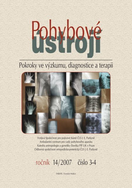
Obrázek na titulní straně časopisu demonstruje charakteristické RTG příznaky diastrofické<br />
dysplazie (DD), a to: oploštělé epifýzy, opoždění osifikace epifýz hlavic femurů,<br />
poloměsíčitý tvar epifyzárních osifikačních center, neúplná osifikace laterální poloviny<br />
distální epifýzy femuru. Krčky femurů jsou krátké a široké, široké jsou všechny metafýzy<br />
dlouhých kostí, které jsou významně zkrácené. Nepravidelné deformity a zkrácení všech<br />
metakarpů, metatarsů a článků prstů, 1. metakarp bývá u dětí oválný. Ploché a široké epifýzy<br />
metakarpů mohou být jediným příznakem u mírných forem DD. U dětí bývají deltovité<br />
deformity distálních metafýz femurů a radií. Na páteři často <strong>pro</strong>greduje dorsolumbální<br />
kyfoskolióza a krční kyfóza. Obratlová těla jsou nepravidelně deformována a jsou relativně<br />
dobře vyvinutá. Platyspondylie nevylučuje však diagnózu DD. V dolní lumbální krajině bývá<br />
mírné zúžení interpedikulární vzdálenosti.<br />
Na obrázku jsou vyobrazeny typické radiografické dysplastické změny pozorované<br />
na ruce, noze, kyčelních a kolenních kloubech i páteři u pacientů s DD v různém věku,<br />
kteří jsou nebo byli léčeni v Ambulantním centru <strong>pro</strong> vady pohybového aparátu v Praze.<br />
Ruka<br />
Nepravidelné zkrácení a deformity metakarpů a falang s rozšířením metafýz a plochými<br />
epifýzami jsou patrny v na snímcích dětí i dospělých. Nalevo obrázku jsou pod sebou<br />
RTG levé ruky, nahoře na RTG ruky ve věku 2,5 roku je patrna dislokace metakarpofalangeálního<br />
kloubu palce, oválný deltovitý tvar 1. metakarpu a výrazné zúžení <strong>pro</strong>ximálních<br />
interfalangeálních kloubů, které svědčí <strong>pro</strong> symfalangismu. Níže umístěné obrázky –11 a 9<br />
let – ukazují velmi ploché a nepravidelné epifýzy radia a ulny.<br />
Páteř<br />
Vlevo dole je RTG přednoží 9letého pacienta, kde je výrazné equinovarosní postavení,<br />
metatarsy jsou krátké s širokými metafýzami, zobrazené epifýzy jsou velmi ploché.<br />
Kyčle<br />
Nahoře a u<strong>pro</strong>střed obrázku jsou kyčelní klouby 4 pacientů ve věku – zleva 2,5; 6,5<br />
a 5,5 roku, u<strong>pro</strong>střed zleva 9 a 11 let. Krčky jsou krátké a široké, epifýzy značně oploštělé,<br />
s věkem <strong>pro</strong>gredují abdukční a flekční kontraktury.<br />
Kolena<br />
Dole na obrázku je RTG kolen 3 pacientů ve věku – zleva 12,5 a 2,5 roku, kde je mírné<br />
oploštění epifýz, na obrázku vlevo je laterální luxace čéšky, na obrázku vpravo – 9 let – je<br />
těžká deformita kolenního kloubu i bérce, který je varosní a rotován dovnitř.<br />
POHYBOVÉ ÚSTROJÍ, ročník 14, 2007, č. 3+4 169
Páteř<br />
V pravé části obrázku nahoře na snímku v transorální (13,5 roku) a bočné (24 let) <strong>pro</strong>jekci<br />
je dobře vytvořený dens epistrophei, mírné oploštění obratlových těl s ventrálními<br />
osteofyty. Vpravo u<strong>pro</strong>střed je na snímku krční páteře v předozadní <strong>pro</strong>jekci –11 let – zobrazena<br />
spina bifida occulta dolních krčních obratlů, vpravo dole je ukázáno mírné zúžení<br />
páteřního kanálu bederní páteře – 6,5 roku, na pravém okraji obrázku dole je ne zcela<br />
<strong>pro</strong> diagnózu typický obraz platyspondylie dorsolumbální páteře se známkami předčasné<br />
spondylózy – 20 let.<br />
Dědičnost<br />
Dědičnost je autosomálně recesivní (AR) s velmi širokou variabilitou exprese postižení.<br />
Prenatální diagnostika je možná v 16. týdnu gravidity na základě zjištění krátkých končetin,<br />
pedes equinovari, ulnární deviace rukou a stopařských palců. Z biopsie choriových klků lze<br />
<strong>pro</strong>kázat mutace v genu sulfátového transportéru diastrofické dysplazie (DTDST). Mutace<br />
postihují různé domény DTDST genu, což vysvětluje širokou variabilitu exprese fenotypu.<br />
170<br />
LOCOMOTOR SYSTEM vol. 14, 2007, No. 3+4
Mutace znemožňuje buněčnou inkorporaci sulfátu a nedostatečná sulfatace <strong>pro</strong>teoglykanů<br />
vede k narušení tvorby matrix chrupavky.<br />
Klinická symptomatologie<br />
Krátká postava a krátké končetiny, mnohočetné kloubní kontraktury zvláště ramenních,<br />
loketních, kyčelních a interfalangeálních kloubů. Hypermobilní vysoko nasedající palce<br />
rukou (tzv. stopařský palec). Chybění volární ohybové rýhy <strong>pro</strong>ximálních interfalangeálních<br />
kloubů prstů rukou označuje fibrosní někdy i kostěný symfalangismus. Pedes equinovari<br />
s větším meziprstním <strong>pro</strong>storem mezi 1. a 2. prstem. Nepravidelně pseudocystické<br />
zduření ušních boltců (patrné od prvních dnů do 3 měsíců věku) později květákovitá<br />
deformita ušních boltců. Rozštěp patra bývá zjištěn u 50 % případů. U většiny dětí <strong>pro</strong>greduje<br />
kyfoskolióza.<br />
Průběh<br />
Perinatální a kojenecká úmrtnost je zvýšená. Nevzniknou-li vážné komplikace z důvodů<br />
deformit páteře, je životní <strong>pro</strong>gnóza normální. Progrese cervikální kyfózy může vést ke<br />
kompresi míchy s rozvojem kvadruplegie a letálním koncem Generalizované postižení<br />
mesenchymu vede v období růstu k <strong>pro</strong>gresi kyfoskoliózy páteře, kontraktur a těžkým equinovarosním<br />
deformitám nohou, které vzdorují ortopedickému léčení – kontraktury recidivují.<br />
V průběhu růstu je třeba individuálně kombinovat konzervativní a operační léčení<br />
kontraktur a deformit dlouhých kostí. Míšní komprese a <strong>pro</strong>grese krční kyfózy je indikací<br />
k operačnímu léčení v raném věku. Předčasná osteoartróza kyčelních kloubů vzniká v důsledku<br />
těžké epifyzární dysplazie a <strong>pro</strong>gredujících kontraktur s růstem. V indikovaných případech<br />
se řeší po skončení růstu kloubní náhradou. Z dentálních anomálií jsou nejčastější<br />
hypodoncie, komprese zubů a malokluse. Výška dospělých mužů je v rozmezí 114–158 cm<br />
(průměr 136 cm) a 98–143 cm u žen (s průměrem 129 cm).<br />
Pro kostní dysplazie obecně platí, že <strong>pro</strong> určení diagnózy je diagnosticky cenné hodnocení<br />
dysplastických změn epifýz, metafýz a obratlů na RTG snímcích zhotovených v období<br />
růstu. Dysplastické změny kyčelních kloubů a celého skeletu u pacientů s DD jsou <strong>pro</strong><br />
potvrzení diagnózy typické od narození do dospělosti.<br />
Doc. MUDr. Ivo Mařík, CSc.<br />
Ambulantní centrum <strong>pro</strong> vady pohybového aparátu<br />
Olšanská 7<br />
130 00 <strong>Praha</strong> 3<br />
E-mail: ambul_centrum@volný.cz<br />
POHYBOVÉ ÚSTROJÍ, ročník 14, 2007, č. 3+4 171
172<br />
PŮVODNÍ PRÁCE ● ORIGINAL PAPERS<br />
GROWTH DYNAMICS OF SOMATIC TRAITS OF<br />
LINEAR BODY PROPORCIONALITY IN PRESENT<br />
CHILDREN AT THE AGE OF 6 TO 15 YEARS<br />
SEDLAK P. 1 , BLÁHA P. 1 , JIROUTOVÁ L. 1 , BRABEC M. 2 ,<br />
VIGNEROVÁ J. 2<br />
1 Department of Anthropology and Human Genetics, Faculty of Science,<br />
Charles University, Prague, Czech Republic<br />
2 National Institute of Public Health, Prague, Czech Republic<br />
SUMMARY<br />
This study deals with <strong>pro</strong>blems of evaluating tendencies in the growth dynamics of<br />
parameters of linear somatic <strong>pro</strong>portionality. Its results are based on analyses of average<br />
velocity curves of human body height, sitting height, subischial length and the length of<br />
upper extremity. The velocity curves were have been constructed from data collected by<br />
a recent semilongitudinal study enquiring into the growth dynamics of Czech children at<br />
the age from 6 to 15 years by means of applying a model of linear regression with mixed<br />
effects. This model made it possible to establish average characteristics for periods of mid-<br />
-growth spurt and pubertal spurt. Timings and durations of growth phases were compared<br />
with the results achieved by the Zurich longitudinal study with regard to the influence of<br />
the secular trend in growth dynamics.<br />
Klíčová slova: growth dynamics, growth velocity curves, growth models, semilongitudinal<br />
study<br />
INTRODUCTION<br />
Various parts of a child’s body do not<br />
grow at the same speed during the <strong>pro</strong>cess<br />
of human ontogenesis, when they pass<br />
different periods of somatic development<br />
they assume their own specific pace of<br />
growth changes. If one part of body under-<br />
goes a dynamic growth, another part may<br />
be found in a state of growth tranquillity.<br />
This phenomenon is characterized by<br />
the rule of ‘periodicity and alternation’,<br />
i.e. periods of faster growth are regularly<br />
superseded by periods of slow-down<br />
growth. A phenotype manifestation of this<br />
<strong>pro</strong>cess is seen in changes of the child’s<br />
LOCOMOTOR SYSTEM vol. 14, 2007, No. 3+4
somatic <strong>pro</strong>portions. On the basis of characteristic<br />
specific traits of growth dynamics<br />
and development the <strong>pro</strong>cess of child<br />
ontogeny may be divided into several successive<br />
stages constituting milestones of<br />
the standard periodisation of child age (5).<br />
Growth curves are constructed with<br />
respect to data obtained by longitudinal<br />
growth studies that <strong>pro</strong>vide figures concerning<br />
an individual course of growth with<br />
regard to an interindividual variability in<br />
a given population. Average growth curves<br />
of a given trait then serve as a normative<br />
for evaluating its growth velocity in the<br />
diagnostics of growth disorders. Regular<br />
measurements of elementary somatic features<br />
such as body height may signal abnormalities<br />
and represent a limiting criterion<br />
for diagnostic treatment.<br />
The basic presupposition of longitudinal<br />
studies is a long-term observation of the<br />
same group of individuals and their repeated<br />
follow-up examinations in precisely<br />
defined intervals. This implies a number of<br />
complications including <strong>pro</strong>jects exhibiting<br />
high demands for organisation. The latter<br />
consist in limited frequencies of examined<br />
groups and a gradual loss of their representative<br />
character owing to irresponsible<br />
individuals, descending mostly from lower<br />
social classes, who are not willing to come<br />
to repeated follow-up examinations.<br />
Another <strong>pro</strong>blem is represented by<br />
the danger that some data lose their upto-date<br />
topicality, because after long-term<br />
observations the output results generalise<br />
figures concerning child population that<br />
were collected more than twenty years<br />
ago and the research does not take into<br />
consideration the role of long-term secular<br />
changes. Information concerning growth<br />
dynamics of somatic traits or the start of<br />
course of pubertal growth acceleration<br />
therefore may lose its validity for recent<br />
child population at the time of presenting<br />
scientific results.<br />
Such deficiencies of longitudinal studies<br />
may be solved by semilongitudinal<br />
observations conceived in such a manner<br />
that various age groups of individuals are<br />
subjected to short longitudinal examinations<br />
that take place simultaneously in the<br />
same time interval. After finishing a single<br />
study it is necessary to ensure that different<br />
age groups exhibit neat mutual boundaries<br />
and links. The advantage of correctly<br />
conceived follow-up semilongitudinal<br />
observation is the possibility of collecting<br />
a representative sample of data in a short<br />
interval of time. If we choose ap<strong>pro</strong>priate<br />
mathematical models, data obtained<br />
in this way may be applied conveniently<br />
for constructing growth curves of somatic<br />
parameters (14, 16).<br />
MATERIAL AND METHODS<br />
In statistical sense, the data from the<br />
our study certainly fall into the broad category<br />
of longitudinal studies (in the sense<br />
that they contain repeated measurements<br />
of different individuals). We use word<br />
semilongitudinal to denote the particular<br />
design used to distinguish it from the most<br />
simple longitudinal design where observations<br />
are repeated (or planned to be<br />
repeated to be precise) at the same set of<br />
times for all individuals (except perhaps<br />
for random-dropout-related irregularities),<br />
which is often referred to as “the longitudinal<br />
study” in anthropology or other applied<br />
disciplines). Our design is planned so that<br />
it has different sets of time points at which<br />
repeated measurements are taken for different<br />
groups of individuals (for different<br />
POHYBOVÉ ÚSTROJÍ, ročník 14, 2007, č. 3+4 173
irth cohorts, actually). Study was carried<br />
out from September 1997 till October 2000<br />
under the auspices of the grant <strong>pro</strong>ject IGA<br />
MZ CR assigned by the National Institute<br />
of Public Health in Prague (1). As a whole,<br />
a sample of 1925 children (990 boys and<br />
935 girls) at 20 elementary schools in four<br />
regions of the Czech Republic from 6 to 15<br />
years of age were examined. On the basis<br />
of detailed information <strong>pro</strong>vided to parents<br />
and their positive ap<strong>pro</strong>val it was possible<br />
to include children that attended school<br />
forms 1, 3, 5 and 7 at elementary schools<br />
in September 1997. Every child was examined<br />
six times at most. The final evaluation<br />
of results involved only individuals that<br />
passed a thorough-going examination at<br />
least three times. As is clear from Table 1,<br />
groups of children examined were divided<br />
with respect to sex, region and grades of<br />
elementary schools.<br />
In every individual the examination<br />
was concentrated on measuring 29 somatic<br />
features. Beside body height and body mass<br />
it included 4 measured variables of height,<br />
4 of breadth, 7 measurements of girth characteristics<br />
of the trunk and extremities,<br />
2 leg <strong>pro</strong>portions and 5 head dimensions<br />
as well as 5 measurements of skinfold<br />
thickness. Moreover, there were two <strong>pro</strong>jective<br />
measures calculated – the length<br />
of the upper extremity and the subischial<br />
174<br />
length (calculated as a subtraction of sitting<br />
height from standing body height).<br />
Our paper present growth velocity curves<br />
of somatic traits of linear <strong>pro</strong>portionality<br />
and the evaluation of mutual relations in<br />
their growth dynamics.<br />
Every individual <strong>pro</strong>band <strong>pro</strong>vided at<br />
least 3 and at most 6 data for <strong>pro</strong>cessing<br />
a given somatic parameter. All the<br />
data were fitted by a smooth (polynomial)<br />
model (12, 15). In order to account for<br />
longitudianal character of the data (i.e.<br />
for correlation among repeated measurements<br />
of the same individual) as well<br />
as for its heteroscedasticity, we employed<br />
linear mixed model, allowing for random,<br />
or individual-specific linear terms (random<br />
intercept and slope). Among other<br />
features, estimate of the mean “distance<br />
curve” (i.e. measurement versus time, e.g.<br />
height versus time) was obtained. The first<br />
derivative of the equation of the distance<br />
curve helped us to obtain the shape of the<br />
resulting velocity curve.<br />
Obtaining real results presupposed<br />
taking into account the fact that points<br />
of measured values represented repeated<br />
examinations of the same sample of individuals.<br />
Secondly, it required realising that<br />
the number of measurements in various<br />
individuals was different. Finally, we chose<br />
‘the model of linear regression with<br />
Class of the primary school 1. 3. 5. 7. Total<br />
Sex ♂ ♀ ♂ ♀ ♂ ♀ ♂ ♀ ♂ ♀<br />
Prague 67 79 69 66 85 61 80 45 301 251<br />
Central Bohemia 68 47 29 21 47 29 16 19 160 116<br />
České Budějovice 89 88 61 76 70 72 61 79 281 315<br />
Olomouc 62 68 65 65 62 61 59 59 248 253<br />
Total<br />
286 282<br />
568<br />
224 228<br />
452<br />
264 223<br />
487<br />
216 202<br />
418<br />
990 935<br />
1925<br />
Table 1: Numbers of examined children<br />
LOCOMOTOR SYSTEM vol. 14, 2007, No. 3+4
mixed effects’ (2, 6, 7, 20). This model<br />
is able to give an accurate description of<br />
straight-line dependence as well as the<br />
dependence of a bent curve type if we<br />
choose a polynomial of sufficiently high<br />
degrees. The model that we have chosen<br />
takes into consideration various sources of<br />
variability in observed traits and decomposes<br />
their overall variability into two types:<br />
on one hand the variability due to random<br />
errors of measurement and on the other<br />
hand fluctuation associated with an individual<br />
variability of every <strong>pro</strong>band’s parameters<br />
(18). This model enables us to make<br />
a combined use of both data measured in<br />
individuals with a complete line-up of measurements<br />
and data obtained from individuals<br />
who passed only a lower number of<br />
examinations (2, 3, 4).<br />
growth velocity [units/year]<br />
1,5<br />
1,4<br />
1,3<br />
1,2<br />
1,1<br />
1,0<br />
0,9<br />
0,8<br />
0,7<br />
0,6<br />
0,5<br />
0,4<br />
T1<br />
T2 T3 T4<br />
By means of the model described it<br />
was possible to establish characteristics<br />
of average growth curves of somatic traits<br />
with an even growth dynamic, that show<br />
significant pubertal spurt (PS) and variability<br />
partial growth acceleration in prepubertal<br />
period, mid-growth spurt (MGS)<br />
(13, 17, 21). The dynamics of both spurts<br />
may be described by means of points of<br />
local maximums and minimums of growth<br />
velocity and points, where growth velocity<br />
begins to bend down or up in the sense of<br />
maximal acceleration or deceleration. This<br />
is how it is possible to get for every spurt 4<br />
points labelled as T1–T4 for MGS and T6–<br />
T9 for PS (8, 9, 10, 11, 22, 23). T1 and T6<br />
are points of minimal growth velocity before<br />
a given spurt, T3 and T8 are points of<br />
maximal growth velocity in a given spurt.<br />
0,3 0 1 2 3 4 5 6 7 8 9 10 11 12 13 14 15 16 17 18 19 20<br />
Figure 1: Model of growth velocity curve (according to Sheehy et al., 1999)<br />
T6<br />
T7<br />
age [years]<br />
POHYBOVÉ ÚSTROJÍ, ročník 14, 2007, č. 3+4 175<br />
T8<br />
T9
The points T2 and T7 are characteristics of<br />
maximum acceleration of growth velocity<br />
in a given spurt, while the points T4 and T9<br />
determine its maximum deceleration. The<br />
points T5 and T10 representing a return to<br />
minimum growth velocity before a given<br />
spurt are not determined by this model.<br />
The presence of single points is given by<br />
the individual growth dynamics of a given<br />
parameter (Figure 1).<br />
When comparative figures are constructed,<br />
the first indispensable step to do<br />
consists in transposing parameters measured<br />
in different units into scales that enable<br />
their mutual comparison. As an optimal<br />
solution it was necessary to chose the<br />
so-called ‘method of standardisation’,<br />
which is based on calculating the quotient<br />
of differences between obtained mathematical<br />
quantities. Their mutual relation is<br />
expressed by the following equation:<br />
x 0→1 = (y der – y min )<br />
(y max – y min )<br />
If x is a positive number ranging in the<br />
interval from 0 to 1, y der is the derivative of<br />
the average value of the marker measured<br />
in a given age category, y min is the minimal<br />
value of a given marker on the whole<br />
scale of the sample observed and y max is<br />
its the corresponding maximal value. This<br />
method makes it possible to carry out an<br />
objective unbiased comparison of seemingly<br />
incomparable somatic markers that<br />
are correlated by deeper growth correspondences<br />
(2, 3).<br />
RESULTS<br />
The development of features with an<br />
even well-balanced growth dynamics:<br />
176<br />
Body height – Boys<br />
(Table 2a, Figure 2a)<br />
The beginning of the MGS of body<br />
height in boys was found at the average<br />
age of 7.2 years (T1), the culminating peak<br />
phase was recorded at the age of 7.9 years<br />
(T3). MGS was finished in the year 9.0 of<br />
age (T4), with the average time of duration<br />
amounting to 1.8 year. After the deceleration<br />
phase of MGS we could observe<br />
a marked decline of growth velocity until<br />
the beginning of PS in the year 10.1 of age<br />
(T6). On average most boys reached the<br />
peak of pubertal growth velocity (T8) at<br />
the age of 13.3 years (7.6 cm/year), i.e. the<br />
year 1.4 after the age of maximal acceleration<br />
of body height (T7). The period<br />
of the acceleration phase of PS lasted 3.2<br />
years with an average height increment<br />
amounting to 20.3 cm. Owing to the age<br />
structure of our sample of children it was<br />
not possible to record the point of maximal<br />
deceleration of growth velocity (T9)<br />
in the period of PS in boys.<br />
Body height – Girls<br />
(Table 2b, Figure 2b)<br />
Owing to the age composition of our<br />
sample and an earlier start of MGS, our<br />
study does not record the beginning of the<br />
MGS of body height. The culminating peak<br />
of growth velocity of MGS (T3) lay in the<br />
same age as in boys (the year 7.2 of age). The<br />
duration of the deceleration phase of MGS<br />
and the further decrease of growth velocity<br />
in girls until the beginning of PS (T6–T3)<br />
was more than 1 year shorter than in boys.<br />
The PS of body height began in the year 8.5<br />
of age (T6) with the culminating peak at the<br />
year 11.2 of age (T8) (average growth velocity<br />
amounting to 6.6 cm/year). The highest<br />
LOCOMOTOR SYSTEM vol. 14, 2007, No. 3+4
Age<br />
(years)<br />
Body height<br />
(cm)<br />
Velocity<br />
(cm/year)<br />
Differences<br />
(years)<br />
Differences<br />
(cm)<br />
Differences<br />
(cm/year)<br />
T A H V Ty–Tx ∆A ∆H ∆V<br />
T1 7,14 125,8 5,78 T2–T1 0,31 1,8 0,03<br />
T2 7,45 127,6 5,81 T3–T1 0,72 4,2 0,07<br />
T3 7,86 130,0 5,85 T3–T2 0,41 2,4 0,04<br />
T4 9,02 136,6 5,48 T4–T3 1,16 6,6 –0,37<br />
T6 10,09 142,2 5,12 T4–T2 1,57 9,0 –0,33<br />
T7 11,88 152,2 6,42 T6–T4 1,07 5,6 –0,36<br />
T8 13,32 162,5 7,56 T6–T3 2,23 12,2 –0,73<br />
Table 2a: Characteristics of growth velocity curve – body height, boys<br />
growth velocity [cm/year]<br />
8,2<br />
7,4<br />
6,6<br />
5,8<br />
5,0<br />
4,2<br />
3,4<br />
2,6<br />
1,8<br />
T1<br />
T2<br />
T3<br />
T4<br />
T6<br />
T7–T6 1,79 10,0 1,30<br />
T8–T7 1,44 10,3 1,14<br />
T8–T6 3,23 20,3 2,44<br />
1,0<br />
6 7 8 9 10 11 12 13 14 15 16<br />
age [years]<br />
Figure 2a: Mean velocity curve of body height – boys<br />
POHYBOVÉ ÚSTROJÍ, ročník 14, 2007, č. 3+4 177<br />
T7<br />
T8
growth velocity [cm/year]<br />
8,2<br />
7,4<br />
6,6<br />
5,8<br />
5,0<br />
4,2<br />
3,4<br />
2,6<br />
1,8<br />
1,0<br />
6 7 8 9 10 11 12 13 14 15 16<br />
age [years]<br />
178<br />
Age<br />
(years)<br />
Body height<br />
(cm)<br />
T3<br />
T4<br />
T6<br />
Velocity<br />
(cm/year)<br />
T7<br />
Figure 2b: Mean velocity curve of body height – girls<br />
T8<br />
Differences<br />
(years)<br />
LOCOMOTOR SYSTEM vol. 14, 2007, No. 3+4<br />
Differences<br />
(cm)<br />
T9<br />
Differences<br />
(cm/year)<br />
T A H V Ty–Tx ∆A ∆H ∆V<br />
T3 7,23 125,5 5,83 T4–T3 0,53 3,1 –0,10<br />
T4 7,76 128,6 5,73 T6–T4 0,73 4,1 –0,13<br />
T6 8,49 132,7 5,60 T6–T3 1,26 7,2 –0,23<br />
T7 9,90 140,8 6,13 T7–T6 1,41 8,7 0,53<br />
T8 11,17 149,0 6,62 T8–T7 1,27 8,2 0,50<br />
T9 13,78 163,5 3,53 T8–T6 2,68 16,3 1,03<br />
Table 2b: Characteristics of growth velocity curve – body height, girls<br />
T9–T8 2,61 14,5 –3,10<br />
T9–T7 3,88 22,6 –2,60
increment of body height was recorded in<br />
the acceleration phase of PS (16.3 cm). Girls<br />
reached the point of maximal growth velocity<br />
deceleration in PS (T9) 2.6 years later at<br />
the average age of 13.8 years.<br />
Intersexual difference<br />
The height in MGS and its development<br />
on the average velocity growth curve of<br />
body height seem to be almost equal in<br />
both sexes. Its start and end is, however,<br />
earlier in girls and a similar development<br />
is observed also in PS. The deceleration<br />
phase of MGS in girls is shorter and softer<br />
than in boys. Girls do not tackle such<br />
a dramatic decline of growth velocity and<br />
confirm the well-known trend determining<br />
that the deceleration phase of MGS<br />
in girls is almost immediately followed by<br />
the acceleration phase of PS. Boys undergo<br />
a much more marked decrease of growth<br />
velocity after the period of MGS, their average<br />
increments at the point T6 sink to the<br />
lowest minimum in the period of whole<br />
childhood. The beginning of the pubertal<br />
spurt of body height therefore starts<br />
1.5 year later than in girls. The average<br />
intersexual difference of their peak height<br />
velocity (T8) amounts to 2 years. In the PS<br />
boys generally undergo a more intensive<br />
growth with a longer term of duration.<br />
Girls reached lower average values of growth<br />
velocity and their velocity curve was less<br />
steep in the acceleration phase as well as<br />
deceleration phase of PS.<br />
Sitting height – Boys<br />
(Table 3a, Figure 3a)<br />
In the MGS the growth acceleration of<br />
sitting height precedes the acceleration of<br />
body height by 0.7 years. The intensity of<br />
growth spurt is weaker, its average growth<br />
velocity at the peak of MGS (T3) is twice as<br />
lower than that in body height. The deceleration<br />
phase of the MGS in sitting height is<br />
slower, it brings a temporary compensation<br />
of trends in the development of the growth<br />
velocity of both parameters, which is manifested<br />
in reaching the minimal growth velocity<br />
at almost the same age before PS (T6).<br />
The start of pubertal acceleration therefore<br />
displays a simultaneous <strong>pro</strong>gress at the age<br />
of 10 years, but the dynamics of PS is different.<br />
The average increments of the growth<br />
velocity of sitting height are <strong>pro</strong>portionally<br />
lower than in body height, but the acceleration<br />
phase of PS shows a considerably<br />
longer period of duration. During the PS<br />
(T8) most boys reach the peak growth velocity<br />
of sitting height towards 14 of age, i.e.<br />
more than 0.6 year after the pubertal peak<br />
of their body height.<br />
Sitting height – Girls<br />
(Table 3b, Figure 3b)<br />
The development of growth velocity in<br />
the MGS in girls bears much similarity to<br />
the tendencies observed in boys. However,<br />
the deceleration phase of MGS is steep and<br />
shorter here, the point of minimal growth<br />
velocity before PS is reached earlier than in<br />
boys (T6 = 8.7 years). This trend is fully in<br />
accord with the development of the growth<br />
velocity of body height, because the age of<br />
the start of PS of sitting height is in relative<br />
correspondence with the beginning of the<br />
PS of body height. It also represents an<br />
almost fluent continuation of the deceleration<br />
of MGS without a previous marked<br />
decrease of growth velocity. The PS of<br />
sitting height exhibits a steep acceleration<br />
phase and on average it reaches the peak<br />
growth velocity 0.7 year later after the peak<br />
POHYBOVÉ ÚSTROJÍ, ročník 14, 2007, č. 3+4 179
growth velocity [cm/year]<br />
4,2<br />
3,8<br />
3,4<br />
3,0<br />
2,6<br />
2,2<br />
1,8<br />
1,4<br />
1,0<br />
6 7 8 9 10 11 12 13 14 15 16<br />
age [years]<br />
Figure 3a: Mean velocity curve of sitting height – boys<br />
180<br />
Age<br />
(years)<br />
Body height<br />
(cm)<br />
T3<br />
Velocity<br />
(cm/year)<br />
T4<br />
T6<br />
Differences<br />
(years)<br />
T7<br />
LOCOMOTOR SYSTEM vol. 14, 2007, No. 3+4<br />
Differences<br />
(cm)<br />
T8<br />
Differences<br />
(cm/year)<br />
T A H V Ty–Tx ∆A ∆H ∆V<br />
T3 7,16 68,5 2,53 T4–T3 1,49 3,6 –0,26<br />
T4 8,65 71,7 2,30 T6–T4 1,29 2,7 –0,24<br />
T6 9,94 74,4 2,04 T6–T3 2,78 6,4 –0,50<br />
T7 12,23 80,0 3,10 T7–T6 2,29 5,5 1,07<br />
T8 13,90 86,0 3,97 T8–T7 1,67 6,1 0,87<br />
T8–T6 3,96 11,6 1,93<br />
Table 3a: Characteristics of growth velocity curve – sitting height, boys
growth velocity [cm/year]<br />
4,2<br />
3,8<br />
3,4<br />
3,0<br />
2,6<br />
2,2<br />
1,8<br />
1,4<br />
Age<br />
(years)<br />
Body height<br />
(cm)<br />
T2<br />
T3<br />
Velocity<br />
(cm/year)<br />
T4<br />
T6<br />
Differences<br />
(years)<br />
1,0<br />
6 7 8 9 10 11 12 13 14 15 16<br />
age [years]<br />
Figure 3b: Mean velocity curve of body sitting – girls<br />
T7<br />
Differences<br />
(cm)<br />
POHYBOVÉ ÚSTROJÍ, ročník 14, 2007, č. 3+4 181<br />
T8<br />
Differences<br />
(cm/year)<br />
T A H V Ty–Tx ∆A ∆H ∆V<br />
T3 7,14 67,4 2,63 T4–T3 0,64 1,6 –0,15<br />
T4 7,78 69,1 2,48 T6–T4 0,93 2,2 –0,20<br />
T6 8,71 71,2 2,28 T6–T3 1,57 3,8 –0,35<br />
T7 10,28 75,1 2,83 T7–T6 1,57 3,9 0,55<br />
T8 11,85 80,2 3,40 T8–T7 1,57 5,0 0,56<br />
T9 13,95 86,4 2,23 T8–T6 3,14 8,9 1,12<br />
T9–T8 2,10 6,3 –1,17<br />
T9–T7 3,67 11,3 –0,61<br />
Table 3b: Characteristics of growth velocity curve – sitting height, girls
growth velocity [cm/year]<br />
4,2<br />
3,8<br />
3,4<br />
3,0<br />
2,6<br />
2,2<br />
1,8<br />
1,4<br />
1,0<br />
6 7 8 9 10 11 12 13 14 15 16<br />
age [years]<br />
Figure 4a: Mean velocity curve of subischial length – boys<br />
182<br />
Age<br />
(years)<br />
Body height<br />
(cm)<br />
T2 T3<br />
Velocity<br />
(cm/year)<br />
T4<br />
T6<br />
Differences<br />
(years)<br />
T7<br />
T8<br />
LOCOMOTOR SYSTEM vol. 14, 2007, No. 3+4<br />
Differences<br />
(cm)<br />
Differences<br />
(cm/year)<br />
T A H V Ty–Tx ∆A ∆H ∆V<br />
T2 7,57 59,3 3,32 T3–T2 0,61 2,1 0,19<br />
T3 8,18 61,3 3,37 T4–T3 0,98 3,3 –0,14<br />
T4 9,16 64,6 3,23 T4–T2 1,59 5,3 –0,08<br />
T6 10,16 67,7 3,09 T6–T4 1,00 3,1 –0,14<br />
T7 11,64 72,5 3,47 T6–T3 1,98 6,4 –0,29<br />
T8 12,82 76,8 3,08 T7–T6 1,48 4,8 0,38<br />
T8–T7 1,18 4,3 0,33<br />
T8–T6 2,66 9,1 0,72<br />
Table 4a: Characteristics of growth velocity curve – subischial length, boys
growth velocity [cm/year]<br />
4,0<br />
3,6<br />
3,2<br />
2,8<br />
2,4<br />
2,0<br />
1,6<br />
1,2<br />
0,8<br />
0,4<br />
Age<br />
(years)<br />
Body height<br />
(cm)<br />
T3 T4 T6<br />
Velocity<br />
(cm/year)<br />
T7<br />
T8<br />
Differences<br />
(years)<br />
Differences<br />
(cm)<br />
0,0<br />
6 7 8 9 10 11 12 13 14 15 16<br />
age [years]<br />
Figure 4b: Mean velocity curve of subischial length – girls<br />
POHYBOVÉ ÚSTROJÍ, ročník 14, 2007, č. 3+4 183<br />
T9<br />
Differences<br />
(cm/year)<br />
T A H V Ty–Tx ∆A ∆H ∆V<br />
T3 7,57 58,9 3,35 T4–T3 0,50 1,7 –0,02<br />
T4 8,07 60,6 3,33 T6–T4 0,59 2,0 –0,02<br />
T6 8,66 62,5 3,31 T6–T3 1,09 3,6 –0,04<br />
T7 9,68 65,9 3,39 T7–T6 1,02 3,4 0,09<br />
T8 10,48 68,7 3,47 T8–T7 0,80 2,8 0,07<br />
T9 13,12 76,3 1,73 T8–T6 1,82 6,2 0,16<br />
T9–T8 2,64 7,6 –1,73<br />
T9–T7 3,44 10,4 –1,66<br />
Table 4b: Characteristics of growth velocity curve – subischial length, girls
growth velocity [cm/year]<br />
4,0<br />
3,6<br />
3,2<br />
2,8<br />
2,4<br />
2,0<br />
1,6<br />
1,2<br />
0,8<br />
0,4<br />
0,0<br />
6 7 8 9 10 11 12 13 14 15 16<br />
age [years]<br />
Figure 5a: Mean velocity curve of upper extremity length – boys<br />
184<br />
Age<br />
(years)<br />
Body height<br />
(cm)<br />
T2<br />
T3<br />
Velocity<br />
(cm/year)<br />
T4<br />
T6<br />
Differences<br />
(years)<br />
T7<br />
T8<br />
LOCOMOTOR SYSTEM vol. 14, 2007, No. 3+4<br />
Differences<br />
(cm)<br />
Differences<br />
(cm/year)<br />
T A H V Ty–Tx ∆A ∆H ∆V<br />
T2 7,13 54,0 2,59 T3–T2 0,78 2,1 0,10<br />
T3 7,91 56,1 2,69 T4–T3 1,08 2,8 –0,17<br />
T4 8,99 59,0 2,52 T4–T2 1,86 4,9 –0,07<br />
T6 10,09 61,6 2,36 T6–T4 1,10 2,7 –0,17<br />
T7 11,88 66,2 2,92 T6–T3 2,18 5,5 –0,34<br />
T8 13,31 70,8 3,41 T7–T6 1,79 4,6 0,57<br />
T8–T7 1,43 4,6 0,49<br />
T8–T6 3,22 9,2 1,05<br />
Table 5a: Characteristics of growth velocity curve – upper extremity length, boys
growth velocity [cm/year]<br />
4,0<br />
3,6<br />
3,2<br />
2,8<br />
2,4<br />
2,0<br />
1,6<br />
1,2<br />
0,8<br />
0,4<br />
Age<br />
(years)<br />
Body height<br />
(cm)<br />
T3<br />
T4<br />
Velocity<br />
(cm/year)<br />
T6<br />
T7<br />
0,0<br />
6 7 8 9 10 11 12 13 14 15 16<br />
age [years]<br />
Figure 5b: Mean velocity curve of upper extremity length – girls<br />
T8<br />
Differences<br />
(years)<br />
Differences<br />
(cm)<br />
POHYBOVÉ ÚSTROJÍ, ročník 14, 2007, č. 3+4 185<br />
T9<br />
Differences<br />
(cm/year)<br />
T A H V Ty–Tx ∆A ∆H ∆V<br />
T3 7,23 53,4 2,70 T4–T3 0,64 1,7 –0,11<br />
T4 7,87 55,1 2,59 T6–T4 0,96 2,4 –0,14<br />
T6 8,83 57,5 2,45 T6–T3 1,60 4,1 –0,25<br />
T7 10,07 60,7 2,67 T7–T6 1,24 3,1 0,21<br />
T8 11,25 64,0 2,88 T7–T4 2,20 5,5 0,07<br />
T9 13,75 70,0 1,45 T8–T7 1,18 3,3 0,21<br />
T8–T6 2,42 6,4 0,42<br />
T9–T8 2,50 6,0 –1,42<br />
T9–T7 3,68 9,3 –1,21<br />
Table 5b: Characteristics of growth velocity curve – upper extremity length, girls
height velocity of body height. The total<br />
length of the PS of sitting height in girls<br />
lasted 3.7 years and its average increment<br />
amounted to 11.0 cm.<br />
Intersexual difference<br />
The velocity curve of sitting height<br />
displays an analogous course of development<br />
in both spurts and both sexes. The<br />
peak of MGS was reached at the same<br />
age (T3 = 7.1 years). Owing to a steeper<br />
deceleration phase and a lesser decrease<br />
of growth velocity girls on average begin<br />
their PS of sitting height about 1.5 years<br />
interval of deviation division<br />
1,1<br />
1,0<br />
0,9<br />
0,8<br />
0,7<br />
0,6<br />
0,5<br />
0,4<br />
0,3<br />
0,2<br />
0,1<br />
0,0<br />
186<br />
Boys<br />
earlier than boys. The acceleration phase<br />
of PS is steeper and remarkably longer in<br />
boys. The difference in reaching maximum<br />
growth velocity of PS (T8) is 2 years.<br />
Subischial length – Boys<br />
(Table 4a, Figure 4a)<br />
Boys reached the maximum average velocity<br />
in MGS at the age of 8.2 years (T3), i.e.<br />
precisely 1 year after reaching the peak of<br />
MGS of sitting height and 0.7 year after reaching<br />
the peak of body height. The point of<br />
average minimal velocity before PS (T6) was<br />
reached at the age of 10.2 years and the cul-<br />
TV-VVS AZDAZ VVS TV<br />
-0,1<br />
6 7 8 9 10 11 12 13 14 15 16<br />
age [years]<br />
Figure 6a: Comparation of growth dynamics of linear traits – boys<br />
LOCOMOTOR SYSTEM vol. 14, 2007, No. 3+4
minating peak of PS at the age of 12.8 years.<br />
The average growth velocity of subischial<br />
length reaches values that are equal to those<br />
of sitting height in all phases of growth.<br />
Subischial length – Girls<br />
(Table 4b, Figure 4b)<br />
In girls the peak of MGS was reached at<br />
the average age of 7.6 years (T3). The average<br />
age of the beginning of pubertal acceleration<br />
of subischial length (T6 = 8.7 years)<br />
coincided with the start of the PS of body<br />
height. However, girls reached maximal<br />
pubertal velocity (T8) and subsequently<br />
interval of deviation division<br />
1,1<br />
1,0<br />
0,9<br />
0,8<br />
0,7<br />
0,6<br />
0,5<br />
0,4<br />
0,3<br />
0,2<br />
0,1<br />
0,0<br />
Girls<br />
also the point of its maximal deceleration<br />
at the age of 10.5 years, i.e. 0.7 year sooner<br />
than in body height parameters.<br />
Intersexual difference<br />
In agreement with other length parameters<br />
the MGS of subischial length starts<br />
sooner in girls and their advance exhibits<br />
an intersexual age difference ranging in the<br />
interval from 0.6 to 1.0 year. The same trend<br />
is observed also in the PS. On average the<br />
start of PS (T6) in girls begins 1.5 years sooner<br />
than in boys and its peak (T8) is reached<br />
2 years earlier. Although the values of the<br />
TV-VVS AZDAZ VVS TV<br />
-0,1<br />
6 7 8 9 10 11 12 13 14 15 16<br />
age [years]<br />
Figure 6b: Comparation of growth dynamics of linear traits – girls<br />
POHYBOVÉ ÚSTROJÍ, ročník 14, 2007, č. 3+4 187
growth velocity of subischial length are very<br />
similar in both sexes, girls reach lower average<br />
increments in all phases of development.<br />
The length of upper extremity<br />
The growth dynamics of upper extremities<br />
is equal in both sexes to the development<br />
of the growth velocity of body height.<br />
Boys<br />
(Table 5a, Figure 5a)<br />
The peak of the average growth velocity<br />
of upper extremity in the MGS (T3)<br />
comes in boys at the age of 7.9 years. In<br />
accord with the PS of body height their PS<br />
(T6) begins at the age of 10.1 years. Boys<br />
reach the peak of PS at the age equal to the<br />
maximum of pubertal acceleration of body<br />
height (T8 =13.3 years).<br />
Girls<br />
(Table 5b, Figure 5b)<br />
In girls and boys we observe a remarkable<br />
agreement in the growth dynamics<br />
of the length of upper extremity and total<br />
body height in the period of both partial<br />
growth accelerations. On average girls<br />
reach the peak of MGS (T3) at the age of 7.2<br />
years. Their PS starts at the age of 8.8 years<br />
(T6) and its culminating peak (T8) comes<br />
at the age of 11.3 years. Similar characteristics<br />
are shown also by the deceleration<br />
phase of the average growth curve.<br />
Intersexual difference<br />
When we consider intersexual difference<br />
between girls and boys, the length of<br />
upper extremity exhibits the same <strong>pro</strong>portionality<br />
of acceleration in various phases<br />
188<br />
of the average growth curve as in other<br />
linear parameters.<br />
Evaluation of mutual relations<br />
The evaluation of mutual relations in the<br />
dynamics of growth <strong>pro</strong>ves that on average<br />
boys start the pubertal growth acceleration<br />
(point T6) of their markers of linear <strong>pro</strong>portionality<br />
almost at the same time. Subischial<br />
length is the first marker to reach the peak<br />
height velocity (T8), whereas sitting height<br />
is the last parameter to reach its peak of<br />
maximum growth. Its delay is expressed<br />
by the difference of one year. The PHV of<br />
somatic height is found in the middle of<br />
peaks of both segments with a <strong>pro</strong>portional<br />
difference of 0.5 years. The length of upper<br />
extremity in boys exhibits a close correspondence<br />
to the course of growth dynamics<br />
of somatic height. Figure 6a demonstrates<br />
almost absolute agreement in the beginning<br />
and the end of stages of development.<br />
Also in girls we may observe considerable<br />
agreement between the upper and<br />
the lower segment of the body as far as<br />
the beginning of the pubertal spurt is concerned<br />
(Figure 6b). The body height starts<br />
its growth acceleration 0.2 years earlier.<br />
The growth dynamics of these markers is<br />
in accord with observations made among<br />
boys. As a rule, the peak of pubertal acceleration<br />
(T8) of subischial length comes 1.4<br />
years earlier than the peak of PS of sitting<br />
height. The PHV of body height is found<br />
again in the middle with <strong>pro</strong>portional difference<br />
of 0.7 years. The length of upper<br />
extremity begins its PS at the same time<br />
in both somatic segments, i.e. 0.3 years<br />
after the body height started its acceleration.<br />
The peak of growth velocity in PS is<br />
reached in accord with the PHV of somatic<br />
height (11.2 years), i.e. 0.7 years after the<br />
LOCOMOTOR SYSTEM vol. 14, 2007, No. 3+4
peak of the upper segment and 0.6 years<br />
before the peak of the upper segment.<br />
Girls first reach the point of maximal<br />
deceleration of their growth velocity in PS<br />
(T9) in the length of the lower body segment.<br />
On the other hand, the last markers<br />
to reach this point are their body height<br />
and the length of upper extremity.<br />
DISCUSSION<br />
The <strong>pro</strong>blems of constructing models<br />
with mixed effects belong to the field of<br />
study that is regarded as the fastest-growing<br />
branches of contemporary statistics from<br />
the viewpoint of general theory as well<br />
as practical applications. One of the most<br />
important sources of motivation for its<br />
development is seen in enquiries into the<br />
growth dynamics of somatic parameters<br />
on the basis of semilongitudinal studies.<br />
Their obvious advantage is that are by far<br />
more feasible in present-day practice and<br />
conditions than longitudinal studies.<br />
It is interesting to compare values of<br />
age in different points of average velocity<br />
curves of altitudinal and longitudinal<br />
Boys T3 T6 T7 T8<br />
Parameter SMLG LG dif. SMLG LG dif. SMLG LG dif. SMLG LG dif.<br />
height 7,9 7,0 0,9 10,1 11,1 1,0 11,9 13,0 1,1 13,3 14,0 0,7<br />
sitting height 7,2 6,7 0,5 9,9 11,2 1,3 12,2 13,1 0,9 13,9 14,2 0,3<br />
subischial length 8,2 7,6 0,6 10,2 11,0 0,8 11,6 12,6 1,0 12,8 13,7 0,9<br />
upper extrem. length 7,9 7,2 0,7 10,1 10,7 0,6 11,9 12,5 0,6 13,3 13,8 0,5<br />
Table 6a: Boys – comparation of timings of growth phases – semilongitudinal study (SMLG) 1997–<br />
1999, Zürich longitudinal study (LG) 1954–1976, means and differences in years<br />
Girls T3 T6 T7<br />
Parameter SMLG LG dif. SMLG LG dif. SMLG LG dif.<br />
height 7,2 6,9 0,3 8,5 9,8 1,3 9,9 11,2 1,3<br />
sitting height 7,1 6,6 0,5 8,7 9,7 1,0 10,3 11,3 1,0<br />
subischial length 7,6 7,2 0,4 8,7 9,4 0,7 9,7 10,6 0,9<br />
upper extrem. length 7,2 6,7 0,5 8,8 9,6 0,8 10,1 11,0 0,9<br />
Girls T8 T9<br />
Parameter SMLG LG dif. SMLG LG dif.<br />
height 11,2 12,2 1,0 13,8 13,5 0,3<br />
sitting height 11,9 12,4 0,5 14,0 13,9 0,1<br />
subischial length 10,5 11,6 1,1 13,1 13,3 0,2<br />
upper extrem. length 11,3 12,1 0,8 13,8 13,7 0,1<br />
Table 6b: Girls – comparation of timings of growth phases – semilongitudinal study (SMLG) 1997–<br />
1999, Zürich longitudinal study (LG) 1954–1976, means and differences in years<br />
POHYBOVÉ ÚSTROJÍ, ročník 14, 2007, č. 3+4 189
Boys T4–T2 T6–T4 T7–T6 T8–T7<br />
Parameter SMLG LG dif. SMLG LG dif. SMLG LG dif. SMLG LG dif.<br />
height 1,6 2,0 0,4 1,1 2,9 1,8 1,8 1,9 0,1 1,4 0,9 0,5<br />
sitting height x 1,7 x 1,3 3,5 2,2 2,3 2,0 0,3 1,7 1,1 0,6<br />
subischial length 1,6 2,1 0,5 1,0 2,2 1,2 1,5 1,7 0,2 1,2 1,1 0,1<br />
upper extrem. length 1,9 1,9 0,0 1,1 2,3 1,2 1,8 1,8 0,0 1,4 1,3 0,1<br />
Table 7a: Boys – comparation of durations of growth phases – semilongitudinal study (SMLG)<br />
1997–1999, Zürich longitudinal study (LG) 1954–1976, means and differences in years, boys<br />
Girls T6–T4 T7–T6 T8–T7<br />
Parameter SMLG LG dif. SMLG LG dif. SMLG LG dif.<br />
height 0,7 1,6 0,9 1,4 1,4 0,0 1,3 1,0 0,3<br />
sitting height 0,9 2,1 1,2 1,6 1,6 0,0 1,6 1,1 0,5<br />
subischial length 0,6 1,2 0,6 1,0 1,1 0,1 0,8 1,0 0,2<br />
upper extrem. length 1,0 1,8 0,8 1,2 1,4 0,2 1,2 1,1 0,1<br />
Girls T9–T8 T9–T6<br />
Parameter SMLG LG dif. SMLG LG dif.<br />
height 2,6 1,3 1,3 5,3 3,7 1,6<br />
sitting height 2,1 1,5 0,6 5,3 4,1 1,2<br />
subischial length 2,6 1,7 0,9 4,4 3,9 0,5<br />
upper extrem. length 2,5 1,6 0,9 5,0 4,1 0,9<br />
Table 7b: Girls – comparation of durations of growth phases – semilongitudinal study (SMLG)<br />
1997–1999, Zürich longitudinal study (LG) 1954–1976, means and differences in years<br />
parameters with results presented by the<br />
Zürich longitudinal study from the years<br />
1954–1980 (19), especially in respect to<br />
parameters of body height, sitting height,<br />
subischial length and the length of upper<br />
extremity (table 6a, 6b). When we estimate<br />
<strong>pro</strong>files of partial spurts recorded<br />
in the study – MGS, PS, we may notice<br />
conspicuous agreement in the timing of<br />
the peak of MGS (T3) in both samples of<br />
examined individuals. The positive difference<br />
in the Czech sample vacillating around<br />
190<br />
the value of 0.5 year is of no significance,<br />
it is caused by a flatter and broader peak<br />
of growth waves in average velocity curves<br />
and at the same time by a considerably high<br />
interindividual variability of MGS. Results<br />
allow us to conclude that on average the<br />
present-day populations of children reach<br />
top velocities of MGS at an age comparable<br />
to populations of children 30 years ago.<br />
What has changed is the development in<br />
the period that follows (table 7a, 7b). The<br />
deceleration phase of MGS is considerably<br />
LOCOMOTOR SYSTEM vol. 14, 2007, No. 3+4
shorter in children of the Czech sample, and<br />
so is also the interval between the point of<br />
the maximal deceleration of MGS and the<br />
point of minimal growth velocity before<br />
PS (T6–T4), namely, from 0.7 do 1.3 years<br />
of age. Subsequent point of inflection (T7,<br />
T9) and the point of the peak growth velocity<br />
of PS (T8 = PHV) exhibit a <strong>pro</strong>portional<br />
difference around one year of age, shown in<br />
the lower age of the Czech sample of children.<br />
The sequence of characteristics of the<br />
course of the pubertal growth wave is very<br />
similar in boys and girls. While the length of<br />
the starting phase of PS (T7–T6) in children<br />
of both sexes is in concord with the Zürich<br />
study, the duration of the acceleration phase<br />
is longer. In the Czech sample the PS starting<br />
phase of body height and sitting height<br />
is on average 0.5 year longer. The length<br />
of upper extremity and subischial length<br />
show no differences. In the subsequent<br />
phase of growth curve, i.e. in the period of<br />
deceleration of growth velocity (T9–T8)<br />
it is possible to notice a considerable difference.<br />
The deceleration phase of PS was<br />
considerably longer in Czech girls, in case<br />
of body height it was almost twice as long,<br />
which was manifested in the total length of<br />
duration of their PS. Observations were carried<br />
out only in the sample of girls, because<br />
in the age categories of boys examined the<br />
point of maximal deceleration period of PS<br />
was not recorded.<br />
CONCLUSION<br />
Semilongitudinal follow-up studies <strong>pro</strong>vide<br />
various up-to-date evidence on human<br />
growth and development, and if they are<br />
supported by conveniently selected mathematical<br />
models, they may be used also as<br />
data of longitudinal type for a long-term<br />
research. They allow us to obtain average<br />
values of the characteristics of velocity<br />
curves of somatic traits studied and enable<br />
us to describe also different phases of their<br />
growth waves.<br />
The long period, needed for completing<br />
longitudinal research, makes it impossible<br />
to publish up-to-date growth data in a given<br />
population. The valuable results of the<br />
Zürich study give detailed characteristics<br />
of curves of velocity and acceleration for<br />
many somatic parameters, yet particular<br />
values of a given trait and age characteristics<br />
of velocity curves nowadays lose validity<br />
owing to the influence of the positive<br />
secular trend. This influence is primarily<br />
seen in the earlier start of PS in present-day<br />
children shifted to lower age categories.<br />
Acknowledgement<br />
This work was supported by the Ministri<br />
of Education, Youth and Sport (Project No.<br />
MSM 0021620843).<br />
REFERENCES<br />
1. BLÁHA, P., KREJČOVSKÝ, L., JIROUTOVÁ,<br />
L. et al.: Somatic development of contemporary<br />
czech children – semilongitudinal study.<br />
Universitas Carolina and National Institute of<br />
Public Health, Prague, 2006, 345 s.<br />
2. BRABEC, M.: The models of linear regression<br />
with mixed effects, Data analysis 99/II,<br />
Trilobyte, Pardubice, 1999, 89 s.<br />
3. BRABEC, M.: State-space model for individual<br />
growth curves, Folia Fac. Sci. Nat. Univ.<br />
Masaryk. Brunensis, Mathematica 15, Brno, 2004.<br />
4. BRABEC, M., HERMANUSSEN, M.: 2005.<br />
Statistical modeling of relationship between the<br />
life-history events and children growth <strong>pro</strong>cess,<br />
Monatsschrift Kinderheilkd., 153, 2005, s. 1019.<br />
POHYBOVÉ ÚSTROJÍ, ročník 14, 2007, č. 3+4 191
5. CAMERON, N., TANNER, J. M.,<br />
WHITEHOUSE, R. H.: A longitudinal analysis of<br />
the growth of limb segments in adolescence,<br />
Ann. Hum. Biol., 9, 1982, č. 2, s. 211–220.<br />
6. COX, D. R., HALL, P.: Estimation in a simple<br />
random effects model with non-normal distributions,<br />
Biom., 89, 2002, č. 4, s. 831–840.<br />
7. GASSER, T., MÜLLER, H. G., KÖHLER, W. et<br />
al.: Non-parametric regsession analysis of growth<br />
curves, Ann. Statistics., 12, 1984, s. 210–229.<br />
8. GASSER, T., KNEIP, A., BINDING, A. et.<br />
al.: Flexible methods for non-parametric fitting<br />
of individual and sample growth curves. In:<br />
TANNER, J. M.: ed. Auxology . London: Smith-<br />
Gordon, 1989 s. 23–30.<br />
9. GASSER, T., KNEIP, A., ZIEGLER, P. et al.:<br />
A method for determining the dynamics and<br />
intensity of average growth, Ann. Hum. Biol., 17,<br />
1990, č. 5, s. 459–574.<br />
10. GASSER, T., KNEIP, A., BINDING, A. et al.:<br />
The dynamics of linear growth in distance, velocity<br />
and acceleration, Ann. Hum. Biol., 18, 1991,<br />
č. 2, s. 187–205.<br />
11. GASSER, T., SHEEHY, A., LARGO, R. H.:<br />
Statistical characterization of the pubertal growth,<br />
Ann. Hum. Biol., 28, 2001, č. 4, s. 395–402.<br />
12. HANSEN, B., CORTINA-BORJA, M.,<br />
RATCLIFFE, S. G.: Assessing non-linear estimation<br />
<strong>pro</strong>cedures for human growth models, Ann.<br />
Hum. Biol., 30, 2003, č. 1, s. 80–96.<br />
13. HERMANUSSEN, M., GRASEDYCK, L.,<br />
KROMEYER-HAUSCHILD, K. et al.: Growth<br />
tracks in pre-pubertal children. Ann. Hum. Biol.,<br />
29, 2002, č. 6, s. 667–676.<br />
14. KARLBERG, J., FRYER, J. G., ENGSTRÖM, I.,<br />
KARLBEG, P.:Analysis of linear growth using a mathematical<br />
model. II. From 3 to 21 years of age.<br />
Acta Paed. Scand., 337, 1987, Suppl., s. 12–29.<br />
15. LAMPL, M., JOHNSON, M. L.: Wrinkles induced<br />
by the use of smoothing <strong>pro</strong>cedures applied<br />
to serial growth data. Ann. Hum. Biol., 25, 1998,<br />
č. 3, s. 187–202.<br />
192<br />
16. LEDFORD, A. W., COLE, T. J.: Mathematical<br />
models of growth in stature throughout childhood.<br />
Ann. Hum. Biol., 25, 1998, č. 2, s. 101–115.<br />
17. MOLINARI, L., LARGO, R. H., PRADER, A.:<br />
Analysis of the growth spurt at age seven (mid-<br />
-growth spurt). Helv. Paed. Acta, 35, 1980, s.<br />
325–334.<br />
18. MOLINARI, L., GASSER, T.: The human<br />
growth curve: distance, velocity and acceleration.<br />
In: HAUSPIE, R. C., CAMERON, N.,<br />
MOLINARI, L.: eds. Methods in Human Growth<br />
Research. Cambridge: Cambridge University<br />
Press, 2004, s. 27–54<br />
19. PRADER,A., LARGO, R. H., MOLINARI, L.,<br />
ISSLER, C.: Physical growth of Swiss children<br />
from birth to 20 years of age. First Zürich<br />
Longitudinal Growth Study. Helv. Paed. Acta, 52,<br />
1989, Suppl., s. 1–125.<br />
20. PINHEIRO, J. C., BATES, D. M.: Mixed-effects<br />
models in S and S-Plus. New York: Springer, 2000.<br />
21. SHEEHY, A., GASSER, T, MOLINARI, L.,<br />
LARGO, R. H.: An analysis of variance of the<br />
pubertal and mid-growth spurts for length and<br />
width. Ann. Hum. Biol., 26, 1999, č. 4, s. 309–331.<br />
22. SHEEHY, A., GASSER, T, MOLINARI, L.,<br />
LARGO, R. H.: Contribution of growth phases<br />
to adult size. Ann. Hum. Biol., 27, 2000, č. 3,<br />
s. 281–298.<br />
23. SHEEHY, A., GASSER, T, LARGO, R. H., MOLI<br />
NARI, L.: Short-term and long-term variability of<br />
standard deviation scores for size in children.<br />
Ann. Hum. Biol., 29, 2002, č. 2, s. 202–218.<br />
Author’s address:<br />
RNDr. Petr Sedlak, Ph.D.<br />
Charles University, Faculty of Science<br />
Department of Anthropology and Human<br />
Genetics<br />
Viničná 7, 128 44 Prague 2<br />
e-mail: petr.sedlak-uk-prf@seznam.cz<br />
LOCOMOTOR SYSTEM vol. 14, 2007, No. 3+4
SOUBORNÉ REFERÁTY ● REVIEWS<br />
NOSOLOGIE A KLASIFIKACE GENETICKÝCH<br />
KOSTERNÍCH PORUCH – REVIZE 2006<br />
SUPERTI-FURGA A., UNGER S. A NOSOLOGICKÁ SKUPINA<br />
International Skeletal Dysplasia Society<br />
Smyslem sdělení skupiny pracovníků, vedených autorkami z Pediatrické kliniky a Centra<br />
<strong>pro</strong> lidskou genetiku Univerzity ve Freiburgu (NSR) je informace a nové revizi „Nosologie<br />
konstitučních poruch kostry“, která zahrnula nově poznané poruchy a respektuje nové<br />
molekulární a patogenetické pojetí. Pro začlenění poruch bylo rozhodujícím hlediskem<br />
(1) významné kosterní postižení, odpovídající definici kosterních dysplazií, metabolických<br />
kosterních poruch, dysostóz a kostních malformací a/nebo redukčních syndromů,<br />
(2) publikace a uvedení v MIM, dále (3) ověřený, nebo vysoce pravděpodobný genetický<br />
podklad jakož i (4) nosologická autonomie, potvrzená molekulární nebo vazebnou analýzou<br />
a/nebo charakteristické diagnostické <strong>pro</strong>jevy a výskyt u početnějších jedinců, nebo<br />
rodin. Zařazeno bylo 372 různých stavů, jež jsou rozděleny do 37 skupin, definovaných<br />
hledisky molekulárními, biochemickými a/nebo rentgenologickými. Z těchto stavů je 215<br />
přidruženo k jednomu nebo více než 140 různým genům. Nosologický status byl definován<br />
jako definitivní (tj. poznaná mutace nebo locus), pravděpodobný (známky rodokmenové)<br />
nebo „bona fide“ (početná pozorování a zřetelná diagnostická hlediska, ale dosud bez<br />
rodokmenového ověření, nebo potvrzení locusu). Počet uznaných genetických poruch<br />
s výraznou kosterní složkou roste a rozlišení mezi dysplaziemi, metabolickými kosterními<br />
poruchami, dysostózami a malformačními syndromy se stírá. Pro potřeby klasifikace se<br />
integrují patogenetická a molekulární hlediska s morfologickými, ale poruchy se nadále<br />
identifikují podle klinických <strong>pro</strong>jevů a rentgenologického vzhledu. Molekulární průkazy<br />
vedou k potvrzení individuálních jednotek a formování nových skupin, ale dovolují také<br />
ohraničení příbuzných, byť odlišných jednotek a dokládají dříve netušenou heterogenitu<br />
molekulárních mechanizmů. Molekulární důkaz tedy nezjednodušuje Nosologii a očekává<br />
se další nárůst počtu jednotek i komplexnosti. Publikací aktuálního přehledu poznaných<br />
jednotek s postižením kostry a zjištěných genetických defektů, může nová Nosologie<br />
pomoci v klinické diagnostice, usnadnit poznání nových jednotek, podpořit a směrovat<br />
výzkum v kosterní biologii a genetických poruchách.<br />
POHYBOVÉ ÚSTROJÍ, ročník 14, 2007, č. 3+4 193
ÚVOD<br />
Po objevu početných chromozomálních<br />
odchylek lékařská genetika zažila<br />
počátkem šedesátých let prudkou expanzi,<br />
která vrcholila v konferencích o vrozených<br />
vadách (Birth Defekt Conferences). Krátce<br />
poté hromadící se doklady o značné heterogenitě<br />
kosterních poruch podnítily skupiny<br />
expertů z různých zemí, aby vytvořily<br />
nomenklaturu, která by ujednotila názvy<br />
toho, co bylo označeno jako „Konstituční (či<br />
Intrinzické kostní poruchy“ (INCDB 1970;<br />
NCDB 171 a, b; McKusick a Scott, 1971).<br />
Záměrem „Nomenklatury“ bylo shromáždit<br />
znalce v oboru radiologie, klinické genetice<br />
a pediatrii tak, aby souhlasili s označením<br />
a klasifikací kosterních poruch,<br />
syndromů a metabolických chorob, které<br />
byly rapidně v krátkém čase popsány. Od<br />
doby kdy byla v r. 1970 publikována prvá<br />
Nomenklatura, se mnoho změnilo a tak<br />
byly připraveny revize v rocích 1977, 1983,<br />
1992, 1997 a 2001 (INCDB, 1978, 1983;<br />
INCO 1998; Hall, 2002). Elektronické <strong>pro</strong>středky<br />
mimořádně zrychlily postup, jímž<br />
lze veřejnost seznámit s novými pozorováními<br />
a jejich výsledky; znalost molekulárního<br />
základu poruch se rozrostla natolik,<br />
že vyvolávající gen je znám u přibližně<br />
poloviny z takřka 400 poruch, které jsou<br />
dnes zavzaty. V důsledku bohatství dostupných<br />
dat o klinických a radiologických<br />
<strong>pro</strong>jevech, způsobu dědičnosti – a v mnoha<br />
případech – molekulárním podkladě,<br />
stanovení nomenklatury a klasifikace<br />
každé poruchy se dnes dá nazvat „nosologií“,<br />
zatímco pojem „konstituční“ by měl<br />
být nahrazen označením „genetické“. Po<br />
založení International Skeletal Dysplasia<br />
Society v roce 1999, a aby bylo možné se<br />
vyrovnat s rostoucí komplexitou informací,<br />
byla revizemi Nosologie pověřena sku-<br />
194<br />
pina znalců, jmenovaných v rámci ISDS<br />
ad hoc, aby se zajistila vyvážená spoluúčast<br />
zkušeností klinických, radiologických<br />
a molekulárních.<br />
METODIKA<br />
Skupina Nosologie v rámci Internatinonal<br />
Skeletal Dyplasia Society se sešla<br />
v srpnu 2005 k revizi vydání Nosologie<br />
z r. 2001 (Hall 2002). V předchozích měsících<br />
kurátoři (obvykle dva až tři u každé<br />
kategorie poruch) byli ustanoveni s posláním,<br />
aby <strong>pro</strong>šli nejnovějším písemnictvím<br />
a <strong>pro</strong>diskutovali možné změny ještě před<br />
zasedáním. Během zasedání bylo dasaženo<br />
souhlasu se změnami a písemné záznamy<br />
byly dány do běhu tak, aby v nich<br />
bylo možné <strong>pro</strong>vést opravy po zasedání.<br />
Hlediska při začlenění jednotlivých poruch<br />
byla:<br />
(1) Významné kosterní postižení, odpovídající<br />
definici kostní dysplazie, dysostózy<br />
a kostních malformací a/nebo redukčních<br />
syndromů.<br />
(2) Publikace a/nebo zařazení do OMIN<br />
(což znamená, že pozorování by neměla<br />
být začleněna do Nosologie, dokud<br />
ne<strong>pro</strong>jdou recenzním řízením),<br />
(3) Genetický podklad se má opírat o rodokmen,<br />
nebo je velmi pravděpodobný<br />
z homogenity fenotypu v nepříbuzných<br />
rodinách,<br />
(4) Nosologická autonomie je potvrzena<br />
analýzou molekulární, nebo vazebnou<br />
a/nebo zřetelnými diagnostickými rysy<br />
a pozorováními u mnoha jedinců nebo<br />
rodin.<br />
LOCOMOTOR SYSTEM vol. 14, 2007, No. 3+4
VÝSLEDKY<br />
V celku 372 různých stavů bylo začleněno<br />
a umístěno v 37 skupinách podle molekulárních<br />
biochemických a/nebo radiologických<br />
hledisek. Z těchto afekcí bylo 215<br />
spojeno s jedním nebo více než 140 různými<br />
geny. Nosologický status byl klasifikován<br />
jako definitivní (byla potvrzena mutace<br />
nebo lokus), pravděpodobný (rodokmenové<br />
známky), nebo bona fide (početná<br />
pozorování a zřetelná diagnostická kriteria,<br />
ale dosud nepotvrzen rodokmen nebo<br />
locus). Výsledky jsou uvedeny v tabulce<br />
číslo 1. Uvnitř skupiny byly uspořádány<br />
choroby se známou molekulární bází před<br />
těmi, u nichž jsou doklady méně zřetelné;<br />
dohromady ale byly shrnuty variety téže<br />
choroby. Pojetí tabulky číslo 1 se vztahuje<br />
ke vstupům OMIN.<br />
DISKUSE<br />
Prvé hledisko, tj. definice „signifikantního<br />
kosterního postižení“ je zatíženo jistým<br />
stupněm subjektivity. Revize Nosologie<br />
z roku 2001 začínala se zařazením souboru<br />
dysostóz. Současná revize jde mnohem<br />
dále se zahrnutím stavů, jako jsou dysostózy<br />
nebo malformační syndromy, které<br />
mají kosterní složku. Katalog MIM obsahuje<br />
mnohé vstupy, zahrnující kosterní postižení<br />
a rozhodnutí o zařazení, či vyloučení na<br />
podkladě toho, co je „signifikantní“ postižení,<br />
může být přibližné.<br />
Podobná hlediska platí i o kriteriu<br />
počtu – „nosologická autonomie“. Je posuzovaná<br />
odchylka nezávislá nosologická jednotka,<br />
nebo snad varieta některé již existující<br />
jednotky? Jsou diagnostická hlediska<br />
natolik specifická, aby dovolila přesnou<br />
diagnózu? Může být genetický základ při-<br />
jat s rozumným přesvědčením? Především<br />
mezi poruchami, které až dosud nebyly<br />
definovány molekulárním potvrzením,<br />
zůstává nosologická autonomie předmětem<br />
jisté libovůle. Pro tyto poruchy je rozhodujícím<br />
významem diskuse v nosologické<br />
komisi, v níž lze harmonizovat mínění jednotlivců<br />
a je-li to třeba, korigovat je kolektivní<br />
odborností. Je relativně velký počet<br />
poruch, které jsou vyjmenovány v MIM, ale<br />
u nichž se shledalo, že neodpovídají hlediskům<br />
vřazení a to většinou <strong>pro</strong> malý počet<br />
pozorování, nebo kvůli chybějící rysům,<br />
jež by dovolovaly jednoznačné oddělení<br />
od jiných odchylek. Je pravděpodobné, že<br />
v budoucnosti dovolí vřazení těchto jednotek<br />
další pozorování, nebo molekulární<br />
vysvětlení; buďto jako samostatných jednotek,<br />
nebo jako „variet“ již existujících<br />
stavů. V tomto smyslu Nosologie dokládá,<br />
že mnoho věcí dosud neznáme.<br />
Uspořádání odchylek do skupin bylo<br />
<strong>pro</strong>ti roku 2001 podstatně změněno (Hall<br />
2002). Bylo formulováno více skupin, opírajících<br />
se o postižení společné molekuly<br />
nebo biochemický mechanismus (skupiny<br />
1–6). Některé skupiny se opírají o anatomické<br />
lokality rentgenologických změn<br />
(skupiny 7–16). Skupiny 17–19 jsou definovány<br />
podle makroskopických hledisek<br />
a klinických znaků (ohyby kostí, štíhlost<br />
kostí, mnohočetné luxce). Skupiny 20–25<br />
a 27 respektují <strong>pro</strong>jevy mineralizace (zvýšená,<br />
nebo snížená kostní hutnota, poruchy<br />
mineralizace ostrůvkovitého vzhledu, osteolýza).<br />
Skupina 26 shrnuje velkou skupinu<br />
lysozomálních poruch s postižením kostry.<br />
Skupina 28 obsahuje choroby s tzv. abnormálním<br />
vývojem kosterních složek, jako<br />
jsou exostózy, enchondromy, ektopické<br />
kalcifikace. Tato skupina je značně heterogenní<br />
a bude v budoucnosti vyžadovat<br />
revizi přeskupení, v závislosti na nových<br />
POHYBOVÉ ÚSTROJÍ, ročník 14, 2007, č. 3+4 195
molekulárních údajích. Skupiny 29–37 jsou<br />
posléze určeny <strong>pro</strong> dysostózy (se skupinou<br />
29, která obsahuje kleidokraniální dysplazii<br />
jako obecně známý příklad přechodu mezi<br />
dysplazií a dysostózou), sledující opět anatomická<br />
hlediska (kranium, obličej, axiální<br />
kostra, končetiny) s přídatnými hledisky,<br />
která zohledňují principy embryonálního<br />
vývoje, jako je redukce končetin nebo hypoplazie<br />
(<strong>pro</strong>ximo-distální růst) versus diferenciace<br />
konečných částí a členění prstů<br />
nebo utváření kloubů. Nakonec se změnily<br />
veškeré římské číslice na arabské, aby se<br />
usnadnilo elektronické vyhledávání.<br />
Kritika předchozí verze Nosologie se<br />
snesla na její „hybridní“ pojetí v tom smyslu,<br />
že se nedrží systémového pojetí, aby byla<br />
buď klinická, nebo molekulární. Je pravdou,<br />
že posláním Nosologie primárně není,<br />
aby sloužila jako diagnostická pomůcka;<br />
v tomto směru mohou být užitečnější jiná<br />
sdělení (Unger, 2002; Offiah a Hall, 2003).<br />
S podobným hlediskem se jiná sdělení více<br />
orientovala na molekulární aspekty genetických<br />
kostních chorob (Hermanns a Lee,<br />
2001; Superti-Furga et. al., 2001; Kornak<br />
a Mundlos, 2003). Je však možná koexistence<br />
Nosologie s jinými klasifikacemi,<br />
opírajícími se o klinický nebo radiologický<br />
přístup, s diagnostickým zaměřením<br />
jež se opírá o molekulární bázi a děje při<br />
jejich postižení a doufá se, že elektronické<br />
<strong>pro</strong>středky usnadní převody mezi různými<br />
klasifikacemi a jejich spolupůsobení,<br />
podle potřeby. Jde o snažení založit systém<br />
webového typu, který by umožnil studie<br />
v databázích o molekulárních defektech,<br />
dějích a klinických příznacích.<br />
Přes daná omezení je Nosologie schopna<br />
poskytnout rychlou pomoc i orientaci<br />
na tomto komplikovaném poli. Pro klinika,<br />
který usiluje o diagnózu, může už<br />
<strong>pro</strong>stý seznam nemocí sestavený podle<br />
196<br />
jejich hlavních symptomů pomoci ve<br />
volbě dalších vyšetření a konzultací u odpovědných<br />
zdrojů. Rozmezí mezi kostními<br />
dysplaziemi a dysostózami, se stávají neostřejšími,<br />
stejně tak u metabolických a molekulárních<br />
afekcí a vrozených vad, takže<br />
diagnostický <strong>pro</strong>ces vyžaduje znalosti, přesahující<br />
meze jednotlivých subspecialit.<br />
Experti Nosologie nabízí rychlé osvěžení<br />
paměti o řadě diferenciálních diagnóz <strong>pro</strong><br />
danou poruchu. V některých případech je<br />
Nosologie seznamem běžně uznávaných<br />
poruch a představuje standard, vůči němuž<br />
má být možná „nová“ porucha porovnána.<br />
A posléze, Nosologie nabízí katalog genů,<br />
které se podílejí na kosterním vývoji a homeostáze<br />
a měla by být předmětem zájmu<br />
a inspirací <strong>pro</strong> všechny, kdo pracují v kostní<br />
biologii a medicíně.<br />
LITERATURA<br />
1. HALL C.M. 2002. International nosology<br />
and classification of constitutional disorders of<br />
bone 2001).Am. J. Med. Genet. 113: 65–77.<br />
2. Hermanns P., Lee B. 2001. Transcriptional<br />
dysregulation in skeletal malformation syndromes.<br />
Am. J. Med.Genet. 106: 258–271.<br />
3. INCDB. 1970. International nomenclature<br />
of consitutional diseases of bones. Ann.Radiol.<br />
(Paris) 13:455–461<br />
4. INCDB. 1978. International nomenclature<br />
of constitutional diseases of bone. Revision –<br />
May, 1977. J. Pediatr. 93: 614–616.<br />
5. INCDB. 1983. International nomenclature<br />
of constitutional diseases of bone. Revision,<br />
May, 1983. Ann. Radiol (Paris) 26: 457–462.<br />
6. INCO 1998. International nomenclature<br />
and classification of the osteochondrodysplasias<br />
(1997). Am.J.Med.Genet. 79: 376–382.<br />
(pokračovaní na straně 212)<br />
LOCOMOTOR SYSTEM vol. 14, 2007, No. 3+4
POHYBOVÉ ÚSTROJÍ, ročník 14, 2007, č. 3+4 197
198<br />
LOCOMOTOR SYSTEM vol. 14, 2007, No. 3+4
POHYBOVÉ ÚSTROJÍ, ročník 14, 2007, č. 3+4 199
200<br />
LOCOMOTOR SYSTEM vol. 14, 2007, No. 3+4
POHYBOVÉ ÚSTROJÍ, ročník 14, 2007, č. 3+4 201
202<br />
LOCOMOTOR SYSTEM vol. 14, 2007, No. 3+4
POHYBOVÉ ÚSTROJÍ, ročník 14, 2007, č. 3+4 203
204<br />
LOCOMOTOR SYSTEM vol. 14, 2007, No. 3+4
POHYBOVÉ ÚSTROJÍ, ročník 14, 2007, č. 3+4 205
206<br />
LOCOMOTOR SYSTEM vol. 14, 2007, No. 3+4
POHYBOVÉ ÚSTROJÍ, ročník 14, 2007, č. 3+4 207
208<br />
LOCOMOTOR SYSTEM vol. 14, 2007, No. 3+4
POHYBOVÉ ÚSTROJÍ, ročník 14, 2007, č. 3+4 209
210<br />
LOCOMOTOR SYSTEM vol. 14, 2007, No. 3+4
POHYBOVÉ ÚSTROJÍ, ročník 14, 2007, č. 3+4 211
7. KORNAK U., MUNDLOS S. 2003. Genetic<br />
disorders of the skeleton: A developmental<br />
ap<strong>pro</strong>ach. Am.Hum.Genet. 73: 447–474.<br />
8. MCKUSICK V.A., SCOTT C.I. 1971<br />
Nomenclature for constitutional disorders of<br />
bone. J. Bone Joint Surg. Am. 53: 978–986.<br />
9. NCDB 1971a. Nomenclature for constitutional<br />
(instrinsic) diseases of bones. Pediatrics 47:<br />
431–434.<br />
10. NCDB 1971b. Nomenclature of the constitutional<br />
(intrinsic) diseases of bone. Radiology<br />
99: 699–702.<br />
11. OFFIAH A.C., HALL C.M. 2003. Radiological<br />
diagnosis of the constitutional disorders of<br />
bone. As easy as A,B,C? Pediatr. Radiol. 33:<br />
153–161.<br />
12. SUPERTI-FURGA A., BONAFE L. RIMOIN<br />
D.L. Molecural–pathogenetic classification of<br />
genetic disorders of the skeleton. Am.J.Med.<br />
Genet. 106: 282–293.<br />
13. UNGER S. 2002. A genetic ap<strong>pro</strong>ach to the<br />
diagnosis of skeletal dysplasia. Clin. Orthop.<br />
Relat. Res. 401: 32–38.<br />
Podle článku: Superti-Furga A, Unger S, and<br />
the Nosology Group of the International<br />
Skeletal Dysplasia Society. 2007. Nosology and<br />
Classification of Genetic Skeletal Disorders: 2006<br />
Revision. Am.J. Med. Genet. Part A 143A:1–18.<br />
Zpracoval:<br />
<strong>pro</strong>f. MUDr. Jaromír Kolář, DrSc.<br />
Adresa: Přesličková 5<br />
106 00 <strong>Praha</strong> 10<br />
212<br />
LOCOMOTOR SYSTEM vol. 14, 2007, No. 3+4
SOUHRN<br />
Lékařská a klinická genetika představují obory potřebné <strong>pro</strong> širší lékařskou<br />
veřejnost, <strong>pro</strong> laboratorní pracovníky v přírodovědných a zdravotnických oborech.<br />
Předkládaná další část je základem a repetitoriem <strong>pro</strong> výuku studentů<br />
lékařských fakult a biologických oborů. Odpovídá zkušenostem autora s výukou<br />
studentů magisterského studia i bakalářských směrů (fyzioterapie, stomatologické<br />
genetiky, antropogenetiky, lékařské genetiky, zdravotních a radiolologických<br />
laborantů, studujících ošetřovatelství a optometrii), ale i dalších. V následující<br />
kapitole jsou uvedena základní fakta cytologie, oganizace buňky se zřetelem k buněčné<br />
organizaci hmoty.<br />
Klíčová slova: buňka – cytologie – organely – metabolismus – mebrány<br />
SUMMARY<br />
SOUBORNÉ REFERÁTY ● REVIEWS<br />
LÉKAŘSKÁ A KLINICKÁ GENETIKA, 2. ČÁST<br />
MEDICAL AND CLINICAL GENETICS, PART 2<br />
KUKLÍK M.<br />
Genetická ambulance – ambulantní centrum <strong>Praha</strong> 3, Olšanská 7<br />
Ústav biologie a lékařské genetiky 2. LF UK <strong>Praha</strong><br />
Ústav biologie a lékařské genetiky CBO, 3. LF UK <strong>Praha</strong><br />
Stomatologická klinika 1. LF UK UK <strong>Praha</strong><br />
Ústav péče o matku a dítě, <strong>Praha</strong><br />
Katedra antropologie a genetiky člověka Přírodovědecké fakulty UK <strong>Praha</strong><br />
Medical and clinical genetics is very important for physician and other medical workers<br />
(e.g. physiotherapy, stomatology). This part of overwiev talk about cytology, organels<br />
and membrans.<br />
Key words: cell – cytology – organels – the metabolismus – membrans<br />
POHYBOVÉ ÚSTROJÍ, ročník 14, 2007, č. 3+4 213
4. BUNĚČNÁ ORGANIZACE<br />
ŽIVÉ HMOTY<br />
Buňky jsou nejmenšími útvary živé<br />
hmoty schopné samostatné existence<br />
a rozmnožování.<br />
Existují také neúplné, nebuněčné formy<br />
života (virusy), které jsou však ve svém<br />
rozmnožování závislé na buňkách a jejich<br />
<strong>pro</strong>teosyntetickém aparátu. Buňky jsou<br />
tak nesporně nejmenšími organizovanými<br />
formami života schopné samostatného<br />
života.<br />
Virusy označujeme jako buněčné parazity,<br />
nejsou schopni samostatného života,<br />
používají <strong>pro</strong>teosyntetický aparát buňky<br />
hostitelské.<br />
Buňky nejsou pozorovatelné pouhým<br />
okem a k jejich studiu je nutné mikroskopické<br />
sledování. Jsou to otevřené systémy,<br />
které si s okolím a zevním <strong>pro</strong>středím<br />
vyměňují látky, energii a informace. Pro<br />
tuto výměnu je důležitý povrch buňky,<br />
představovaný buněčnou membránou,<br />
<strong>pro</strong>bíhá přes ni tok látek. Cytoplasmatické<br />
organely jsou nezbytné <strong>pro</strong> energetickou<br />
výměnu. Pro činnost <strong>pro</strong>teosyntetického<br />
aparátu jsou nezbytné informace z jádra.<br />
Buňka je se svým okolím ve vztahu<br />
dynamické rovnováhy – tzv. steady state.<br />
Metabolické vztahy v buňkách, které jsou<br />
závislé na přísunu kyslíku jsou přirovnávány<br />
k hoření. Jsou však i buňky které existují<br />
na úrovni anearobního metabolismu – viz<br />
dále.<br />
Buněčná teorie je základním postulátem<br />
biologie, V r. 1871 vyslovil Rudolf Virchow<br />
teorii, že každá žijící buňka vznikla z buňky<br />
předcházející. (Omnis cellula ex cellula).<br />
Tento pruský patolog si první povšiml<br />
významu buněčných změn v patologii lidských<br />
chorob a spolu s K. Rokitanským, českým<br />
anatomem položili tak základy exakt-<br />
214<br />
ního lékařství, dodnes platné tzv. buněčné<br />
patologie. Buněčné změny se ukázaly<br />
velice charakteristické i u geneticky podmíněných<br />
chorob, jak si ukážeme dále.<br />
Buňky jsou velmi rozmanitými útvary,<br />
které mají ovšem vždy společný stavební<br />
plán a společná kriteria. Ve skutečnosti neexistuje<br />
nějaká univerzální buňky, ale obecné<br />
schéma vytvořené obecnou biologií.<br />
Spíše než morfologií, musíme se nejdříve<br />
zabývat definicí a obecnými znaky buňky.<br />
Definice buňky nepředpokládá jinou formu<br />
života než na principu buněčné organizace.<br />
Studium buňky se nyní obrací na úroveň<br />
molekulární, mluví se o tzv. o molekulární<br />
biologii, molekulární cytologii a genetice.<br />
Nauka o buňce – cytologie, užívá převážně<br />
optických zobrazovacích metod, např.<br />
optické mikroskopie ve viditelném světle,<br />
ultrafialovém a ínfračerveném světle,<br />
mikroskopie fázově kontrastní, interferenční,<br />
fluorescenční, polarizační, rentgenové<br />
i elektronové, z nichž každá má specifický<br />
význam a ovšem také omezení. Použití různého<br />
spektra délky elektromagnetického<br />
vlnění znamená také různou rozlišovací<br />
schopnost. Nejpodrobnější sledování jde<br />
na úroveň tzv. rentgenové difrakce, které<br />
umožňuje i popis jednotlivých makromolekul.<br />
Sledování buněk vyžaduje ovšem určité<br />
zpracování tzv. preparátů, které s sebou<br />
nutně nese vznik tzv. artefaktů. Slovo<br />
artefakt, známé v trochu jiném významu<br />
jako synonymum uměleckých výtvorů,<br />
znamená vznik jiných útvarů v preparované,<br />
neživé buňce, odlišných než v živé<br />
buňce. To je určité riziko tzv. redukcionistické<br />
biologie. Existují ovšem i pozorování<br />
živých buněk, tzv. mikrokinematografie.<br />
Abychom překonali určité riziko chybných<br />
interpretací pozorování, je nutno výše uvedené<br />
metody kombinovat.<br />
LOCOMOTOR SYSTEM vol. 14, 2007, No. 3+4
Buňka je základní organizační, strukturní<br />
a funkční jednotkou živých soustav.<br />
Za určitých situací může zastávat funkci<br />
celého organizmu (jde o skupinu jednobuněčných<br />
organizmů).<br />
Buňky, přes veškeré rozdíly u jednotlivých<br />
typů organismů a jejich tkání mají univerzální<br />
skladbu (jak u <strong>pro</strong>karyot – jednodušších<br />
buněk, tak i u eukaryot – které mají<br />
pravé jádro, které mají jádro jako distinktní<br />
organelu). Univerzální skladba je nacházena<br />
jak u jednobuněčných, tak i mnohobuněčných<br />
organizmů, rostlin a živočichů.<br />
K buňkám patří především:<br />
1. vlastní genetický a <strong>pro</strong>teosyntetický<br />
aparát<br />
2. vlastní systém metabolismu vytvářející<br />
a využívající makroergické<br />
vazby<br />
Prokaryotická buňka je původnější typ<br />
buněk, jednodušší, známý u baktérií.<br />
Eukaryotická buňka je pozdější a složitější<br />
typ, má více organel, má typické<br />
jádro měnící se v průběhu dělení na chromozomy.<br />
4.1 Strukturaeukaryotické buňky<br />
Organely<br />
4.1.1 Jádro<br />
Plasma jako základní hmota, obsah<br />
buňky, někdy též nazývaná cytosol obsahuje<br />
rozličné organely. Jádro ja hlavní, nadřazenou<br />
organelou buňky.<br />
Velikost jádra závisí na řadě faktorů, na<br />
stupni vývoje, intenzitě látkové přeměny.<br />
V průběhu dělení se jádro vytrácí a zůstávají<br />
z něho distinktní útvary, chromozomy.<br />
U polyploidních buněk obsahujících<br />
vícenásobné zmnožené sady chromozomů<br />
je jádro větší a zároveň je větší celkový<br />
objem buněk, takže je zachován celkový<br />
nukleoplasmatický poměr. Nitro jádra tvořeno<br />
zejména chromozomy, barvitelnými<br />
zejména zásaditými, bazickými barvivy,<br />
na rozdíl od odlišně barvitelné cytoplasmy<br />
buňky.<br />
Jádro je ohraničeno jadernou membránou,<br />
která jako všechny membrány v buňce<br />
je dvojitě konturována. Je vybavena průduchy,<br />
milipory. Chromozomy jádra tvoří<br />
nukleo<strong>pro</strong>teiny s DNA. DNA slouží jako<br />
templát, předloha <strong>pro</strong> různých typů RNA<br />
(ribonukleové kyseliny), především messenger<br />
m(RNA).<br />
Místem přepisu DNA na RNA jsou jadérka<br />
– (jadérko = nukleolus, popř. větší počet<br />
jadérek v buňce), jsou zároveň ukazatelem<br />
<strong>pro</strong>teosyntézy v buňce, tj. indikačním<br />
místem syntézy <strong>pro</strong>teinů v buňce. Samotná<br />
syntéza <strong>pro</strong>teinů <strong>pro</strong>bíhá v ribozómech.<br />
Buňky, které mají momentální větší <strong>pro</strong>teosyntézu<br />
a tudíž přepis z DNA na RNA, mají<br />
více jadérek.<br />
4.1.2 Chloroplasty (plastidy)<br />
Vyskytují se pouze u rostlinných buněk<br />
a některých jednobuněčných. Některé<br />
jejich strukturální a funkční charakteristiky<br />
jsou analogické jako u mitochodrií,<br />
avšak určité <strong>pro</strong>cesy u nich <strong>pro</strong>bíhají jakoby<br />
obráceně. Mají specifickou vnitřní strukturu.<br />
Mají opět dvojitou membránu, jejíž<br />
vnitřní vrstva je zřasena do útvarů zvaných<br />
thylakoidy. Celková stavba thylakoidů<br />
připomíná váleček složený z mincí. Jsou<br />
místem fotosyntézy, <strong>pro</strong>dukce kyslíku,<br />
neboť obsahují rostlinné barvivo chlorofyl.<br />
Na chlorofylu a fotosyntéze je závislý<br />
celý aerobní život na Zemi. Existuje několik<br />
typů různě dokonalých typů chlorofylu<br />
podle fylogenetického zařazení. Na tomto<br />
POHYBOVÉ ÚSTROJÍ, ročník 14, 2007, č. 3+4 215
místě je třeba připomenout, že kyslíková<br />
atmosféra existuje na Zemi až po vzniku<br />
fotosyntézy !<br />
V chloroplastech na thylakoidech<br />
dochází k transformaci zářivé energie,<br />
zpravidla sluneční, ale i uměle vytvořené<br />
na vnitřní, volnou energii dvou příslušných<br />
makroergických sloučenin adenosintrifosfátu<br />
a nikotinadenindihydrofosfátu. Při<br />
této reakci vzniká buď elementární kyslík<br />
nebo síra či její anion u sirných bakterií,<br />
fotosyntetizujících na bázi sirovodíků a síry<br />
(analogie vody a kyslíku).<br />
Chloroplasty obsahují též určité množství<br />
vlastní DNA a chovají se jako samostatně<br />
se dělící, autonomní organely. Mohou<br />
být nositeli tzv. cytoplasmatické dědičnosti,<br />
která se řídí jinými zákonitostmi než<br />
mendelovská dědičnost.<br />
Podobně jako mitochondrie (viz dále)<br />
vykazují chloroplasty stejně jako další plastidy<br />
známky původně samostatného organismu<br />
<strong>pro</strong>karyotického původu.<br />
Chloroplasty jsou vlastně pouze jednou<br />
z forem tzv. plastidů, rostlinných organel<br />
obsahujících rozličná barviva, z nichž<br />
nejvýznamnější je samozřejmě chlorofyl.<br />
Jejich geneze vychází od bezbarvých mladých<br />
plastidů. Ty jsou nazývány leukoplasty,<br />
neobsahují ještě žádné barvivo.<br />
Později obsahují chlorofyl, ale i některá<br />
jiná barviva, která v chloroplastech jsou<br />
překryta zelenou barvou chlorofylu, ale<br />
v některých jiných plastidech, tzv. chromoplastech<br />
mohou být jeho hlavní součástí<br />
a příčinou odlišného zabarvení listu.<br />
Z ostatních barviv je významný především<br />
xanthin, způsobující žlutavý kolorit<br />
listů, některá barviva jsou poříčinou rudého<br />
zabarvení listů (např. červený javor<br />
obsahuje též chlorofyl, ale výsledná barva<br />
listu je daná překrývajícím červeným barvivem)<br />
a podobně. Konečné zabarvení listu<br />
216<br />
je výsledkem přítomnosti toho kterého barviva<br />
v plastidu a může se v průběhu sezony<br />
měnit. Listy které jsou v průběhu jara a léta<br />
zelené, jsou v důsledku zástavy syntezy<br />
chlorofylu na podzim žlutavě a červeně<br />
zabarvené přítomností různých, především<br />
xanthinových barviv.<br />
Dědičnost barvy listů vykazuje všechny<br />
znaky cytoplasmatické dědičnosti. Tak<br />
např. u jevu, kterému říkáme panašování<br />
listů je pozorován jev, kdy část plastidů<br />
zůstává i u dospělých listů ve stavu leukoplastů,<br />
bezbarvých organel, které jsou<br />
příčinou bělavého zabarvení u listů rostliny<br />
Tradescantia (vodénka, běžná pokojová<br />
rostlina). Podobně bychom mohli<br />
jmenovat jiné příklady žlutavě a zeleně<br />
strakatých (tzv. panašovaných) listů u jiných<br />
rostlinných species.<br />
4.1.3 Mitochondrie<br />
Byly popsány v letech 1886 – 1997<br />
Bertholdem, Koellikerem a Bendou. Jejich<br />
morfologie je velice rozmanitá, rozměry<br />
většinou 7 x 0,5 mikrometrů. Mají enzymaticky<br />
vybavenou dvojitou membránu,<br />
jejíž dvě vrstvy mají nesmírný význam: rozděluje<br />
se na vnější a vnitřní vrstvu. Jejich<br />
<strong>pro</strong>storová organizace umožňuje správný<br />
a zdárný průběh enzymatických reakcí.<br />
Je to jednak aerobní fosforylace, která<br />
je typická <strong>pro</strong> vnitřní membránu, která<br />
je zřasena do lamel – neboli krist. Prostor<br />
ohraničný vnitřní membránou je vyplněn<br />
tzv. matrix (základní, matečnou hmotou).<br />
Mitochondrie transformují chemický potenciál<br />
některých sloučenin (cukrů) na volnou,<br />
okamžitě využitelnou energii adenosintrifosfátu<br />
(ATP).<br />
Aerobní fosforylace není nic jiného<br />
než tzv. biochemický dýchací řetězec<br />
daný spřažením, spojením elektronového<br />
LOCOMOTOR SYSTEM vol. 14, 2007, No. 3+4
přenosu (dýchání) se syntézou ATP. Za<br />
určitých okolností může dýchání existovat,<br />
<strong>pro</strong>bíhat i bez syntézy ATP.<br />
Bakterie jako jednodušší, <strong>pro</strong>karyotní<br />
organismy nemají žádné mitochondrie, tyto<br />
zmíněné mitochondriální funkce přebírají<br />
jejich buněčné membrány. V ostatních<br />
živých systémech jsou mitochondrie vytvořeny<br />
jako samostatné organely.<br />
Mitochondrie obsahují též enzymy citrátového<br />
(Krebsova) cyklu, místem jeho<br />
realizace v buňce jsou právě mitochondrie.<br />
Vnitřní membrána má význam <strong>pro</strong> realizaci<br />
Krebsova cyklu.<br />
Důležitost membrán mitochondrií<br />
vyplývá též z faktu, že aerobní fosforylaci<br />
nelze re<strong>pro</strong>dukovat mimo místo struktury<br />
membrán. Transport elektronů se realizuje<br />
na vnitřních membránách.<br />
K poznání funkce mitochondrií a jeji<br />
závislosti na struktuře slouží jejich experimentální<br />
rozbíjení ultrazvukem na tzv.<br />
elektron transportující partikule. Při určitém<br />
stupni rozbití nejsou nejprve schopny<br />
Krebsova cyklu, při dalším stupni nejsou<br />
schopny ani aerobní fosforylace.<br />
Mitochondrie jsou též místem tzv. beta<br />
oxidace mastných kyselin, tzv. Lynnenovy<br />
spirály. Mitochondrie jsou autonomními<br />
organelami, obsahují vlastní kruhovou<br />
DNA, mitochondriální DNA je zřejmě bakteriálního<br />
původu a dělí se samostatně.<br />
Mitochondrie mají poměrně krátkou životnost,<br />
jejich poločas je 5 až 10 dní.<br />
Další charakteristiky mitochondriální<br />
souvisí s tzv. mitochondriální nebo cytoplasmatickou<br />
dědičností. Tento typ dědičnosti<br />
bývá označován jako matroklinní typ<br />
dědičnosti. Zatímco chloroplasty slouží<br />
k akumulaci energie, mitochodrie slouží<br />
k jejímu uvolňování. Obě organely mají<br />
význačné vztahy k energetickému metabolismu<br />
buňky.<br />
Vzhledem k autonomnímu charakteru<br />
těchto organel se předpokládá jejich původ<br />
jako původně nitrobuněčných <strong>pro</strong>karyotických<br />
parazitů, kteří začali kooperovat<br />
s hostitelským organismem eukaryotické<br />
buňky (tzv. endosymbiotická teorie Linn<br />
Marguliusové,“mitochondria was ingestid,<br />
but not digestid“). Struktura mitochondrií,<br />
vlastní <strong>pro</strong>teosyntetická aktivita a kruhová<br />
DNA svědčí <strong>pro</strong> jejich <strong>pro</strong>karyotický<br />
původ.<br />
4.1.4 Lysozómy<br />
Objevil je Christian de Duve, <strong>pro</strong>fesor<br />
lovaňské univerzity v Belgii. Lysozómy<br />
jsou sídlem hydrolytických enzymů, tzv.<br />
hydroláz – což je obecné a ne zcela přesné<br />
označení skupiny enzymů štěpících<br />
makromolekuly polysacharidů, <strong>pro</strong>teinů,<br />
nukleových kyselin a některých menších<br />
lipidů. Jestliže dříve uvedené mitochondrie<br />
bývají označovány buněčnými elektrárnami,<br />
lysozómy pak můžeme nazvat zažívacím<br />
traktem buňky. Jako všechny organely<br />
jsou vybaveny univerzální jednotkovou<br />
membránou, dvojitě konturovanou (unit<br />
membrane). Poškození této membrány<br />
může vést až k jejímu prasknutí a autolytickému<br />
poškození buňky (samonatrávení).<br />
Lysozómy mají vztah k fagocytóze<br />
a pinocytóze (viz dále), což je podmíněno<br />
jejich schopností splývat s membránovými<br />
strukturami (fagocytovaný materiál ohraničený<br />
buněčnou membránou splývá<br />
s lysozómem v tzv. fagolysozóm).<br />
Z početných hydrolytických enzymů<br />
obsažených v lysozómu jmenujeme např.<br />
ribonukleázu, <strong>pro</strong>teázu, fosfatázu.<br />
Existují geneticky podmíněné defekty<br />
lysozomálních enzymů, které označujeme<br />
jako lysozomální střádací choroby (lysosomal<br />
storage disease). Jejich společnou<br />
POHYBOVÉ ÚSTROJÍ, ročník 14, 2007, č. 3+4 217
charakteristikou je z valné části autosomálně<br />
recesivně dědičný metabolický blok<br />
(vzácně X-recesivně dědičný typ se týká<br />
syndromu Hunter mukopolysacharidózy)<br />
podmiňující hromadění neodbouraného<br />
metabolitu (intermediární <strong>pro</strong>dukt) v lysozómech.<br />
To má obecně za následek zvětšování<br />
rozměrů lysozómu, takto patologicky<br />
změněný lysozóm zabírá valnou část buňky<br />
– cytoplasmatického <strong>pro</strong>storu. Takto změněné<br />
buňky pak svým vzhledem připomínají<br />
spíše buňky rostlinné s vakuolami, zde<br />
se však nejedná o pravé vakuoly, které jsou<br />
výsadou rostlinných buněk jako shromaždiště<br />
odpadových látek, ale o pseudovakuoly<br />
z patologicky přetvořených lysozómů.<br />
Lysozomální střádací choroby se dále<br />
člení do skupin podle charakteru chybějího<br />
nebo defektního enzymu – jako jsou např.<br />
poruchy z hromadění patologicky změněného<br />
glykogenu – glykogenózy, kterých<br />
je několik typů podle toho kde v kterém<br />
orgánu k střádání dochází (např. kardiální<br />
typ glykogenozy, tzv. Pompeho typ, popř.<br />
muskulární či hepatální typ). Vzhledem<br />
buněk postižených glykogenózou má tzv.<br />
vodojasnou cytoplasmu, typickou opět<br />
<strong>pro</strong> některé rostlinné buňky, připomínají<br />
tedy buňky rostlinné.<br />
Další skupinou chorob charakteru<br />
lysozomálních střádacích chorob jsou tzv.<br />
mukopolysacharidózy (MPS), typické<br />
střádáním kyselých mukopolysacharidů,<br />
v novější nomenklatuře nazývané též glykosaminoglykany<br />
(GAG). Existuje celá řada<br />
typů a variant mukopolysacharidóz, vždy se<br />
jedná o poškození lysozómů, rozbor fenotypických<br />
charakteristik by byl již mimo<br />
rámec buněčné biologie a bude uveden na<br />
jiném místě.<br />
Existuje Hurlerův typ mukopolysacharidozy,<br />
Hunterův typ mukopolysacharidózy,<br />
San – Filippo, Morquio, Maroteaux – Lamy,<br />
218<br />
Scheiův typ a řada dalších vzácnějších,<br />
všechny jsou charakterizovány vylučováním<br />
patologických metabolitů charakteru buď<br />
a nebo dermatan – sulfátu, heparan-sulfátu,<br />
k. hyaluronové a keratansulfátu, podle<br />
toho o jaký typ MPS se jedná. Příbuznou,<br />
fenotypicky podobnou skupinou jsou tzv.<br />
mukolipidózy (ML).<br />
1.1.5 Centrosom (centriol)<br />
Je to organela, která má vztah k dělením<br />
buněk. Objevil ji Vejdovský, podstatnou<br />
součástí je centriol. Zahajuje dělení<br />
buňky, v mitoze se rozděluje na dva <strong>pro</strong>tilehlé<br />
centrioly. Základem centriolu je 11<br />
vláknová struktura, kdy je přítomna 9 periferních<br />
vláken a 2 centrální. Je již znám<br />
vztah centrosomu k ostatním pohybovým<br />
(kinetickým) buněčným strukturám v normě<br />
i v patologii (nepohyblivé spermie,<br />
situs viscerum inversus s nepohyblivými<br />
řasinkami apod.).<br />
Analogická 11 vláknová struktura je<br />
známa u bazálních tělísek – kinetosomů<br />
u bičíků a brv.<br />
1.1.6 Cytoskelet (endoplasmatické<br />
retikulum)<br />
Vnitřní <strong>pro</strong>stor buňky je složitě strukturovaný,<br />
jedná se o jakousi vnitřní kostru.<br />
Termín endoplasmatické retikulum představuje<br />
určité buněčné mřížoví, udržující<br />
heterogennost cytoplasmy. Vnitřní<br />
uspořádání tohoto systému je však ve stavu<br />
kinetické rovnováhy s ostatními látkami<br />
cytoplasmy. Endoplasmatické retikulum<br />
plynule navazuje na jadernou membránu<br />
a a představuje množinu chodbiček – cisteren,<br />
obalených membránou. Kromě<br />
membrán jspou zde přítomny kanálky jako<br />
jejich pokračování. Někteří badatelů před-<br />
LOCOMOTOR SYSTEM vol. 14, 2007, No. 3+4
pokládají vznik (genezi) cytoskeletu a endoplasmatického<br />
retikula odštěpováním<br />
z jaderné membrány.<br />
Tyto membrány jsou buď hladké nebo<br />
granulární struktury. Granule (zrníčka)<br />
zde nejsou nic jiného než ribozómy, místa<br />
<strong>pro</strong>teosyntézy. U buněk, kde se syntetizují<br />
<strong>pro</strong>teiny je větší podíl granulárního<br />
endoplazmatického retikula (jaterní buňky,<br />
buňky pankreatické). Význam endoplazmatického<br />
retikula (ER) spočívá v obrovském<br />
zvětšení vnitřního povrchu buňky, v optimálním<br />
uspořádání metabolických drah,<br />
tak aby správný enzym byl na správném<br />
místě ve správný čas.<br />
Endoplasmatické retikulum je mikrokybernetický<br />
systém schopný dynamické<br />
reakce na podněty zvenčí. Složitost endoplasmatického<br />
retikula je samozřejmě větší<br />
u fylogeneticky vyšších, složitějších organismů.<br />
Endoplasmatické retikulum je místem<br />
<strong>pro</strong>teosyntézy a látky zde <strong>pro</strong>dukované<br />
(<strong>pro</strong>peptidy) jsou transportovány dále na<br />
periferii do zv. Golgiho aparátu. Obě organely<br />
jsou tudíž ve funkční naváznosti.<br />
1.1.7 Golgiho komplex<br />
Golgiho komplex (aparát) má úzký<br />
vztah k endoplasmatickému retikulu. Při<br />
jeho výstavbě je opět uplatňován tzv. membránový<br />
princip. Proteiny syntetizované<br />
v endoplazmatickém retikulu se uskladňují<br />
v Golgiho komplexu, <strong>pro</strong>bíhají zde další<br />
tzv. posttranslanční upravy, např. vazba<br />
sacharidů, vznikají zde glyko<strong>pro</strong>teiny, <strong>pro</strong>teohormony<br />
a imunoglobuliny, bílkoviny<br />
pojiva a <strong>pro</strong>enzymy (připojuje se např.<br />
kovová složka, <strong>pro</strong>stetická skupina u tzv.<br />
metalo<strong>pro</strong>teináz).<br />
1.1.8 Peroxisomy<br />
Jsou nověji popsanými organelami,<br />
majícími význam v některých oxidačních<br />
reakcích mastných kyselin. Podobně jako<br />
u lysozomů byla vyčleněna skupina lysosomálních<br />
onemocnění, i zde existuje skupina<br />
onemocnění peroxisomálních, především<br />
v oblasti nervového systému (neurologická<br />
skupina onemocnění). Jde např. o poreuchy<br />
oxidace mastných kyselin.<br />
1.1.9 Biologické membrány<br />
Jsou základní strukturní složkou buněk.<br />
Představa o složení a struktuře membrán<br />
<strong>pro</strong>dělala dlouhou historii. Původní model<br />
z 1. poloviny 20. století podle Danielliho<br />
a Dawsona byl nahrazen modelem fluidně<br />
mosaikovým, který se nyní uplatňuje ve<br />
výkladu řady <strong>pro</strong>cesů látkové a energetické<br />
výměny buňky, včetně signalizace.<br />
Povrchovou membránu buňky tvoří<br />
dvojvrstva fosfolipidů, jejichž součástí<br />
jsou tzv. integrální <strong>pro</strong>teiny. Z membrán<br />
vyčnívají rozličné receptory, molekulární<br />
čidla, která vážou molekuly specifických<br />
látek – hormonů, růstových faktorů apod.<br />
Membrány mají vztah k metabolickým<br />
<strong>pro</strong>cesům jako je difúze, facilitovaná<br />
difúze a aktivní transport, dále mají vztah<br />
k pinocytóze a endocytóze, vpravování<br />
tekutých a pevných látek do nitra buňky.<br />
Srovnejme též tuto situaci s funkcí lysozómů.<br />
Specifickou funkcí související s činností<br />
membrán je tzv, fagocytóza, uplatňující se<br />
v boji <strong>pro</strong>ti infekcím.<br />
Studiem fagocytózy se zabýval jako první<br />
Ilja Mečnikov v 19. a 20. století, ruský badatel<br />
žijící ve Francii v Paříži. Pro pochopení<br />
mechanismu fagocytozy se v posledních<br />
letech ukázala kromě fluidně – mozaikového<br />
pojetí membrán též důležitou metoda<br />
POHYBOVÉ ÚSTROJÍ, ročník 14, 2007, č. 3+4 219
konečných elementů používaných obecně<br />
k studiu některých biomechanických jevů.<br />
Studium některých struktur, jevů a funkcí<br />
v biomechanice tak přechází na molekulární<br />
úroveň.<br />
Membrány mají vztah k dalším metabolickým<br />
<strong>pro</strong>cesům jako je difúze, facilitovaná<br />
difúze a aktivní transport.<br />
Na buněčných membránách mitochondrií<br />
a chloroplastů dochází k transformaci<br />
energie. Na membráně mitochondrií je<br />
to posun <strong>pro</strong>tonů z vnitřku mitochondrie<br />
ven, na membráně chloroplastů pohlcování<br />
fotonů do chlorofylu.<br />
5. HISTORICKÝ VÝVOJ<br />
POZNÁNÍ V BIOLOGII,<br />
ZNEUŽITÍ BIOLOGIE<br />
A GENETIKY, CENTRÁL-<br />
NÍ DOGMA BIOLOGIE<br />
5.1 Úvod do biologie<br />
Biologie je považována za nauku o životě,<br />
o vlastnostech života. Prvé znalosti<br />
tzv. biologického charakteru nacházíme<br />
u starých řeckých filozofů. Samotný termín<br />
biologie byl použit poprvé francouzským<br />
vědcem Jean Baptister Pierre Antoine de<br />
Monet Chevalierem de Lamarckem <strong>pro</strong><br />
soubor vědomostí o živých bytostech v r.<br />
1801.<br />
Otázka po podstatě života je otázkou<br />
filozofickou, nikoliv empirickou. V této<br />
otázce řeší biologie jednotlivé přídomky<br />
zkušenosti o životě, tj. tzv. empirické atributy.<br />
Je to např. otázka jak vznikl život,<br />
<strong>pro</strong>č stejné plodí zase stejné, jak se organismy<br />
vyvíjejí (ontogeneze).<br />
Biologie je charakterizována jednotlivými<br />
součástmi, jednotlivými podobory jako<br />
220<br />
je anatomie, histologie, fyziologie a vlastně<br />
celá medicina jako aplikovaná biologie.<br />
Platí přitom, že medicina jako obor je součástí<br />
biologie, nikoli naopak. V současné<br />
době nazrálo v biologii k řešení několik<br />
základních <strong>pro</strong>blémů. Stav současné biologie<br />
„nové biologie“ je analogický situaci ve<br />
fyzice, která byla na přelomu 19. a 20. století<br />
před objevem základů atomové fyziky.<br />
Klasickou biologii spojuje s novými poznatky<br />
genetiky tzv. molekulární biologie.<br />
Lékařská biologie se zabývá především<br />
molekulární biologií buňky a obecnými<br />
<strong>pro</strong>blémy jako jsou otázky evoluce, základy<br />
imunologie, vztahy mezi organismy a <strong>pro</strong>středím,<br />
biologickou <strong>pro</strong>blematikou populací<br />
a zejména základy obecné genetiky<br />
s aplikací na člověka.<br />
5.1.1 Historická cesta vývoje<br />
biologie<br />
Pozorováním přírody a zvířat se zabýval<br />
již starověký filozof Aristoteles. Přes veškeré<br />
zásluhy se ve svých popisech dopouštěl<br />
četných chyb. Tvrdil např. že žena má méně<br />
zubů než muž, že mouchy mají pouze čtyři<br />
končetiny apod. Tyto nesmysly se pak tradovaly<br />
i ve středověku. Aristoteles (žil v letech<br />
384 – 322 př.n.l.) se ve svých popisech<br />
dopouštěl četných chyb. Říkal si hrdě „pán<br />
těch, kteří vědí“.<br />
Vyučoval v přírodě na vycházce, byl<br />
zakladatelem tzv. školy peripatetické (vyučování<br />
při chůzi). Byl synem tráckého<br />
lékaře (Thrakie – krajina na hranicích<br />
dnešního jižního Bulharska a Řecka). Měl<br />
význačný vztah k přírodě. Tvrdil, že všechno<br />
co vzniká od přírody má látku. Co vzniká<br />
má tzv. přirozenost určitou tj. jedná<br />
se např. o rostlinu či živočicha. Všímal<br />
si i druhové specificity – mínil tím tzv.<br />
stejnotvarou přirozenost (je např. zřejmé<br />
LOCOMOTOR SYSTEM vol. 14, 2007, No. 3+4
že člověk plodí vždy člověka). Všímal si<br />
i vnitrodruhové variability: „Je zřejmo, že<br />
u mnohých bytostí to, co plodí, jest takové<br />
jako to, co je tím plozeno, že však s tím<br />
není totožné.“ Říká doslova: „Plodí-li např.<br />
kůň mezka, jest to jenom zjev, který je<br />
<strong>pro</strong>ti přírodě. Nejbližší rod, který by byl<br />
společný <strong>pro</strong> koně a mezka, nemá zvláštního<br />
jména.<br />
Člověk je podle Aristotela tvor společenský,<br />
zoon politikon. Člověk je od přirozenosti<br />
živočich státotvorný. Mezi živočichy<br />
člověk jediný má řeč, kdežto hlas je vlastní<br />
i jiným živočichům. Jejich přirozenost<br />
dospěla totiž tak daleko, že mají pocity<br />
libosti a nelibosti a dovedou si je sdělovat.<br />
Zvláštnosti člověka <strong>pro</strong>ti jiným živočichům<br />
je, že on jediný má cit <strong>pro</strong> rozlišování<br />
dobra a zla, práva a bezpráví.<br />
Řecká přírodní filozofie kladla důraz<br />
především na jednotlivé podmínky existence<br />
života.<br />
Tak např. Thales Milétský žijící v letech<br />
660 až 550 př. n.l. si prvý všiml významu<br />
vody <strong>pro</strong> život. Aniximenes si zase<br />
prvý uvědomil význam vzduchu <strong>pro</strong> život.<br />
Anaximandros žijící v letech 611 až 548 př.<br />
n.l. vyslovil domněnku, že člověk pochází<br />
z živočichů, organismy jako takové se<br />
podle něho yvinuly z jednodušších forem.<br />
Herakleitos byl zakladatelem dialektického<br />
pojetí v nazírání na přírodu –<br />
razil zásadu „pantha rei“ – vše plyne, je<br />
autorem výroku že nevstoupíš dvakrát<br />
do stejné řeky. Názorově je blízký jonským<br />
(přírodním) filozofům. Herakleitos<br />
shrnuje své názory v díle“De rerum natura“<br />
– „O podstatě přírody“. Stalo se klasikou<br />
a dodnes je překládáno a je k dispozici<br />
i v českém vydání. Uvědomoval si také<br />
nedostatečnost smyslových vjemů. Popsal<br />
smyslové klamy a dokázal tak, že naše představa<br />
o okolním světě může být značně<br />
zkreslená. Je autorem výroku, že „hůl do<br />
vody ponořená, zdá se býti zalomená“.<br />
Optické klamy mají význam v řadě medicinských<br />
oborů, např. v rentgenologii je<br />
známo, že dlouhá kost se silnější kompaktou<br />
ve <strong>sro</strong>vnání se stejně dlouhou kostí ale<br />
tenčí kompaktou se zdá být subjektivně<br />
kratší. Stejně tak vidění různých složitých<br />
obrazců v optice a oftalmologii může podléhat<br />
různým tzv. zrakovým klamům. Toho<br />
využívají tzv. optotypy při vyšetřování poruchy<br />
barvocitu či barvosleposti. Je známo<br />
např., že stejně dlouhá úsečka zakončená<br />
šipkami obrácenými dovnitř se zdá být<br />
subjektivně paradoxně menší než ta, která<br />
je při objektivně stejné délce zakončena<br />
šipkami obrácenými zevně. Ta se zdá být<br />
naopak paradoxně větší.<br />
Atomistickou školu představovali<br />
Leukippos a Demokritos, kteří předpokládal<br />
existenci nehmotných částic, atomů,jež<br />
tvoří základ veškeré, tedy i živé hmoty.<br />
Jejich pojetí atomů bylo však podstatně<br />
odlišné od dnešního chápaní v atomové<br />
fyzice.<br />
Empedokles /490 až 430 př.n.l./ se snažil<br />
o vysvětlení přírody, kterou pokládal za<br />
pravdivé jsoucno, láska a nenávist podle<br />
něj oživují hmotu. Je zastáncem přírodní<br />
filozofie.<br />
Anaxagoras z Klazomen /500 až 428<br />
př.n.l./ byl zastáncem mechanistického<br />
pojetí přírody a života.<br />
Platon – učitel Aristotelův žil v letech<br />
429 až 347 př.n.l., idealistický filozof,<br />
který byl <strong>pro</strong>slulý svým učením o anamnesis<br />
– rozpomínání duše, která pobývala<br />
v světě idejí, jakémsi ideálním světě, kde<br />
jsou určena pravidla. Duše je nesmrtelná<br />
o schopná převtělování. Stvoření světa<br />
rozebírá v díle Timaios, psaného formou<br />
dialogu, rozhovoru dvou osob o daném<br />
<strong>pro</strong>blému. Formou dialogu píše i v jiných<br />
POHYBOVÉ ÚSTROJÍ, ročník 14, 2007, č. 3+4 221
dílech. Stvoření světa je podle Platóna<br />
výsledkem dění přírody a bytí ideí.<br />
Termín anamnesis převzala současná<br />
medicina, kdy termín anamnéza je<br />
označováno předchorobí, popis historie<br />
pacienta a jiných <strong>pro</strong>jevů předcházejích<br />
samotnému vzniku choroby (viz termíny<br />
osobní a rodinná anamnéza).<br />
Tzv. křesťanská filozofie vychází především<br />
z Aristotela: Tomáš Akvinský (1225–<br />
1274) definuje duši jako formu těla, tj.<br />
utvářející princip organismu. U živých<br />
organismů rozlišuje tři hierarchické stupně<br />
uspořádání – tzv. trojí formu – jsou to<br />
jednak rostliny nadané organickou silou<br />
vegetační, živočichové s duší vnímající,<br />
pamatující a reagující a člověk, jehož duše<br />
se vyznačuje funkcemi vegetačními i senzitivními<br />
a navíc ještě rozumem a svobodnou<br />
vůlí. Analogické myšlenky nacházíme<br />
mnohem později u Goetha, který se snažil<br />
vystihnout rozdílnost živé a neživé přírody<br />
následujícím aforismem: „Nerosty rostou,<br />
rostliny rostou a žijí, živočichové rostou,<br />
žijí a cítí.“ O Goethovi a jeho přínosu <strong>pro</strong><br />
přírodní vědy bude pojednáno dále.<br />
Z uvedeného příkladu vyplývá, že počet<br />
základních filozoficko-biologických názorů<br />
není nevyčerpatelný, jednotlivé základní<br />
myšlenky a teorie, hypotéze, ideje se v průběhu<br />
historického poznání lidstva opakují.<br />
Je to např. vitalistické pojetí biologie,<br />
které původně předpokládalo existenci<br />
nehmotné síly, tzv. vis vitalis, životní síly.<br />
Vitalistické pojetí biologie doznalo rozvoje<br />
v 19. století. Vis vitalis byla zde pojímána<br />
jako tzv. entelechie (Aristotelův termín).<br />
Živé organismy nemohou zde být pojímány<br />
jako stroje, neboť kromě mechanických<br />
faktorů v nich působí ještě jiné, vitáýlní,<br />
specifické a zvláštní právě <strong>pro</strong> jiné bytosti.<br />
Dalším základním směrem je tzv. racionalistický<br />
dualismus. Za jeho zaklada-<br />
222<br />
tele můžeme považovat René Descarta<br />
(1596–1650), V jeho dualistickém pojetí<br />
organismu, těla a duše, lze hovořit o jakémsi<br />
paralelismu těla a ducha (duše),<br />
navzájem nezávislém, podvěsek mozkový<br />
(hypofýza) pak umožňuje jejich vzájemné<br />
působení. Mechanistický pohled na<br />
svět zahrnuje i oblast biologie v celé šíři.<br />
Zvířata pokládá (necitelně, pozn. autora)<br />
za pouhé stroje, <strong>pro</strong>tože prý nemají duši.<br />
Descartes je objevitelem reflexů – bezděčných<br />
pohybových reakcí organismů na<br />
popud. Ovlivnil řadu pozdějších filozofických<br />
směrů, zejména pak vitalismus Henri<br />
Bergsona.<br />
K racionalistickém dualismu lze řadit<br />
i opozdější příbuzný směr tzv. psychofyzický<br />
paralelismus německého fyzika Fechnera<br />
(žijícího v 19. století), který si jako prvý<br />
všiml logaritmické závislosti vjemu a počitku<br />
u smyslového vnímání (zrak, chuť, čich,<br />
sluch apod.)<br />
Závislost vjemu na počitku není<br />
přímo úměrná, ale lze ji vyjádřit v logaritmické<br />
škále přirozených logaritmů<br />
(Napierových).<br />
Ze zkušenosti vychází filozofický směr<br />
empirismus, reprezentovaný především<br />
Francisem Baconem (1561 – 1626), který je<br />
zakladatelem metody induktivní. Tato se<br />
dnes běžně uplatňuje v přírodních vědách.<br />
Jde vlastně o aplikaci opakovaných analogií.<br />
Např. pozorování jevu nepřítomnosti<br />
chlorofylu v houbách, což nás opravňuje<br />
při dostatečném počtu pozorování k tvrzení,<br />
že v houbách se chlorofyl nevyskytuje.<br />
Opakovaná pozorování na statisticky<br />
postačujících vzorcích tak umožňují z analogií<br />
vyvodit zobecnění, abstrakci, závěr.<br />
Baconova metoda a jeho zájem o experimentální<br />
zkoumání přírody znamenaly<br />
dosti převratný přínos <strong>pro</strong> rozvoj přírodních<br />
věd. Analogie se dodnes ukazuje jako<br />
LOCOMOTOR SYSTEM vol. 14, 2007, No. 3+4
nejplodnější metoda ve vědě (např. aplikace<br />
biotechnologických analogií z živé<br />
přírody na technicky pokročilejší aparatury).<br />
(Francis Bacon Nové Organon, nakl.<br />
Svoboda, <strong>Praha</strong>, 1974, s. 251).<br />
Charles Linné nar. 1707 – 1778, švédský<br />
badatel, botanik, působící a pochovaný<br />
v Uppsale byl systematik, snažil se o klasifikaci<br />
živočichů a rostlin.<br />
Výsledkem jeho snažení byl v podstatě<br />
dodnes platný plán či schéma klasifikace<br />
živočichů a rostlin. Každý organismus má<br />
jméno rodové a druhové. Nad ním existuje<br />
ovšem další klasifikační skupinové řazení.<br />
Linné nepřipouštěl možnost vývoje a změn<br />
jednotlivých druhů, tj. evoluce, domníval<br />
se, že tolik je druhů, kolik jich od počátku<br />
bylo stvořeno věčným tvůrcem. Druhy byly<br />
v jeho pojetí neměnné, jednou <strong>pro</strong>vždy stanovené.<br />
Linneova klasifikace nebyla v rozporu<br />
s prvou kapitolou bible.<br />
Pokrok v biologii byl v podstatě<br />
umožněn technickým pokrokem.<br />
Příkladem může být pokrok optiky a vynález<br />
mikroskopu. Anthony van Lewenhoek<br />
pak koncem 17. století postupně zdokonaleným<br />
mikroskopem objevil oběh krve<br />
v kapilárách, spermie, bakterie a prvoky.<br />
Theodor Schwann 1847 objevil existenci<br />
tkání, které definoval jako soubor stejných<br />
buněk (stejného charakteru) a schopných<br />
dělení.<br />
Pozitivistická filozofie je reperezentována<br />
zakladatelem tohoto směru Augustem<br />
Comtem (1798 – 1857). Domnívá se, že<br />
pohým pozorováním a zjišťováním fakt<br />
lze dojít k poznání zákonů, kterými se<br />
řídí skutečnost, „pozitivní filozofie“ se tak<br />
snaží dojít k zákonitosti přírodních jevů<br />
a obrátit lidský duch ke skutečnosti a pozorováním<br />
a experimentem.<br />
Vědy klasifikuje podle složititosti předmětu<br />
jejich zkoumání. Tento <strong>pro</strong>blém byl<br />
později rozpracován dalšími autory. Byl<br />
to např první prezident Československé<br />
republiky <strong>pro</strong>f. T.G. Masaryk, v Rusku<br />
akdemik Kedrov aj. Podle exaktnosti<br />
následuje pořadí oborů podle Comteho od<br />
matematiky, astronomie, fyziky, chemie,<br />
biologie, sociologie.<br />
Subjektivistická iracionální<br />
filozofie<br />
Je reprezentována Arturem Schopenhaurem<br />
(1788–1860). Vystudoval medicinu<br />
a filozofii. V letech 1814 – 1818 napsal<br />
své stěžejní dílo „Die Welt als Wille und<br />
Vorstellung“ (Svět jako vůle a představa).<br />
Předpokládá v něm existenci jakési<br />
vůle k životu, která působí v říši organické<br />
i anorganické, a to v různé podobě<br />
– vytváří např. krystaly, rostlině dává<br />
růst, působí jako přítažlivost zemská, oživuje<br />
člověka. Jevy v přírodě se různí podle<br />
toho, jak v nich vůle po životě uplatňuje<br />
svoji snahu. Svět a život je <strong>pro</strong>jevem vůle.<br />
Pomocí nervového systému, mozku a intelektu<br />
vzniká v člověku svět jako představa.<br />
Tato představa se samozřejmě značně<br />
liší od reálné skutečnosti. Poznání je <strong>pro</strong>duktem<br />
vůle k životu.<br />
V pesimistickém pojetí života dle<br />
Arturta Schopenhauerta shledáváme určité<br />
iracionální prvky.<br />
„Lidé se podobají hodinkovým strokjům,<br />
které se natahují a jdou aniž vědí<br />
<strong>pro</strong>č. A pokaždé, když byl člověk zplozen<br />
a narozen, jsou hodiny lidského života<br />
znovu nataženy, aby opakovaly opět<br />
svou kolovrátkovou hru, již bezpočtukát<br />
ohranou, větu za větou, takt za taktem<br />
s nevýznamnými variacemi. Každý člověk,<br />
každá lidská tvář a její běh života je<br />
jenom o krátký sen více než nekonečný<br />
přírodní duch a než vytrvalá vůle k ži-<br />
POHYBOVÉ ÚSTROJÍ, ročník 14, 2007, č. 3+4 223
votu. Přírodní duch jej hravě rýsuje na<br />
nekonečný list času a <strong>pro</strong>storu a nechává<br />
mizivě krátkou chvíli existovat a pak<br />
vymaže, aby učinil místo dalším. Každý<br />
z těchto pomíjivých útvarů, těchto povrchních<br />
nápadů musí být zaplacen celou vůlí<br />
k životu, ve vší její prudkosti s mnohými<br />
a hlubokými bolestmi a nakonec s dlouho<br />
obávanou a konečně nastavší trpkou<br />
smrtí. Proto nás pohled na mrtvolu činí<br />
náhle tak vážnými.<br />
Život každého jednotlivce je, přehlédneme-li<br />
jej vcelku a všeobecně a vyzvedneme-li<br />
jenom nejvýznamnější rysy, vlastně<br />
vždy jenom tragédií, ale v jednotlivostech<br />
má charakter veselohry. Neboť ruch a dřina<br />
dne, neustálé škádlení okamžiku, přání<br />
a obavy týdne, nehody v každé hodině, jež<br />
působí náhoda vždy zaměřená na čtveráctví,<br />
jsou samé veseloherní scény.<br />
Avšak nikdy nesplněná přání, zmařená<br />
úsilí, naděje nemilosrdně rozdupané<br />
osudem, nešťastné bludy celého života se<br />
vzrůstajícím utrpením a smrtí na konci<br />
vždy dávají truchlohru.“<br />
V umění spatřuje člověk typy jsoucna,<br />
pravzory přírody, Platonovy ideje věcí.<br />
Fridriech Nietzsche /1844 – 1900/ je<br />
dalším reprezentantem iracionální subjektivistické<br />
filozofie. Jeho nejvýznamnější<br />
dílo „So sprach Zarathustra“ navazuje<br />
na filozofii Schopenhauerovy, rozvíjí dále<br />
a specificky učení o vůli k životu. V díle<br />
„Radostná věda“ vydaném, v r. 1882<br />
hovoří o podstatě světa, kde vládne pud<br />
moci a poznatky moderní přírodovědy.<br />
Organismus vzniká akumulací síly a život<br />
jedince je případem obecného snažení po<br />
nejvyšším pocitu moci. Lidské vědomí je<br />
<strong>pro</strong>duktem životních <strong>pro</strong>cesů. Duševno je<br />
výrazem osobnosti, jež vyplývá z těla.<br />
V zmíněném díle „So sprach<br />
Zarathustra“ /1883/ vychází ze zkresle-<br />
224<br />
ných vývojových teorií tehdejší přírodovědy<br />
a filozofie. Snaží se o vytvoření nové<br />
kultury a nového člověka nezatíženého<br />
nedostatky minulosti – nový vývojový stupeň<br />
člověka, kterého nazývá nadčlověkem.<br />
Nadčlověk uznává jen biologické hodnoty,<br />
podle nich přehodnocuje všechny hodnoty<br />
ostatní.<br />
Vývoj chápe Nietzsche jako věčný boj<br />
sil <strong>pro</strong>ti sobě. Hybnou silou vývoje světa<br />
je mu životní pud vytvářející nové a nové<br />
formy života.<br />
Nietzschova filozofie dala kromě jiných<br />
důležitých faktorů vznik fašismu a návratu<br />
k iracionalismu v Německu v první<br />
polovině dvacátého století.<br />
Vitalismus a neovitalismus<br />
Představitelé vitalismu hrají významnou<br />
roli v biologii dosud neboť vitalismus<br />
a neovitalismus přežívají v určité podobě<br />
ještě v současné době.<br />
Patří sem např. Henri Bergson (1859–<br />
1941), jehož hlavní a stěžejní dílo „Vývoj<br />
tvořivý“ dokončené v r. 1907 hodnotí život<br />
jako nejvyšší hodnotu. Pojetí života jako<br />
životního vzmachu je u Bergsona naturalistického<br />
rázu jako u Schopenhauera.<br />
Protiklad času a <strong>pro</strong>sto se obráží v <strong>pro</strong>tikladu<br />
těla a¨duše, hmoty a vědomí.<br />
Rozum slouží k poznání praktických věcí,<br />
ale není schopen poznání teoretické pravdy.<br />
Život je tak při rozboru redukován<br />
na mechanismus. Elan vital je základní<br />
silou všeho jsoucna, žene vývoj v přírodě<br />
kupředu. Vývoj druhů ukazuje, že tento<br />
životní vzmach není nějakou cílevědomou<br />
silou, která by vedla vývoj druhů po<br />
cestě jednotné vzestupné linie dále, ale že<br />
není ani silou určovanou náhodou, která<br />
by vytvářela vývoj druhů ve formě lomené<br />
linie. Tento životní <strong>pro</strong>ud se ve vývo-<br />
LOCOMOTOR SYSTEM vol. 14, 2007, No. 3+4
ji druhů podle okolností větví a vytváří<br />
takové organické formy života, které sice<br />
nejsou nutné, ale možné v daných podmínkách.<br />
Za každých podmínek je několik<br />
možných řešení dané úlohy života. Elan<br />
vital se <strong>pro</strong>jevuje jako instinkt a intelekt,<br />
který dosahuje vrcholu v člověku.<br />
Dalším představitelem vitalismu je<br />
Hans Driesch, jako německý biolog 19. století,<br />
který <strong>pro</strong>váděl početné experimenty<br />
na mořských ježovkách. Stal se objeivtelem<br />
tzv. regulačních vajíček, byl <strong>pro</strong>to považován<br />
za význačného vitalistu, žil v letech<br />
1897 – 1941. Ve svých úvahách převzal termín<br />
entelechie – používaný Aristotelem.<br />
Byl neuspokojen darwinismem a mechanistickými<br />
výklady života, obrátil se <strong>pro</strong>to<br />
<strong>pro</strong>ti strojovému pojetí organismu.<br />
Založil moderní vitalismus, který<br />
se opírá o celkovost a účelnost objevovanou<br />
některými biology v živé přírodě.<br />
Organismus se <strong>pro</strong>jevuje jako by byl<br />
homogenní – z části vajíčka se vyvine celý<br />
organismus a část mozku některých zvířat<br />
může vykonávat tytéž úkony jako mozek<br />
celý. V organismu vládne jednota – aktivní<br />
celkovost, která působí v čase a <strong>pro</strong>storu<br />
– síla vitální.<br />
Doplnil přírodovědeckou (lineární)<br />
kauzalitu pojmem účelovosti /entelechie/,<br />
tedy principem teleologickým (účelovým).<br />
Život se vždy vztahuje k určitému cíli, je<br />
určen plánem, který je obsažen v zárodku<br />
každého organického celku,v podobě<br />
informace.<br />
Infomace je hybnou silou vývoje, principem<br />
života.<br />
5.1.2 Biologové 19. a 20. století<br />
Nastínili vývojovou teorii. Patří sem<br />
především Charles Darwin – který vyslovil<br />
teorii o vzniku druhu přírodním výbě-<br />
rem, ale již před ním se začalo ve filozofii<br />
objevovat nové a nebezpečné slovo vývoj.<br />
Učenci z jiných oborů, jako Kant a Laplace,<br />
se domnívali, že naše sluneční soustava<br />
se vyvinula z jakési mlhoviny. Kant<br />
sám mluví o tzv.prapůvodní organizaci, což<br />
nepřipouští postupný vývoj druhů podle<br />
mechanistických principů.<br />
Známý polyhistor Goethe hovořil<br />
o <strong>pro</strong>měňování neboli metamorfóze rostlin,<br />
podle níž k tvarovým rozmanitostem<br />
rostlin dochází postupnou úpravou jednoho<br />
a téhož základního orgánu, tj. listu.<br />
Dnešní botanika mu v tomto ohledu dala<br />
v mnohém za pravdu. První přišel na myšlenku,<br />
že lebka se vyvinula z obratlů, skelet<br />
lbi je derivátem obratlů. I tato myšlenka je<br />
přijata anatomií. S pojmem vývoje operuje<br />
také filozof Hegel.<br />
Erasmus Darwin (1731–1802), děd<br />
známějšího Charlese Darwina, lékař a přírodovědec<br />
první užil asociační teorie k vysvětlení<br />
vývoje v biologii. Zastával mylné<br />
učení, že zmechanizované činnosti organismů<br />
se mohou dědit a přeměnit se tak<br />
v tzv. instinkty. Byl přívržencem přírodního<br />
léčení, přišel na myšlenku ochranného<br />
zbarvení (mimikry) a pohlavního výběru.<br />
Z jeho učení vycházel francouzský přírodovědec<br />
Jean Baptist de Lamarck (viz výše)<br />
1744–1829 – vysvětloval všechnu rozmanitost<br />
neustálým svárem vnějších podmínek<br />
s vnitřními podmínkami organismů. Nové<br />
formy, které se tímto bojem vytvářejí – se<br />
pak upevňují „zvykem a dědičností“, a tak<br />
se přenášejí na další generace.<br />
Charles Darwin, vnuk Erasma Darwina<br />
(1809–1882) vycházel z učení Roberta<br />
Malthuse, který tvrdil že pokrok ve vědě<br />
a v lidském poznání není úměrný rozmnožování<br />
lidstva. Toto učení přenesl do biologie<br />
a do přírody. V přírodě zůstává naživu<br />
jen malé <strong>pro</strong>cento „zplozenců“, tj. zygot<br />
POHYBOVÉ ÚSTROJÍ, ročník 14, 2007, č. 3+4 225
a potomků vlivem tzv. přirozeného výběr<br />
a boje o život.<br />
Přirozený výber znamená, že „statní“<br />
jedinci se snáze uživí než slabí, snáze se<br />
brání, snáze se množí a jejich potomci po<br />
nich dědí jejich individuální přednosti,<br />
odchylky od průměru. Hybnou silou vývoje<br />
je tak utrpení, hlad a smrt.<br />
Darwin a jeho učení nazývané darwinismus<br />
bylo nutně neúplné a nedokonalé.<br />
Bylo doplněno poznatky dalších biologických<br />
oborů a lékařství. Stále je noisným<br />
tématem k diskusi. Ukazuje se, že boj o život<br />
není jediným regulátorem nad<strong>pro</strong>dukce<br />
potomstva. Infekční choroby decimují<br />
příliš vzrostlou populaci daleko účinněji<br />
než zápas jedince <strong>pro</strong>ti jedinci. Představa<br />
přírodního výběru nestačí k vysvětlení<br />
vzniku nových druhů, odporují tomu nové<br />
poznatky genetiky a mendelismu.<br />
Herbert Spencer (1820–1903), současník<br />
Darwinův viděl vývoj nikoliv v boji<br />
o život, ale v nezničitelném pohybu nezničitelné<br />
hmoty.<br />
Formuloval dokonce zákon o ustavičném<br />
přetřiďování (redistribuci) hmoty<br />
a pohybu, což se <strong>pro</strong>jevuje ve dvou nepřátelských<br />
věčných dějích. Je to vývoj a rozklad.<br />
Vývoj je charakterizován především<br />
zocelováním, integrací, přechodem od<br />
volné k vázanější souvislosti. Redistribuce<br />
se podle Spencera <strong>pro</strong>jevuje jak v biologických,<br />
tak i v společenských dějích, jsou<br />
období, kdy se státy spojují a jsou období,<br />
kdy se rozpadají.<br />
Ernst Haeckel (1834–1919) se zabýval<br />
gastrulací vyšších živočichů. Formuloval<br />
základní biogenetický zákon, kdy každý<br />
žijící jedinec, tedy i člověk <strong>pro</strong>dělává před<br />
narozením jako zárodek zkrácený a zjednodušený<br />
vývoj, který <strong>pro</strong>běhl od jednobuněčných<br />
organismů až po dnešní dobu. Ve<br />
vývoji každého jedince se stručně opakuje<br />
226<br />
vývoj jeho předků. Ontogenetický (individuální)<br />
vývoj ze zkrácenou a zjednodušenbou<br />
rekapitulací vývoje fylogenetického<br />
(kmenového).Byl monista, za základ přírody<br />
živé a neživé pokládal tzv. monády,<br />
jakousi obdobu atomů. Ve filozofické terminologii<br />
byl tedy monista, domníval se<br />
že není <strong>pro</strong>tikladu mezi hmotou živou<br />
a neživou.<br />
V německé biologii a filozofii tehdejší<br />
doby stál na opačném pólu než Rudolf<br />
Virchow (viz dále).<br />
K jeho nejvýznačnějším dílům patří:<br />
Die Weltraetsel (Otázky světa), Dile<br />
Lebenswunder (Zázraky života), Generelle<br />
Morphologie (Obecná morfologie).<br />
Málokdo si dnes uvědomuje, že zakladatelem<br />
ekologie jako vědeckého oboru.<br />
Definoval ekologii jako vědu o vzájemných<br />
vztazích organismů s jejich <strong>pro</strong>středím.<br />
Rudolf Virchow, pruský patolog, opakovaně<br />
a zdokonaleně (1858 a 1871) formuloval<br />
buněčnou teorii a tvrdil, že každá buňka<br />
vznikla pouze z buňky předchozí. Tato téze<br />
nebyla dosud přes pokroky moderní „nové“<br />
biologie dosud překonána.<br />
Je nutné zmínit ještě významný biochemický<br />
objev 19. století, kdy německý<br />
biochemik Miescher v r. 1871 izoloval DNA<br />
z jader mrtvých leukocytů, ale nepochopil<br />
její význam a úlohu v dědičnosti tělesných<br />
znaků. Neznal nic o její podrobnější struktuře<br />
a úloze jako informační a replikační<br />
makromolekuly.<br />
5.5.3 Mendelismus jako základ<br />
současné biologie<br />
Johan Gregor Mendel a jeho učení<br />
– tzv. mendelismus představují význačný<br />
odkaz <strong>pro</strong> studium dnešní monogenní<br />
dědičnosti a genetiky všeobecně.<br />
LOCOMOTOR SYSTEM vol. 14, 2007, No. 3+4
Pokusy na rostlině jestřábníku pouhými<br />
statistickými výpočty o dědičnosti znaků<br />
z rodičů na generace následující předběhl<br />
dobu. Johan Gregor Mendel byl německé<br />
národnosti, narodil se však na severní Moravě<br />
ve vesnici Hynčice. Později se stal opatem<br />
augustiniánského kláštera v Brně a zde se<br />
věnoval přírodovědecké činnosti. Jeho publikovaná<br />
práce nedoznala velkého ohlasu<br />
a zapadla. Mendelovy zákonitosti (viz dříve)<br />
byly znovuobjeveny v r. 1900, kdy současně<br />
a nezávilse na sobě badatelé Tschermak<br />
v Rakousku, Hugo de Vries v Holandsku<br />
a Correns v Německu dospěli jinými způsoby,<br />
jinými experimenty na vhodných modelových<br />
organismech ke stejným závěrům<br />
jako Mendel a jeho prvenství uznali.<br />
Dalšími pokračovateli Mendelova učení<br />
bylo August Weismann, který 1892 stanovil,<br />
že podstatou dědičnosti je přenos<br />
specifické jaderné hmoty.<br />
Celý tento úsek biologie byl nazýván<br />
mendelismus – weismannismus – morganismus,<br />
zejména odpůrci těchto poznatků<br />
jako byli šarlatáni <strong>pro</strong>f. Lepešinská a Lysenko,<br />
v jejich pojetí byl tento termín spíše<br />
nadávkou.<br />
Od poznání významu jádra jako nadřazené<br />
organely a nositele dědičnosti v buňce<br />
byl krok k poznání chromozomů. Byl Walter<br />
Sutton, který v r. 1903 popsal chromozomy<br />
jako specifické nositele Mendelových<br />
jednotek dědičnosti, Byla tak materiálně<br />
potrvzena podstata Mendelových statistických<br />
pokusů.<br />
Jednotky dědičnosti ležící na chromozomech<br />
byly později pojmenovány termínem<br />
geny Wilhelmem Johanssenem v r. 1909<br />
Thomas Hunt Morgan v USA <strong>pro</strong>pracoval<br />
chromozomální teorii dědičnosti ve<br />
dvacátých letech dvacátého století na vhodném<br />
modelovém organismu, kterým byla<br />
tzv. banánová muška drosofila melanogas-<br />
ter (u které je snadný chov, nenáročnost<br />
miniaturního organismu, rychlá obměna<br />
generací daná rychlým rozmnožováním,<br />
malý počet chromozomů, vhodné variabilní<br />
a dědičné znaky ve stavbě těla např.<br />
tykadla, křídla,facetového oka a končetin<br />
aj.). Morgan navázal na učení Suttona a <strong>pro</strong>pracoval<br />
chromozomální teorii dědičnosti<br />
na drosofile v dvacátých letech. Objevil tzv.<br />
genovou vazbu (jev, kdy dva znaky uložené<br />
na stejném chromozomu mají tendenci<br />
předávat se společně z jedné buňky do<br />
druhé, z jedné generace do druhé). Objevil<br />
genovou vazbu včetně lineární umístění<br />
genů na chromozomech a dále metodu<br />
jejich mapování pozice na chromozomu.<br />
Barbara Mc Clintocková v r. 1931 zjistila<br />
možnost změny pořadí genů, jejich<br />
rekombinace, zlomy a spojení chromatid<br />
při rekombinaci s chromozomálními segmenty,<br />
které se mohou pořemisťovat, jsou<br />
mobilní, tzv. jumping genes, skákavé geny.<br />
Jsou <strong>pro</strong>kázané nyní i v lidské patologii,<br />
např. v mutacích hemofiliků.<br />
Poznatky genetiky vedly ke vzniku<br />
nového oboru – eugeniky, která si vytkla<br />
za cíl zušlechtění lidstva k dokonalejší<br />
podobě křížením. Jejím zakladatelem byl<br />
Galton, bratranec Darwina, jeho pokračovatel<br />
a spolupracovník. Zabýval se též<br />
biometrikou.<br />
Holismus<br />
Je to moderní filozoficko-biologický<br />
směr vycházejízí z celostního pojetí organismu,<br />
kdy se předpokládá, že celek organismu<br />
je víc než pouhý součet jeho částí, je<br />
to celek integrálního charakteru. Vychází<br />
z neopozitivismu a vitalismu.<br />
Zakladatelem tohoto směru je jihoafrický<br />
politik J. Smuts. Holistické pojetí v biologii<br />
má význam především <strong>pro</strong> fyziology.<br />
POHYBOVÉ ÚSTROJÍ, ročník 14, 2007, č. 3+4 227
V tomto směru je rozpracováno biokybernetické<br />
pojetí organismu. Směr prezentuje<br />
např. Ludwig von Bertalanffy v díle<br />
Člověk, stroj a myšlení.<br />
Název holismus vychází z termínu holos<br />
= celek.<br />
6. ORGANICISMUS<br />
A TEORIE SYSTÉMU<br />
A JEJICH PŘÍNOS PRO<br />
BIOLOGII A GENETIKU<br />
6.1 Historické kořeny teorie<br />
systému<br />
Kybernatika pojímá svět jako organizaci.<br />
Je to rozdíl přístup o<strong>pro</strong>ti jiným vědám.<br />
Ostatní vědy než kybernetika se soustřeďovaly<br />
dosud na svět jako na jednosměrnou<br />
kauzalitu. Věda se dříve zabývala neorganizovanou<br />
komplexitou, tj. složitými statistickými<br />
jevy jako důsledky nahodilých událostí.<br />
Druhý zákon termodynamický vystihuje<br />
chování plynu jako průměrné chování souhrnu<br />
všech molekul.<br />
Problémy organizované složitosti existují<br />
v <strong>pro</strong>blematice organizace atomu.<br />
Fyzika zná přes 100 elementárních částic,<br />
neznanjmí se však přesné zákony jejich<br />
organizace. Strukturální chemie zná organizaci<br />
molekul od jednoduchých ke složitějším,<br />
s nimiž se setkáváme v živém světě.<br />
Moleklujlární biologie pracuje s „organizačními<br />
pojmy“ jako je Watsonův a Crickův<br />
model DNM, genetický kód nebo posloupnost<br />
bílkovin, které daleko překračují biochemické<br />
poznatky posledních letech.<br />
Dle von Bertalanffyho je vlastním předmětem<br />
biologie studium posloupnosti a organizace<br />
částí i <strong>pro</strong>cesů na všech úrovních<br />
živého světa. Organistická biologie je při-<br />
228<br />
jímána jako doplněk k biologii molekulární.<br />
Vznikl tak termín systém jako klíčový<br />
pojem vědeckého výzkumu. Systémový<br />
výzkum sbližuje mnohé specializované soudobé<br />
směry vědeckého bádání.<br />
Teorie systému pomáhá vysvětlit i vznik<br />
života na naší planetě. Jedné se o pohled<br />
na vznik života z poněkud jiného zorného<br />
úhlu, než tomu bylo dosud. Jak bylop<br />
již zmíněno v předchozí kapitole,v historickém<br />
vývoje biologie se objevuje omezený<br />
počet základních koncepcí biologie.<br />
Tyto ideje se objevují v průběhu dějin vždy<br />
znovu, spirálovitě, na stále vyšší úrovni<br />
dokonalosti.<br />
Ve stručnosti můžeme zdůraznit, uvést<br />
tři základní koncepce biologie: mechanistické,<br />
organistické a vitalistické. Jejich<br />
zdroje jsou v Demokritovi, Aristotelovi<br />
a Hippokratovi.<br />
Organistické pojetí může vést až<br />
ke Claude Bernardovi, Goethovi a Peracelsovi.<br />
Systémové pojetí biologie vychází ze<br />
3 zdrojů: 1) z teorie systémů formulované<br />
von Bertalanffym v druhé polovině čtyřicátých<br />
let dvacátého století, 2) z kybernetiky.<br />
3) z praktických požadavků techniky kladené<br />
na člověka.<br />
Biologie se postupně stává exaktní<br />
vědou. Analýza živých systémů <strong>pro</strong>bíhá<br />
stejným způsobem jako v neživé přírodě.<br />
Stavba živé přírody ve <strong>sro</strong>vnání s neživou<br />
je však nesmírně komplikovaná. Již pouhý<br />
<strong>pro</strong>centuální obsah prvků se liší v živé<br />
hmotě od <strong>pro</strong>centuálního zastoupení prvků<br />
v zemské kůře. To je ovšem pouze jenom<br />
jedna z rozdílností. Také strukturas a funkce<br />
živé hmoty jsou uspořádány tak neobyčejně,<br />
žer je to z hlediska termodynamické<br />
rovnmováhy vysoce nepravděpodobné.<br />
Chování živých organismů je předurčeno<br />
jejich funkcemi, chápeme jejich části,<br />
LOCOMOTOR SYSTEM vol. 14, 2007, No. 3+4
<strong>pro</strong>cesy organizované k jejich udržení ve<br />
vývoji. Organismy jsou částí evolučního<br />
systému. Organismy jsou <strong>pro</strong>měnné v čase,<br />
nejsou neměnným systémem. Vyplývá to<br />
z empirického pozorování orgánů, struktur,<br />
<strong>pro</strong>cesů, enzymů a hormonů, týká se<br />
to všech objekltů biologického výzkumu.<br />
Nerespektování tohoto uspořádání vede<br />
k metafyzickým předsudkům.<br />
Kvantová mechanika nepředpokládá<br />
možnost existence života na základě spontánního<br />
a neorganizovaného vývoje hmoty.<br />
Na molekulární úrovni, či na submolekulární,<br />
atomární úrovni necházíme známé<br />
fyzikální a chemické prvky a zákonitosti.<br />
Organizované celky, buňky a organismy se<br />
hůře charakterizují než molekuly a atomy.<br />
Nejsou známy zákonitosti působení mezi<br />
atomy a molekulami a celkem organismu<br />
– komplexního a složitého uspořádání<br />
základních jednotek.<br />
Všechny děje a <strong>pro</strong>cesy směřují k zachování<br />
systému. Stejné fyzikální zákonitosti<br />
platí však <strong>pro</strong> živý organismu (systém)<br />
i neživý organismus, vždy platí stejné zákony<br />
fyziky a chemie. Za určitých okolností<br />
mohou nastat v organismu události, které<br />
jej ničí, <strong>pro</strong>bíhající mimo rámec regulací.<br />
Pro biologa a lékaře je podstatný rozdíl<br />
mezi událostmi uspořádanými k udržení<br />
systému a těmi, který organismus ničí.<br />
Teorie systémů má veůlice blízko k holismu,<br />
uznává, že celek je víc než pouhým<br />
součtem svých částic, je jejich integrálním<br />
spojením. V každém komplexnějším systému<br />
nacházíme vlastnosti, které se nedají<br />
dopředu předpověděr z vlastností jednotek<br />
systém vytvářející. K poznání principů<br />
života je nutné poznání teorie integrace<br />
a organizace i řízení <strong>pro</strong>cesů.<br />
Organismus je organizovanou věcí.<br />
Týkají se ho zákony organizacem celistvosti,<br />
pořadí částí a <strong>pro</strong>cesů, multivari-<br />
ačních interakcí. Organismus stále ještě<br />
představuje <strong>pro</strong>blém tzv. černé skříňky,<br />
při němž jsou známy pouze vstupní a výstupní<br />
parametry.<br />
Molekulární pochody se vyznačují určitým<br />
stupněm volnosti a možnostmi alternativních<br />
řešení. Kvantová teorie umožňuje<br />
pouze jednoznačně pravděpodobnostní<br />
odpověď. Konkretní jednotlivá událost v organismu<br />
nemůže být však dopředu určena.<br />
Živý organimus má v rozhodování vždy<br />
více možností. Biologie struktur je zatím<br />
vyčerpána ve svých možnostech, Víme více<br />
o funkcích než o strukturách, které je podmiňují.<br />
Metodika, která by měla v tomto<br />
ohledu přinést nové poznatky je teorie<br />
řízení, kontroly a informací (informatika).<br />
Zejména informatika bude mít v biologii<br />
v nejbližších letech dominantní úlohu.<br />
Kybernetika přispěla převzetím analogické<br />
terminologie do biologie. V aplikaci<br />
kybernetických pojmů do biologické regulace<br />
se základními pojmy staly zpětná<br />
vazba a informace.<br />
Systémová teorie uznává neredukovatelné<br />
vyšší principy života, překonává<br />
nebo se snaží překonat dogmata vitalistická<br />
a materialistická. Je zásadně v opozici <strong>pro</strong>ti<br />
redukcionismu.<br />
K predikcím v organismu používá však<br />
mechanistického výkladu. Do určité míry<br />
můžeme tak předvídat za definovaných<br />
okolností, jak bude živý systém reragovat.<br />
Z hlediska syntetického pojetí organismu<br />
má molekulární biologie jen omezené možnosti.<br />
Biologie se zabývá systémy mnohem<br />
komplikovanějšími než jsou fyzikální systémy,<br />
které stejně tak jako systémy chemické<br />
nejdou tak daleko jako život sám.<br />
Biologické regulace označuje zpravidla<br />
pojmem homeostáza. Jde v podstatě o řadu<br />
biologických regulací směřujících k udržení<br />
důležitých parametrů na konstatní úrovni.<br />
POHYBOVÉ ÚSTROJÍ, ročník 14, 2007, č. 3+4 229
Příkladem může být udržení acidobasické<br />
rovnováhy organismu, homoiotermie<br />
– strukturálně odpovídající technické<br />
funkci termostatu a může být zobrazena<br />
podobným diagramem.<br />
K zvláštnostem živých systémů patří<br />
jejich mnohonásobná komplexita o<strong>pro</strong>ti<br />
neživým systémům. Existence živého systému<br />
je podmíněna určitým <strong>pro</strong>středím.<br />
Tzv. kruhová kauzalita je typická pomocí<br />
smyčky zpětné vazby. Je to model kybernetický.<br />
Tímto způsobem se pokoušel již<br />
před druhou světovou válkou vysvětlovat<br />
paměť akademik Laufberger. V současnosti<br />
se tak vysvětluje součinnost endokrinních<br />
žláz mezi sebou i s centrálním nervvým<br />
systémem.<br />
Z hlediska termodynamického pojetí<br />
organismů se jedná o tzv. otevřené systémy.<br />
DNA v organismech zajišťuje autore<strong>pro</strong>dukci,<br />
adaptaci na zevní podmínky a jiné<br />
funkce.<br />
Genetický materiál je garantem konzervativní<br />
stability života. Ta je n utná k podržení<br />
informací a jejich předání dalším<br />
generacím. Adapatace organismů představují<br />
určitou oscilaci kolem této základny.<br />
Kvalitativně odlišné plateau lze získat<br />
pouze mutací. Mutace je <strong>pro</strong>jevem lability<br />
živého systému, mutace představují změny<br />
nastřádaných informací.<br />
Všichni zastánci systémové teorie v biologii<br />
uznávají, že nejmenším živým systémem<br />
je buňka. Virusy a fágy kriterium<br />
živého systému nesplňují (jsou to buněční<br />
parazité využívající enzymatických systémů<br />
buňky i dalšího jejího materiálu je<br />
svému <strong>pro</strong>spěchu a ke škodě hostitelské<br />
buňky).<br />
V evolučním <strong>pro</strong>cesu vznikají homeostatické<br />
mechanismy s nervovými a hormonálními<br />
zpětnými vazbami. Jde o síť reakcí<br />
v otevřeném systému regulující metabolis-<br />
230<br />
mus buňky, dýchání, fotosysntézu a další<br />
biologické <strong>pro</strong>cesy.<br />
Organicismus a teorie systému předpokládají<br />
koncept celku, který činí život<br />
specifický, celek je abstrakcí pohybu,<br />
přizpůsobení, příjmu, výdeje a zpracování<br />
informací v podobě hmoty a energie.<br />
Celek zahrnuje růst, množení a reakci na<br />
podněty, atd. Celek je soubor jevů koordinovaně<br />
pracujících.<br />
Porucha koordinace tohoto souboru<br />
jevů se nazývá smrtí individua. Může nastat<br />
snáze u organismů vysoce diferencovaných.<br />
Po smrti individua jako celku mohou<br />
ještě jeho jednotlivé složky po nějakou,<br />
<strong>pro</strong> jednotlivé tyto složky různou dobu<br />
pracovat.<br />
Jednotlivé složky otevřeného systému<br />
jímž celek je, do detailů neznáme. Je-li<br />
tento systém ponechán sám o sobě, překonává<br />
tendenci směřovat do termodynamické<br />
rovnováhy, klesá v něm entropie.<br />
Hranice otevřeného systému jsou však dost<br />
pevné, tento systém nemůže difundovat<br />
do okolí. Hranice otevřeného systému jsou<br />
dostatečně <strong>pro</strong>pustné, částečně <strong>pro</strong>pustné,<br />
selektivně permeabilní. Mohou jimi <strong>pro</strong>cházet<br />
oběma směry určité látky, respektive<br />
energie či informace, což jsou různé formy<br />
jednoho a téhož jevu. Do otevřeného systému<br />
neustále vstupuje zvenčí energie, Živý<br />
oevřený systém přijímá komplexnější a organizovanější<br />
hmotu, než tu kterou vylučuje.<br />
Příjem energeticky bohatých látek stojí<br />
tu v jednotě <strong>pro</strong>tikladů vůči výdeji energeticky<br />
chudých zplodin. Energetický zisk se<br />
využívá k vnitřnímu <strong>pro</strong>vozu systému, <strong>pro</strong><br />
jeho chod. Jde o průběžnou rovnováhu,<br />
ustálený stav (steady state).<br />
Cyklické pochody <strong>pro</strong>bíhají kontinuálně<br />
a jsou podkladem steady state jako<br />
trvalé opakování rozmanitých cyklů. Zde<br />
je na místě představa systémů ozubených<br />
LOCOMOTOR SYSTEM vol. 14, 2007, No. 3+4
koleček v stroji, navzájem do sebe zapadajících.<br />
V podrobnostech můžeme odkázat na<br />
biochemické cykly udržující energetický<br />
metabolismus organismu.<br />
LITERATURA<br />
1. ALBERS B. et al.: Základy buněčné biologie,<br />
Espero Publishing, Ústí n/L, 2000, 630 s., ISBN<br />
80 – 90 – 2906 – 0–4<br />
2. BENEŠ, J.: Člověk, Nakladatelství Mladá<br />
fronta, <strong>Praha</strong>, 1994. 342 s.<br />
3. ČERNÝ, M.: Lékařská genetika. <strong>Praha</strong>: Státní<br />
zdravotnické nakladatelství, 1967, 310 s.<br />
4. FARIA, LIMA DE: Handbook of molecular<br />
cytology: North Holland Publishing Company,<br />
Amsterdam – London 1969, Reprinted 1972,<br />
1508 s.<br />
5. BUCHAR J., DROBNÍK, Z., HADAČ,<br />
E., JANKO, J., KVĚT, J., ROČEK, Z.: Život,<br />
Nakladatelství Mladá fronta, 1987, 477 s.<br />
6. VOET D., VOETOVÁ J.: Biochemie, Victoria<br />
Publishing, <strong>Praha</strong> 1995, 325s.<br />
7. NEČAS, O.: Obecná biologie, <strong>Praha</strong> H and<br />
H, 2000, 400 s.<br />
Adresa autora:<br />
MUDr. Miloslav Kuklík, CSc.<br />
Genetická ambulance<br />
Olšanská 7, <strong>Praha</strong> 3<br />
POHYBOVÉ ÚSTROJÍ, ročník 14, 2007, č. 3+4 231
232<br />
SOUBORNÉ REFERÁTY ● REVIEWS<br />
OSTEOGENESIS IMPERFECTA<br />
BELLEMORE M. C., MUNNS C. F.<br />
The Children’s Hospital at Westmead, Sydney, Australia.<br />
ABSTRACT<br />
Osteogenesis imperfecta is a genetic disorder of increased bone fragility, low bone<br />
mass and other connective tissue manifestations. Seven clinical types are now recognised.<br />
In most patients the disorder is caused by mutations in genes encoding for collagen type 1.<br />
In some types the underlying genetic defect has not yet been detected.<br />
The major aims of therapy in osteogenesis imperfecta are im<strong>pro</strong>ved quality of life<br />
and function. Established surgical treatment of recurrent fractures and <strong>pro</strong>gressive limb<br />
deformities consists of intramedullary rod fixation. Plate fixation is contra-indicated. The<br />
decision whether to use a solid rod or a telescopic rod has until recently been based on<br />
the personal preference of the treating surgeon. The advent of the Fassier-Duval Telescopic<br />
Intramedullary System has been a major advance in the surgical treatment of OI. This<br />
telescopic nail fixation has a simple surgical technique, low complication rate and low revision<br />
rate. Scoliosis surgery is frequently required for children with moderate to severe OI.<br />
The most important therapeutic advance in recent years is the use of bisphosphonate<br />
therapy. Cyclical intravenous bisphosphonate therapy for children with moderate to<br />
severe forms of OI results in increased bone mineral density and increased bone cortical<br />
width. The primary mechanism of action of bisphosphonates is to suppress osteoclastic<br />
bone resorption by reducing the number and activity of osteoclasts on the bone surface.<br />
Bisphosphonate therapy reduces both bone resorption and formation, resulting in decreased<br />
bone remodelling.<br />
The beneficial outcomes of bisphosphonate therapy in OI patients are less bone pain,<br />
decreased fracture rate, less deformity and increased endurance and mobility. In the short<br />
to medium term, bisphosphonate therapy appears safe. An unfavourable outcome of bisphosphonate<br />
therapy is delayed healing osteotomies and possibly fractures.<br />
Bisphosphonate therapy for OI has now been in use for nearly 20 years. The dosage<br />
and timing of therapy needs to be co-ordinated with surgical interventions. The long term<br />
consequences of bisphosphonate therapy are yet to be determined.<br />
Optimal care of children with osteogenesis imperfecta necessities a multi-disciplinary<br />
ap<strong>pro</strong>ach.<br />
Keywords: osteogenesis imperfecta, bisphosphonate, intramedullary rod, telescopic<br />
intramedullary nail<br />
LOCOMOTOR SYSTEM vol. 14, 2007, No. 3+4
INTRODUCTION<br />
Osteogenesis imperfecta is a heritable<br />
disorder characterised by bone fragility<br />
and low bone mass. The effects of this<br />
disorder are sometimes apparent in utero<br />
and are certainly manifest in early childhood<br />
in most cases. Recurrent fractures and<br />
<strong>pro</strong>gressive limb and spine deformities<br />
impact greatly on the wellbeing of affected<br />
children and their families. Our knowledge<br />
of the clinical manifestations, underlying<br />
genetics, bone pathology, treatment modalities<br />
and <strong>pro</strong>gnosis has advanced greatly in<br />
the last 30 years.<br />
CLASSIFICATION<br />
The most widely used classification of<br />
osteogenesis imperfecta is that <strong>pro</strong>posed<br />
by David Sillence in 1979 in which he<br />
described 4 main types (1). This classification<br />
is based on<br />
– the severity of skeletal manifestations<br />
– the presence or absence of extra skeletal<br />
manifestations<br />
– the inheritance pattern<br />
The majority of cases of OI types I–IV<br />
are associated with mutations in the genes<br />
encoding collagen type 1. Collagen type<br />
1 is the main structural <strong>pro</strong>tein of bone,<br />
skin, tendons, dentin and sclera. The collagen<br />
type 1 in OI bones is defective in<br />
quantity and quality. Over 250 mutations in<br />
genes encoding collagen type 1 have been<br />
reported accounting for the great variability<br />
seen in OI (2). Types V–VII have been<br />
added to the classification. Types V–VII<br />
are not associated with mutations in type<br />
I collagen.<br />
Type I<br />
Type I OI is a relatively mild form with<br />
fractures occurring in early childhood.<br />
It has an autosomal dominant pattern of<br />
inheritance. Patients have blue sclerae,<br />
generalised ligament laxity and relatively<br />
normal stature. Dentinogenesis imperfecta<br />
is uncommon in this form. Radiological<br />
features include osteopenia, thin cortices,<br />
fractures, mild bone deformities and<br />
Wormian bones.<br />
Type II<br />
Type II is the most severe form with<br />
multiple fractures occurring in utero and<br />
in the neonatal period. It has an autosomal<br />
dominant pattern of inheritance. Affected<br />
babies are small, hypotonic with dark blue<br />
sclerae. Radiological features include short<br />
wide bones with cortices so thin that they<br />
tend to crumple.<br />
This form of OI is lethal in the perinatal<br />
period, usually because of respiratory failure<br />
resulting from multiple rib fractures.<br />
Type III<br />
Type III is the most severe form compatible<br />
with life. It has an autosomal dominant<br />
pattern of inheritance. Patients have<br />
a triangular facial appearance. They have<br />
very short stature, blue sclerae which become<br />
whiter with age and dentinogenesis<br />
imperfecta. Multiple fractures may occur<br />
in utero and in the neonatal period. The<br />
tendency to fracture persists into adulthood<br />
and is associated with <strong>pro</strong>gressive<br />
deformities in the upper and lower limbs<br />
and spine. Radiological features include<br />
osteopenia, thin cortices, narrow diaphy-<br />
POHYBOVÉ ÚSTROJÍ, ročník 14, 2007, č. 3+4 233
ses and relatively wide metaphyses and<br />
Wormian bones.<br />
Type IV<br />
Type IV is the most common form.<br />
It is heterogeneous in it manifestations<br />
and is likely to be subdivided as more is<br />
learnt about the disorder. Autosomal dominant<br />
and recessive patterns of inheritance<br />
have been identified. Patients have mild to<br />
moderate bone deformities, greyish sclerae,<br />
variable short stature and dentinogenesis<br />
imperfecta. Multiple fractures occur<br />
in early childhood, but the tendency to<br />
fracture decreases with age. Radiological<br />
features include osteopenia, thin cortices,<br />
fractures and Wormian bones.<br />
Type V<br />
Type V is uncommon. It has an autosomal<br />
dominant pattern of inheritance; however<br />
the gene mutation has not yet been identified.<br />
Patients have mild to moderate short<br />
stature and limb deformities are variable.<br />
Sclerae are white and dentition is normal.<br />
The distinguishing radiological features are<br />
fractures with hypertrophic callus, calcification<br />
of the forearm interosseous membrane<br />
which results in loss of forearm rotation and<br />
sometimes dislocation of the radial head.<br />
Wormian bones are also seen (3).<br />
Type VI<br />
Type VI is uncommon. The pattern of<br />
inheritance has not yet been identified.<br />
Bone deformities are moderate to severe,<br />
with significant variation in phenotype.<br />
Limb fractures and vertebral compression<br />
fractures occur frequently. Sclerae are<br />
white and dentition is normal. This type<br />
234<br />
of OI is defined by histological findings on<br />
bone biopsy. Unlike the other forms of OI,<br />
type VI bone has features of osteomalacia<br />
with a marked increase in unmineralised<br />
osteoid. There is however no radiological<br />
signs of rickets and no disturbance in<br />
serum mineral homeostasis. There are no<br />
Wormian bones (4).<br />
Type VII<br />
Type VII is rare. It has been identified<br />
in a consanguineous community of native<br />
Americans in northern Quebec and the<br />
African American community. It has an<br />
autosomal pattern of inheritance and is<br />
due to mutations in cartilage associated<br />
<strong>pro</strong>tein on chromosome 3. It is characterised<br />
by rhizomelic short stature and coxa<br />
vara. The phenotype can vary in severity<br />
however multiple fractures are common<br />
from birth. Facial appearance is normal,<br />
sclerae are white and dentition is normal.<br />
There are no Wormian bones (5).<br />
BONE PATHOLOGY<br />
A combination of the organic and inorganic<br />
abnormalities of OI bone alters its<br />
biomechanical <strong>pro</strong>perties and makes it<br />
brittle. On an organic level, OIM mouse<br />
studies have shown that collagen type 1<br />
has reduced tensile strength compared to<br />
its wild-type counterpart (6). Somewhat<br />
surprisingly, OI bone is harder than normal<br />
bone, with an increased material density.<br />
Despite this, it breaks more readily and<br />
accumulates fatigue damage more easily.<br />
In addition, there is a reduction in the<br />
amount of bone in OI.<br />
Histomorphometric studies have shown<br />
a decrease in core width, cortical thickne-<br />
LOCOMOTOR SYSTEM vol. 14, 2007, No. 3+4
ss, and trabecular number and thickness<br />
in OI bone. Individual osteoblasts <strong>pro</strong>duce<br />
a reduced amount of bone in OI, but<br />
due to their increased number, the bone<br />
formation rate is increased. This does not<br />
lead to a net gain in bone mass however,<br />
because osteoclastic activity is also increased.<br />
Together these findings indicate a high<br />
turnover state with minimal net gain in<br />
bone mass. The increase in bone turnover<br />
is reflected in increased serum and urinary<br />
levels of markers of bone resorption<br />
(deoxypyridinoline and N-telopeptide) and<br />
bone formation (alkaline phosphatase and<br />
osteocalcin). The reduction in core width<br />
seen on trans-iliac bone biopsies translates<br />
into thinner long bones with a reduced<br />
polar moment of inertia, further increasing<br />
the <strong>pro</strong>pensity to fracture (7).<br />
IN SUMMARY<br />
a) Bone Quality<br />
The bone matrix is defective. There is<br />
a relative increase in woven bone and a decrease<br />
in lamellar bone.<br />
b) Bone Quantity<br />
The amount of cortical and trabecular<br />
bone is decreased. Bone cortices are thin,<br />
trabeculae are thin and fewer in number.<br />
Histology shows decreased osteoblastic<br />
activity on the periosteal surface and increased<br />
osteoclastic activity on the endosteal<br />
surface.<br />
c) Bone Geometry<br />
Normal diaphyseal bone is tubular or<br />
pipe-like. In OI the pipe walls are thin<br />
with defective lamination and the diameter<br />
is narrow causing weakness. Recurrent<br />
fractures and the abnormal nature of OI<br />
bone results in <strong>pro</strong>gressive deformity. Bent<br />
bones are inherently weaker and susceptible<br />
to further deformity and fracture.<br />
TREATMENT OF OI<br />
The aim of treatment in OI is to maximize<br />
mobility and other functional capacities.<br />
The optimal treatment ap<strong>pro</strong>ach involves<br />
an interdisciplinary team consisting of orthopaedic<br />
surgeons, physicians, geneticists,<br />
rehabilitation specialists, physiotherapists<br />
and occupational therapists.<br />
SURGICAL TREATMENT<br />
The risk of recurrent fractures and<br />
<strong>pro</strong>gressive deformity can be reduced by<br />
internal metal fixation. The use of intramedullary<br />
nails is the treatment of choice.<br />
Various techniques have been over the last<br />
thirty years. These include:<br />
– stacked Kirschner wires<br />
– solid Sofield rods<br />
– telescopic nails<br />
The standard practice at our institution<br />
for many years has been to use solid Sofield<br />
rods (8). Inserted into the tibia across the<br />
calcaneus and ankle joint; and inserted into<br />
the femur through the knee joint. The placement<br />
of a straight rod in a bent bone<br />
requires correction of malalignment at the<br />
time of fixation. Correction of the bony<br />
deformity in very young children can often<br />
be achieved by closed osteoclasis. In older<br />
children osteoclasis can still be achieved<br />
facilitated by drill holes at the apex of the<br />
POHYBOVÉ ÚSTROJÍ, ročník 14, 2007, č. 3+4 235
deformity. In children who have been treated<br />
with bisphosphonate therapy, the resulting<br />
increased bone density may necessitate<br />
an osteotomy to achieve deformity correction.<br />
Sofield rods are an effective intra-medullary<br />
splint. The fixation technique has a relatively<br />
low complication rate. Disadvantages<br />
are the potential damage to the ankle and/or<br />
knee joint at the time of surgery and the fact<br />
that in time the bone will tend to grow off<br />
the rod with distal bone deformity which<br />
may necessitate rod revision.<br />
Telescopic intramedullary rods have<br />
been available to treat OI for many years<br />
(9, 10). The concept being that the rods<br />
will gradually increase in length with bone<br />
growth, thus avoiding rod revision. However<br />
weaknesses in the instrumentation available<br />
resulted in a relatively high complication<br />
rate (11, 12). The Fassier-Duval telescopic<br />
intramedullary system was developed in<br />
2000 in collaboration with Francois Fassier<br />
at the Shriners Hospital for Children,<br />
Montreal, Canada and Pierre Duval at<br />
Hopital Brome-Missisquoi-Perkins, Quebec,<br />
Canada to overcome these technical difficulties<br />
(13). The intramedullary device has<br />
only 2 components- male and female nails-<br />
each with a thread at one end for fixation in<br />
the <strong>pro</strong>ximal and distal epiphysis of a long<br />
bone. The Fassier–Duval IM nails come in<br />
a range of sizes suited to the femur, tibia and<br />
humerus of short stature paediatric patients<br />
over the age of 18 months.<br />
TREATMENT WITH<br />
BISPHOSPHONATES<br />
A major advance in recent years has<br />
come from the introduction of bisphosphonate<br />
therapy, and cyclic intravenous<br />
pamidronate is now viewed as the “gold<br />
236<br />
standard” for the treatment of children and<br />
adults with moderate to severe OI. To date,<br />
little data is available on the treatment of<br />
mild OI or the use of oral bisphosphonates,<br />
although large multinational placebo-controlled<br />
trials are currently underway to<br />
address both these issues.<br />
Bisphophonates are synthetic analogs<br />
of pyrophosphate that bind to the hydroxyapatite<br />
crystal found in bone. They<br />
act as specific inhibitors of osteoclastic<br />
mediated bone resorption. The use of bisphosphonates<br />
in the treatment of children<br />
with OI was pioneered by Francis Glorieux<br />
and colleagues at the Shriners Hospital for<br />
Children in Montreal, Canada. The first<br />
patient was treated in 1992. The most<br />
extensively studied bisphosphonate in OI<br />
is Pamidronate®, a moderately potent amino-bisphosphonate.<br />
Pamidronate has been<br />
reported to decrease bone pain, enhance<br />
well-being, im<strong>pro</strong>ve mobility and muscle<br />
strength, reduce fracture incidence, increase<br />
long bone cortical thickness, increase<br />
vertebral size with vertebral reshaping,<br />
and increase bone mass and bone mineral<br />
density (15, 16, 17). In an attempt to prevent<br />
growth disturbance and spine and<br />
limb deformity, cyclical intravenous pamidronate<br />
has also been used in babies and<br />
infants with OI. The treatment response<br />
in the younger children was more <strong>pro</strong>nounced<br />
than in the older cohort, but<br />
further follow-up is needed to evaluate if<br />
function, growth, scoliosis and limb deformities<br />
are im<strong>pro</strong>ved. A multicenter international<br />
trial of the potent bisphosphonate,<br />
Zoledronate®, is currently underway to<br />
assess its dosage, efficacy and safety in OI<br />
compared to pamidronate. Zoledronate has<br />
the benefits of being able to be administered<br />
more rapidly and of having a longer<br />
dosing interval than pamidronate, both of<br />
LOCOMOTOR SYSTEM vol. 14, 2007, No. 3+4
which may <strong>pro</strong>ve advantageous to patients<br />
and health care facilities.<br />
Histomorphometry has shown that<br />
the major effects of pamidronate are to<br />
increase cortical thickness and trabecular<br />
number. Trabecular thickness is not increased.<br />
Bone turnover is significantly reduced<br />
with a decrease in both bone resorption<br />
and formation below that of age matched<br />
normal controls. There was also an increase<br />
in residual calcified cartilage within<br />
the bone.<br />
The safety of bisphosphonate therapy<br />
continues to be of concern. In association<br />
with the first infusion, pamidronate lowers<br />
serum calcium concentrations and leads<br />
to an acute ‘flu-like acute phase reaction<br />
(fever, muscle pain, headache and vomiting).<br />
In vitamin D replete individuals receiving<br />
the recommended calcium intake, the<br />
hypocalcaemia is self remitting. And the<br />
‘flu-like symptoms can be minimized by the<br />
administration of acetaminophen (paracetamol)<br />
or anti-inflammatory medication.<br />
Infants with severe OI and pre-existing<br />
respiratory com<strong>pro</strong>mise have been reported<br />
to experience an acute respiratory<br />
distress in associated with the first pamidronate<br />
infusion. This report stresses the<br />
often fragile state of young children with<br />
severe OI and the need for close monitoring<br />
of this cohort during treatment (18).<br />
Pamidronate significantly im<strong>pro</strong>ved<br />
the growth of children and adolescents<br />
with moderate to severe OI compared<br />
to historical controls over a 4 year treatment<br />
period by preventing limb and spine<br />
deformity. Transient uveitis occurs in<br />
ap<strong>pro</strong>ximately 1% of children who receive<br />
pamidronate (19).<br />
At high doses, pamidronate can interfere<br />
with bone modeling and result in<br />
undertubularization of long-bones. In the<br />
growing skeleton, a reduction in bone<br />
modeling results in the accumulation of<br />
mineralized cartilage, which contributes<br />
to the increase in bone density seen with<br />
pamidronate treatment. Further, acute reductions<br />
in remodeling and the persistence of<br />
calcified cartilage in bone, account for the<br />
characteristic sclerotic metaphyseal lines<br />
seen on long-bone radiographs of children<br />
receiving pamidronate therapy. Suppressed<br />
bone remodeling can also interfere with the<br />
repair of microdamage and may account for<br />
the delay in osteotomy healing (20) and<br />
possibly fracture repair seen in children<br />
with OI who receive pamidronate.<br />
Bisphosphonates are contraindicated<br />
during pregnancy, and all females of re<strong>pro</strong>ductive<br />
age should have a negative pregnancy<br />
test before each pamidronate treatment<br />
cycle or before commencing oral<br />
bisphosphonates.<br />
Other issues that can only be addressed<br />
through the continued systematic evaluation<br />
of bisphosphonate therapy in children<br />
and adults with OI are: the long-term benefits<br />
of treatment, the optimal treatment<br />
regime to maximize benefit and minimize<br />
potential long-term side-effects, and the<br />
outcome following cessation of therapy.<br />
IN SUMMARY<br />
1. Cyclic intravenous pamidronate has<br />
good short-term safety and efficacy in<br />
children and adolescents with moderate<br />
to severe osteogenesis imperfecta<br />
and related disorders.<br />
2. Pamidronate therapy should be offered<br />
to children with moderate to severe<br />
OI as defined by: two or more<br />
long-bone fractures per year, and/or<br />
vertebral crush fractures, and/or long-<br />
POHYBOVÉ ÚSTROJÍ, ročník 14, 2007, č. 3+4 237
-bone deformities, and/or children<br />
with OI type III or IV.<br />
3. In severe cases, treatment can be started<br />
during infancy, but these children need<br />
to be monitored very closely, especially<br />
during the first infusion cycle.<br />
4. Treatment continues to be effective<br />
in older teenagers and the upper age<br />
limit of responsiveness still remains to<br />
be defined.<br />
5. Several dosage regimens appear to be<br />
effective.<br />
6. All children treated with a bisphosphonate<br />
should be part of a well run<br />
clinical trial. This will enable the safety<br />
and efficacy of these compounds can<br />
be adequately evaluated.<br />
7. Further study is required before bisphosphonates<br />
can be administered to<br />
all patients with OI on the basis of low<br />
BMD alone.<br />
TEAMWORK<br />
Optimal care of children with osteogenesis<br />
imperfecta necessities a multi-disciplinary<br />
ap<strong>pro</strong>ach.<br />
It is imperative that there are open lines<br />
of communication between team members.<br />
This is especially important around critical<br />
events such as fracture and surgery. With<br />
this in mind, The Children’s Hospital at<br />
Westmead has established an Osteogenesis<br />
Imperfecta Clinic, which includes a paediatric<br />
endocrinologist, two orthopaedic<br />
surgeons, a geneticist, two clinical nurse<br />
practitioners, a physiotherapist, a rehabilitation<br />
physician, an orthotist, a social worker<br />
and a musculoskeletal radiologist. At<br />
one clinic visit the patient can be assessed<br />
by all the therapists involved in his/her<br />
care. The physiotherapist and rehabilita-<br />
238<br />
tion physician play an important role in<br />
assessing gross motor development and,<br />
maintaining patient mobility and planning<br />
rehabilitation following orthopaedic surgery.<br />
Bisphosphonate therapy is prescribed by<br />
the endocrinologist according to an internationally<br />
accepted regime but varied in each<br />
patient based on the results of XRays and<br />
bone density studies. Bisphosphonate therapy<br />
can be titrated around surgical intervention<br />
to decrease the incidence of delayed<br />
union and non-union of osteotomies.<br />
Maintaining weight bearing activities<br />
and early postoperative mobilisation is<br />
aided by fitting orthoses.<br />
The team ap<strong>pro</strong>ach is also an excellent<br />
moral booster for families of OI patients<br />
and it means less disruption to family life<br />
with fewer visits to the hospital.<br />
REFERENCES<br />
1. SILLENCE D.O., SENN A., DANKS D.M.<br />
Genetic heterogenicity in osteogenesis imperfecta.<br />
J Med Genet 1979; 16: 101–116.<br />
2. KUIVANIEMI H., TROMP G., PROCKOP<br />
D.J. Mutations in fibrillar collagens (types I, II,<br />
III, and XI), fibril- associated collagen (type IX),<br />
and network-forming collagen (type X) cause<br />
a spectrum of diseases of bone, cartilage, and<br />
blood vessels. Hum Mutat 1997; 9: 300–315.<br />
3. GLORIEUX F.H., RAUCH F., PLOTKIN H.,<br />
WARD L., TRAVERS R., ROUGHLEY P., et al.<br />
Type V osteogenesis imperfecta: a new form of<br />
brittle bone disease. J Bone Miner Res 2000; 15:<br />
1650–1658.<br />
4. GLORIEUX F.H., WARD L.M., RAUCH F.,<br />
LALIC L., TRAVERS R. Osteogenesis imperfecta<br />
type VI: a form of brittle bone disease with<br />
a mineralization defect. J Bone Miner Res 2002;<br />
17: 30–38.<br />
LOCOMOTOR SYSTEM vol. 14, 2007, No. 3+4
5. WARD L.M., RAUCH F., TRAVERS R.,<br />
CHABOT G., AZOUZ E.M., LALIC L. et al.<br />
Osteogenesis imperfecta type VII: an autosomal<br />
recessive form of brittle bone disease. Bone<br />
2002; 31:12–18.<br />
6. MISOF K., LANDIS W.J., KLAUSHOFER<br />
K., FRATZL P. Collagen from the osteogenesis<br />
imperfecta mouse model (oim) shows reduced<br />
resistance against tensile stress. J Clin Invest<br />
1997; 100: 40–45.<br />
7. RAUCH F., GLORIEUX F.H. Osteogenesis<br />
Imperfecta. The Lancet 2004; 363: 1377–1385.<br />
8. SOFIELD H.A., MILLAR E.A. Fragmentation,<br />
realignment, and intramedullary rod fixation<br />
of deformities of the long bones in children. J<br />
Bone Joint Surg 1959; 41A: 1371–1391.<br />
9. BAILEY R.W., DUBOW H.I. Evolution of the<br />
concept of an extensible nail accommodating<br />
to normal longitudinal bone growth: clinical<br />
considerations and implications. Clin Orthop<br />
1981; 159: 157–170.<br />
10. WILKINSON J.M., SCOTT B.W., CLARKE<br />
A.M. et al. Surgical stabilization of lower limbs<br />
in osteogenesis imperfecta using the Sheffield<br />
telescoping intramedullary rod system. J Bone<br />
Joint Surg 1998; 80B: 999–1004.<br />
11. PORAT S., HELLER E., SEIDMAN D.S.,<br />
MEYER S. Functional results of operation in<br />
osteogenesis imperfecta: elongating and non-<br />
-elongating rods. J Pediatr Orthop 1991; 11:<br />
200–203.<br />
12. JOSEPH B., ROBELLO G., KANT B C. The<br />
choice of intramedullary devices for the femur<br />
and the tibia in osteogenesis imperfecta. 2005;<br />
14B: 311–319.<br />
13. FASSIER F., DUVAL P. New concept for<br />
telescopic rodding in osteogenesis imperfecta:<br />
preliminary results. Annual Meeting Pediatric<br />
Orthopaedic Society of North America; 2–5 May<br />
2001 Mexico <strong>pro</strong>ceedings p. 101.<br />
14. GLORIEUX FH, BISHOP NJ, PLOTKIN H,<br />
CHABOT G, LANOUE G, TRAVERS R. Cyclic<br />
administration of pamidronate in children with<br />
severe osteogenesis imperfecta. N Engl J Med<br />
1998; 339: 947–52.<br />
15. ASTROM E, SODERHALL S. Beneficial<br />
effect of long term intravenous bisphosphonate<br />
treatment of osteogenesis imperfecta. Arch Dis<br />
Child 2002; 86: 356–64.<br />
16. ZACHARIN M, BATEMAN J. Pamidronate<br />
treatment of osteogenesis imperfecta: lack of<br />
correlation between clinical severity, age at<br />
onset of treatment, predicted collagen mutation<br />
and treatment response. J Pediatr Endocrinol<br />
Metab 2002; 15: 163–74.<br />
17. MUNNS C.F., RAUCH F., MIER R.J.,<br />
GLORIEUX F.H. Respiratory distress with pamidronate<br />
treatment in infants with severe osteogenesis<br />
imperfecta. Bone 2004; 35: 231–234.<br />
18. MUNNS C.F.J., RAUCH F., TRAVERS R.,<br />
GLORIEUX F.H. Effects of intravenous pamidronate<br />
treatment in infants with osteogenesis<br />
imperfecta: clinical and histomorphometric outcome.<br />
J Bone Miner Res. 2005; 20: 1235–1243.<br />
19. MUNNS C.F.J., RAUCH F., ZEITLIN L.,<br />
FASSIER F., GLORIEUX F.H. Delayed osteotomy<br />
but not fracture healing in pediatric osteogenesis<br />
imperfecta patients receiving pamidronate. J<br />
Bone Miner Res. 2004; 19: 1779–1786.<br />
Author address:<br />
Prof. Dr. Michael Bellemore. F.R.A.C.S.<br />
Head of Department of Orthopaedic Surgery<br />
The Children‘s Hospital at Westmead, Sydney<br />
Australia<br />
E-mail: mbellemore@michaelbellemore.com<br />
POHYBOVÉ ÚSTROJÍ, ročník 14, 2007, č. 3+4 239
240<br />
KONFERENCE ● CONFERENCE<br />
INTERNATIONAL SCIENTIFIC SYMPOSIUM<br />
TO HONOUR FOR THE 80 TH BIRTHDAY OF<br />
PROF. DR. JACQUES CHÊNEAU, MAY 11–13, 2007,<br />
REGENSBURG<br />
MAŘÍK I., MAŘÍKOVÁ A.<br />
Ambulantní centrum <strong>pro</strong> vady pohybového aparátu<br />
Olšanská 7, 130 00, <strong>Praha</strong> 3<br />
E-mail: ambul_centrum@volny.cz<br />
Ve dnech 11.–13. května 2007 jsme se zúčastnili Mezinárodního symposia uspořádaného<br />
k osmdesátiletému výročí narození <strong>pro</strong>fesora Dr. Jacquese Chêneau, předního světového<br />
odborníka v oblasti ortotiky páteře (korzetoterapii). Sympozium bylo uspořádáno<br />
ortopedickou klinikou University v Regensburgu. Na vědecké a společenské organizaci<br />
Symposia měli zásluhu především asistenti Ortopedické kliniky University v Regensburgu<br />
paní Dr. Med. Gudrun Engels, pan Dr.med. Jan Matussek aj. Dr. Matussek velmi zdařile<br />
konferoval celý odborný sobotní <strong>pro</strong>gram, který se konal v malé posluchárně Kliniky<br />
University (Franz-Josef-Strauss-Allee 11, D-93053 Regensburg). Shrnutí životního díla <strong>pro</strong>fesora<br />
Dr. Jacquese Chêneau poutavě přednesl Univ.-Prof. em. Dr. George Neff z Berlina pod<br />
LOCOMOTOR SYSTEM vol. 14, 2007, No. 3+4
názvem „Ein Leben für die konservative Skoliosebehnadlung“. Mnohaleté zkušenosti s především<br />
konzervativním léčením deformit páteře referovali odborníci z Německa, Polska,<br />
Francie, Rakouska, České republiky, Slovenska, Itálie, Maďarska a Španělska. Po jednotlivých<br />
přednáškách následovala konstruktivní diskuse, do které se nejednou zapojil<br />
J. Chêneau. Oživením <strong>pro</strong>gramu byla účast dvou předškolních dětí ze Švýcarska a Francie,<br />
léčených úspěšně originálním Chêneau korzetem <strong>pro</strong> vrozené deformity páteře (1.při<br />
syndromu kontraktur a 2. při poruše formace a segmentace páteře). Účastníci měli možnost<br />
shlédnout do<strong>pro</strong>vodnou výstavu pozitivních modelů korzetů <strong>pro</strong> korekce tří a více<br />
obloukových skolióz.<br />
Symposium mělo vysokou společenskou úroveň, všichni účastníci se sešli na společné<br />
sobotní večeři v restauraci „Fürstliches Brauhaus Waffnergasse“ a mnozí se zúčastnili organizovaného<br />
nedělního výletu lodí po Dunaji do kláštera „Kloster Weltenburg“.<br />
Dále uvádíme souhrny a abstrakta některých odborných sdělení.<br />
THE BRACE – WORKING MODES,<br />
ONES WITH PROBLEMS, RULES TO<br />
BE RESPECTED, FAILURES MAINLY<br />
COMMITTED<br />
Chêneau J.<br />
39 rue des Chanterelles, 31650 Saint Orens, France<br />
E-mail : Chêneau.jacques@club-internet.fr<br />
There are nine mechanisms of action<br />
of brace, four passive and five active.<br />
Pressures on hollow, concave areas of the<br />
body should be avoided. Pressure part 1<br />
should have its centre as height as the<br />
apex vertebra. The lumbar pressure part<br />
1’ is scarf-shaped and should press only on<br />
the lumbar apex. Never press on several<br />
vertebrae, such as L2 + 3 + 4. The pelvis has<br />
to be pushed from its bulging side, over<br />
and under the bulging iliac crest. Bending<br />
towards right side is got by a pressure<br />
under left armpit. Under the right breast<br />
7_19, although a huge space is managed,<br />
the patient leans softly on the brace part. It<br />
puts the right shoulder backwards and contributes<br />
to expand his hollow back. Never<br />
press under and in front of the right armpit.<br />
Never use brace parts which should cross<br />
over one or both shoulders. Always manage<br />
a 5–6 cm deep space behind the back line<br />
thus allowing derotation and expansion of<br />
hollow back. A <strong>pro</strong>blem on an iliac crest<br />
can be solved by a precise pressure around<br />
the waist 14 and by another pressure on<br />
the lower right part of abdomen 37. A short<br />
brace is indicated when thoracic apex is<br />
Th 6, 7, 11 or 12, and when there is a single<br />
lumbar curve. An apex in L3 needs a free<br />
space 14 on the other side. Most other<br />
failures consist in concave sided pressures,<br />
particularly in the stupid non elective symmetric<br />
belly pressure.<br />
SO-CALLED IDIOPATHIC<br />
SCOLIOSIS. COMPARISON OF<br />
OLD AND NEW CONSERVATIVE<br />
TREATMENT. RESULTS, EXAMPLES,<br />
STATISTICS. RULES OF EARLY<br />
SCREENING<br />
Karski T., Kalakucki J., Karski J., Okonski M.,<br />
Dlugosz M.<br />
Chair and Department of Pediatric Orthopaedics<br />
and Rehabilitation of Skubiszewski Medical<br />
University of Lublin<br />
University Pediatric Hospital, Chodźki St. 2, 20-093<br />
Lublin, Poland<br />
phone/fax 0048 81/741 56 53<br />
POHYBOVÉ ÚSTROJÍ, ročník 14, 2007, č. 3+4 241
Introduction<br />
Until the discovery of the etiology<br />
of the so-called idiopathic scoliosis<br />
(Karski 1995–2006) the rehabilitation treatment<br />
was not successful. Children performed<br />
exercises, wore the corset and many<br />
of them were operated on. Nobody knew<br />
which scoliosis is <strong>pro</strong>gressive and which<br />
is non-<strong>pro</strong>gressive. The indication for “the<br />
strengthening exercises” was the following<br />
chain of thinking: *a sudden appearance<br />
and development of scoliosis, *it means<br />
that the paravertebral muscles are weak,<br />
*so the child was in need to perform the<br />
extension exercises. Unfortunately, after<br />
such exercises the scoliosis: the curves and<br />
the deformation of the trunk and rib hump<br />
were bigger.<br />
Material and Methods. Old and new<br />
exercises for AIS<br />
A. In our material we observed<br />
629 children in three groups:<br />
a. children who were never treated<br />
through rehabilitation exercises before<br />
our first examination, b. children<br />
treated for a short time (3–6 months)<br />
before our first examination, c. children<br />
treated for a longer time (1–2–3<br />
years) before our first examination.<br />
We studied all “strengthening exercises”,<br />
e.g. in the <strong>pro</strong>ne position raising<br />
of the head and the trunk, the same<br />
exercise with the additional weighting<br />
of the arms, elevating of the whole<br />
trunk in the <strong>pro</strong>ne position from the<br />
rehabilitation table.<br />
We prepared a physical-mathematical<br />
formula (<strong>pro</strong>f. Marek Malicki – Polish<br />
Academy of Science) for the forces<br />
acting on the sacro-lumbar junction,<br />
242<br />
the lumbar and thoracic spine. It was<br />
stated that these deforming forces<br />
are four-five-six times bigger as the<br />
weight of the body. They make scoliosis<br />
only worse. The formula is as follows:<br />
F(ro)=F(cor) x sinα x krg / r [full<br />
information in tables and pictures].<br />
Now the old exercises are in Poland<br />
reject from the “conservative <strong>pro</strong>gram<br />
of treatment”. The observations of<br />
authors and also of Malawski (Warsaw,<br />
1994) and the latest of Kotwicki &<br />
Dobosiewicz & Szulc & Rąpała (2003)<br />
about the “harmful effects” of extension<br />
and “strengthening exercises” were<br />
confirmed in many orthopaedic centres<br />
in Poland.<br />
B. Our new rehabilitation exercises are:<br />
– Exercises to remove the contractures<br />
of the right hip – stretching<br />
exercises, “post-isometric stretching<br />
exercises” in <strong>pro</strong>ne position or on<br />
side – easy to elaborate for every<br />
orthopaedic surgeon, rehabilitation<br />
doctor or parents. Additionally we<br />
use termotherapy and hand massage.<br />
– Flexion-extension asymmetric exercises<br />
for spine – (two phases) a) flexion<br />
for 5 – 10 second for stretching<br />
of muscles, capsules and ligaments<br />
and other contracted para-vertebral<br />
soft tissues and than b) extension<br />
– that means active exercises<br />
for muscles. These are exercises to<br />
remove the contractures of the concave<br />
side of scoliosis (Lumbar curve<br />
& Thoracic curve) – bending exercises<br />
to the left foot for treatment of<br />
lumbar scoliosis and bending to the<br />
right foot for treatment of thoracic<br />
scoliosis.<br />
LOCOMOTOR SYSTEM vol. 14, 2007, No. 3+4
– Active sport practice in schools and<br />
in “sport clubs” – stretching exercises<br />
like Karate, Taekwon do, Aikido,<br />
Judo, Kung fu, Tai chi, ballet, dance<br />
etc.<br />
– Special kind of sleep positions.<br />
Foetus position on one side – with<br />
maximally flexed legs and spine<br />
–with knees at the chin – is a very<br />
important <strong>pro</strong>tective position against<br />
scoliosis.<br />
– Free stand position only on the left<br />
leg for all years (previous X-ray examination<br />
of hip joint). This requires<br />
good cooperation with parents –<br />
because the free stand position on<br />
the right leg at many children is very<br />
difficult to change even over half or<br />
one year.<br />
New treatment is very effective in the<br />
second and third (II nd & III rd ) etiopathological<br />
groups of scoliosis and very effective<br />
in the beginning phase of the first (I st )<br />
etiopathological group of scoliosis. The<br />
lecture will give all the details of the new<br />
exercises and inform about “many forms<br />
of <strong>pro</strong>per exercises”. In summary we can<br />
say that in <strong>pro</strong>phylaxis and treatment it is<br />
correct: a) standing position with weight<br />
only on the left leg, b) all active exercises,<br />
while standing, with legs apart, with two<br />
phases, bending to the left leg and then<br />
to the right leg for 5–8 seconds and then<br />
returning to the upright position, c) sleeping<br />
on the side with spine and legs flexed<br />
as in a foetal position, d) exercises while<br />
lying on back and lifting legs as far as on<br />
can toward head, e) sitting in a relaxed<br />
position – not upright, f) active sport exercises,<br />
g) about 20 % of children with scoliosis<br />
(mostly from the I–epg) need corset<br />
therapy. We use mostly Chêneau corset or<br />
Lublin model W-K”.<br />
Material and Methods. Screening<br />
examination for the so-called<br />
idiopathic scoliosis<br />
Screening examination was performed<br />
in kindergartens and primary schools in<br />
2000–2003 (it is still continued). Age of<br />
children 4–6–8 years. There were 436 children<br />
examined in total. In detail 130 children<br />
(66 boys and 64 girls) with danger of<br />
scoliosis were repeatedly examined, including<br />
X-ray and photo documentation. The<br />
applied examination consisted of clinical<br />
tests(1): a) checking of symmetry/asymmetry<br />
of movement of both hips. Especially<br />
of adduction in strait position of both<br />
joints; b) checking of flexion contracture<br />
of right/both hips (Elly-Duncan test, Thom<br />
test, Staheli test); c) checking of shape<br />
of spine in flexion position (during the<br />
Adams/Meyer “bending test for scoliosis”);<br />
d) using the new “side bending test for<br />
scoliosis” (Karski/Lublin); e) looking at the<br />
“habit to stand on free of left or right leg”;<br />
f) looking at the “habit during the sleeping”;<br />
g) body build; h) anomalies – e.g.<br />
spina bifida occulta (yes, no); i) accompanying<br />
illnesses (e.g. rikets); j) sport – yes/<br />
no and radiological and computer tests (2):<br />
a) X-ray pictures, b) computer gait analysis,<br />
c) photographic documentation.<br />
Results<br />
Results of new treatment and of screening<br />
examination will be presented in<br />
tables. In the I st etiopathological group<br />
of scoliosis we observed <strong>pro</strong>gression of<br />
scoliosis only at 13 % of children and in the<br />
II nd etiopathological group only at 3 % of<br />
children.<br />
POHYBOVÉ ÚSTROJÍ, ročník 14, 2007, č. 3+4 243
Conclusion<br />
1. The old “strengthening exercises”<br />
are wrong. They only make scoliosis<br />
bigger. Such a deformity is iatrogenic<br />
deformity (Malawski, Karski,<br />
Kotwicki, Dobosiewicz, Szulc, Rąpała<br />
from Poland, Tomaschewski, Popp<br />
from Germany, Kadas, Fasekas from<br />
Hungary).<br />
2. The new symmetric flexion exercises<br />
and asymmetric flexion-rotation exercises,<br />
made up of two phases, are correct<br />
and constitute good <strong>pro</strong>phylactics<br />
and they are effective in the beginning<br />
of scoliosis.<br />
3. Long term observations of the old and<br />
new rehabilitation exercises confirm<br />
the biomechanical etiology of the so-<br />
-called idiopathic scoliosis and <strong>pro</strong>ve<br />
that the new exercises give effective<br />
results of conservative treatment and<br />
<strong>pro</strong>phylactics.<br />
4. The new screening enables discovery<br />
of scoliosis “in stadium incipiens”.<br />
5. Among 130 children studied according<br />
to rules of new screening we found<br />
scoliosis incipiens or fixed scoliosis in<br />
41 children (31,5 %). At all children we<br />
confirmed scoliosis on X-rays. Parents<br />
of 40 children did not know about<br />
danger of scoliosis at their child.<br />
6. In schools after the screening examination<br />
the PE teachers changed exercises<br />
according to the rules of “neo-<strong>pro</strong>phylaxis”.<br />
7. We should introduce the rules of neo-<br />
-<strong>pro</strong>phylaxis and of new screening in<br />
our orthopaedic management of the<br />
so-called idiopathic scoliosis. These<br />
are: new clinical tests for discovery<br />
of scoliosis and new preventive exercises.<br />
We should remember that there<br />
244<br />
can be “scoliosis without any curves”<br />
but only with stiffness of spine as<br />
result of rotation deformity (confirmation<br />
of our research by <strong>pro</strong>f. Kenneth<br />
Cheung/Hong Kong 2004).<br />
8. Our material shows that ap<strong>pro</strong>x. 13% of<br />
children would require operative ap<strong>pro</strong>ach<br />
and the remaining children require<br />
only neo-<strong>pro</strong>phylaxis and can be cured<br />
with new exercises effectively.<br />
Literature by author<br />
(www.karski.lublin.pl, www.ortopedia.karsk)<br />
A REVIEW OF CONGENITAL AND<br />
SYSTEMIC SPINE DEFORMITIES<br />
Mařík I. 1 , Maříková A. 1 , Černý P. 2<br />
1 Ambulant Centre for Defects of Locomotor<br />
Apparatus, Olšanská 7, 130 00 Prague 3,<br />
Czech Republic,<br />
E-mail: ambul_centrum@volny.cz<br />
2 Ortotika s.r.o., V Úvalu 84, 150 18 <strong>Praha</strong> 5,<br />
areál FN Motol, Czech Republic,<br />
E-mail: pavel@ortotika.cz<br />
Congenital spine deformities (CSD)<br />
from etiopathogenetical point of view are<br />
caused by environmental factors (e.g. infection,<br />
irradiation, chemical toxins, etc) We<br />
distinguish spine defects from failure of formation<br />
that originate in embryonal period,<br />
failure of segmentation of vertebral bodies,<br />
arches or ribs that arise in foetal period or<br />
there are miscellaneous/mixed defects of<br />
spine and/or unclassifiable ones associated<br />
with neural tissue defects (e.g. myelomeningocele,<br />
meningocele, dysraphismus,<br />
diastematomyelia, lipoma, cyst, etc).<br />
The above mentioned congenital spine<br />
deformities cause variable vertebral symptomatology.<br />
On X-rays (CT, MRI), there are<br />
<strong>pro</strong>ved multiple hemivertebrae, butterfly<br />
LOCOMOTOR SYSTEM vol. 14, 2007, No. 3+4
vertebrae, asymmetrical fusions so-called<br />
unsegmented unilateral or bilateral bars<br />
or symmetrical blocks of vertebral bodies,<br />
absent ribs or fused ribs. CSD occurs as<br />
an isolated anomaly and cause scolioses,<br />
kyphoses, lordoses, kyphoscolioses, lordoscolioses,<br />
blocks of vertebral bodies. The<br />
certain localisation of CSD can be pathognomonic<br />
or major component of syndromic<br />
association. It includes spondylocostal and<br />
spondylothoracic dysplasia, ischiovertebral<br />
dysplasia, cerebrofaciothoracic dysplasia or<br />
they are a part of other genetic syndromes,<br />
e.g. Robinow syndrome, Klippel-Feil syndrome,<br />
Sprengel sequence, etc. There are<br />
not rare nosologic units of so-called oculo-<br />
-auriculo-vertebral spectrum with severe<br />
involvement of spine and facial stigmatization,<br />
e.g. Goldenhar syndrome, VACTERL<br />
association and Spondylocarpotarsal synostosis<br />
syndrome where scoliosis is malignantly<br />
<strong>pro</strong>gressive early in life and steadily<br />
during the growth.<br />
Multiple CSD and fusion of ribs are<br />
seldom balanced in their distribution and<br />
result in a severe congenital scoliosis that<br />
is unrelentingly <strong>pro</strong>gressive with subsequent<br />
growth. From the biomechanical<br />
point of view the worst <strong>pro</strong>gression of<br />
congenital scoliosis is caused by combination<br />
of segmented hemivertebra and unsegmented<br />
lateral bar. The worst kyphosis is<br />
consequence of anterior unsegmented bar<br />
and hemivertebra. Isolated hemivertebra<br />
results in a short, relatively mild curvature<br />
that is usually inconspicuous. Progression<br />
of such a curvature is unlikely.<br />
Systemic (developmental) spine deformities<br />
(SSD) are a pathognomonic symptom<br />
of some bone dysplasias (especially<br />
those with predominant involvement of<br />
spine e.g. pseudoachondroplasia, metatropic<br />
dysplasia, spondyloepiphyseal dysplasia<br />
congenita, etc.), metabolic disorders (e.g.<br />
osteomalatic syndrome, idiopathic juvenile<br />
osteoporosis, mucopolysaccharidoses and<br />
oligosaccharidoses, etc.), collagen bone<br />
diseases (e.g. osteogenesis imperfecta,<br />
Marfan and Ehlers-Danlos syndromes) and<br />
some genetic syndromes (e.g. Turner syndrome).<br />
Pathogenesis of SSD is determined<br />
by molecular genetic factors. Severity of<br />
spine defects and/or deformities similarly<br />
like at long bone deformities is influenced<br />
by functional adaptation of bone tissue<br />
according to previously defined mechanisms<br />
of bone remodelling (Utah paradigm by<br />
H. Frost, 1996).<br />
The <strong>pro</strong>gnosis of congenital and systemic<br />
spine deformities in any given child<br />
may be difficult to predict and, therefore,<br />
repeated clinical and radiographic examinations<br />
at regular intervals are required<br />
to choose the most ap<strong>pro</strong>priate form of<br />
treatment. Our strategy is to begin with<br />
physiotherapy, rehabilitation and bracing<br />
as soon as possible and according to the<br />
course of curvature (regular radiographic<br />
follow up especially during growth spurt)<br />
the decision for conservative and/or surgical<br />
treatment is individually made.<br />
An important pathognomonic sign of<br />
SSD as well as CSD is secondary osteoporosis<br />
of any degree. Growth (especially acceleration<br />
of growth in puberty) is the most<br />
suitable period for complex treatment of<br />
SSD and also CSD that instead of mentioned<br />
conservative and surgical treatment<br />
includes monitored medication of suitable<br />
calciotropic drugs. Treatment using calciotropic<br />
drugs should be monitored by<br />
examination of bone metabolism markers<br />
and dual energy densitometry (DXA) with<br />
application of child soft wear (at indicated<br />
cases by histological, histochemical examination<br />
and histomorphometry is carried<br />
POHYBOVÉ ÚSTROJÍ, ročník 14, 2007, č. 3+4 245
out). The aim of comprehensive treatment<br />
of children with CSD and SSD is correction<br />
of bone metabolism and achievement of<br />
individual peak bone mass in adulthood.<br />
Typical deformities of vertebral bodies<br />
and spine both in CSD and SSD and methods<br />
of treatment are documented at short case<br />
reports. Attention is also devoted to recently<br />
described collagenopathy type II called<br />
Czech Dysplasia – metatarsal type.<br />
Key words: congenital spine deformities,<br />
bone dysplasias, metabolic disorders,<br />
collagen bone diseases, functional adaptation<br />
of bones, comprehensive treatment<br />
References<br />
1. CZEIZEL A, TELEGDI L, TUSNÁDY G.<br />
VACTERL-association, pp. 247–280. In: Multiple<br />
congenital abnormalities. Budapest: Akadémiai<br />
Kiadó, 1988, 462 pp.<br />
2. JONES KL. Oculo-auriculo-vertebral<br />
spektrum. In: Smith’s Recognizable Patterns<br />
of Human Malformation. 5th Ed. Philadelphia,<br />
Pennsylvania, Saunders, 1997, p. 642–45.<br />
3. MAŘÍK I. Vater asociace. In: Systémové, končetinové<br />
a kombinované vady skeletu – 2.část:<br />
vybraná kasuistická sdělení. Pohybové ústrojí 8,<br />
2001, č. 3+4, s. 240–48.<br />
4. MAŘÍK I. Systemic, limb and combined<br />
defects of the skeleton – part 2: case reports (in<br />
Czech). Monograph. In: Locomotor System, 8,<br />
2001, No. 3+4, p. 102–256.<br />
5. MAŘÍK I, MARIKOVA O, ZEMKOVA D,<br />
KUKLIK M, KOZLOWSKI K. Dominantly inherited<br />
<strong>pro</strong>gressive pseudorheumatoid dysplasia<br />
with hypoplastic toes. Skeletal Radiol 33, 2004,<br />
p. 157–164.<br />
6. KOZLOWSKI K, MARIK I, MARIKOVA<br />
O, ZEMKOVA D, KUKLIK M. Czech dysplasia<br />
metatarsal type. Am J Med Genet, 2004, 129A,<br />
p. 87–91.<br />
246<br />
7. MARIK I, MARIKOVA O, ZEMKOVA D,<br />
KUKLIK M, KOZLOWSKI K. Czech dysplasia<br />
metatarsal type. Hungarian Radiology, 79, 2005,<br />
No. 2, p. 88–93.<br />
8. MARIK I, KAISSI AA, CHEHIDA FB, SAFI<br />
H, GHACHEM MB, MASEL J, KOZLOWSKI K.<br />
Spondylocarpotarsal synostosis syndrome (not<br />
rare but infrequently recognised syndrome).<br />
Pol J Radiol 71, 2006, 2: pp.100–107.<br />
9. TAYBI H, LACHMAN RS. VATER association,.<br />
In: Radiology of syndromes, metabolic disorders,<br />
and skeletal dysplasias, 4th ed, St. Louis,<br />
Baltimore, Boston: Mosby, 1996, pp. 510–12.<br />
IDIOPATHIC AND CONGENITAL<br />
SCOLIOSES BRACING OF THE<br />
CZECH CHILDREN<br />
Cerny P. 1 , Marik I. 2<br />
1 ORTOTIKA, s.r.o., areal of the Motol Faculty<br />
Hospital in Prague, V Úvalu 84,<br />
150 06 Prague 5<br />
2 Ambulant Centre for Defects of Locomotor<br />
Apparatus, Olsanska 7, 130 00 Prague 3<br />
The orthotic facility of ORTOTIKA,<br />
s.r.o. was established in 1992 with the<br />
possibility to pay medical aids by medical<br />
insurance companies.<br />
During the period from 1992 to 2006,<br />
our orthotic facility treated more than<br />
2,200 patients with scoliosis in total. At<br />
present, we have more than 6,000 X-ray<br />
pictures of scoliosis in our archive. Most<br />
treated patients can be included in the<br />
group of juvenile and adolescent scolioses<br />
with curves between 20° and 50° for all<br />
types of curvatures.<br />
From the beginning of our activities,<br />
we have been using a modified Chêneau<br />
brace of type I, based on the experience<br />
gained during a one-year internship at<br />
a German company, in Werner-Wickers<br />
Klinik in Bad Wildungen in 1991. In Czech<br />
LOCOMOTOR SYSTEM vol. 14, 2007, No. 3+4
Republic was the form of brace modified<br />
according to clinic claims and technology<br />
possibility of our workshop. We have used<br />
lower version of Chêneau brace type I for<br />
chest curves too.<br />
We have developed in 1995 a dynamic<br />
correction spinal orthosis type Cerny, in<br />
2000 a dynamic correction spinal orthosis<br />
for sitting type Kosteas and in 2002 a dynamic<br />
night-time correction spinal orthosis.<br />
The dynamic spinal orthosis of the Cerny<br />
type has connected with a dorsal joint<br />
allowing for inclination and the night-time<br />
adjustment in hyper-correction.<br />
Since 1998, we have been regularly meeting<br />
doctor J. Chêneau in Prague on symposiums<br />
as well as in our specialised facility<br />
where we exchange valuable experience<br />
which cannot be gained otherwise than<br />
by many-year and intensive practicing. It is<br />
admirable and unique that Dr. J. Chêneau<br />
has worked and still works at his age in the<br />
field of scolioses in such a self-sacrificing<br />
and assiduous manner, both in theory and<br />
practice, not only in his homeland but also<br />
all over the world, and he would certainly<br />
deserve gratitude from everyone engaged<br />
in the conservative treatment of scolioses.<br />
ORTHOTIC TREATMENT<br />
OF SCOLIOSIS IN SLOVAKIA<br />
Brozmanová B.², Rehák Ľ.¹, Juríček M.¹, Koreň J.²,<br />
Olexík J.²<br />
1 1 st Department of Orthopedic and<br />
Traumatology, Comenius university,<br />
Bratislava, Slovakia<br />
2 Specialized Hospital for Orthotic and Prosthetic<br />
Bratislava, n.o., Záhradnícka 42, 821 08<br />
Bratislava, Slovakia<br />
Key words: scoliosis, spinal brace, orthosis,<br />
conservative treatment<br />
Background<br />
Orthotic bracing is the most common<br />
non-surgical treatment for scoliosis, either<br />
alone or in combination with exercises.<br />
Efficiency of bracing is controversial (3,<br />
8, 14) – we can find out some negative<br />
reports (5, 6, 12), but also reports where<br />
bracing treatment change the natural history<br />
of scoliosis (11, 15, 19) and reduce the<br />
incidence of surgery (9, 13, 16). However,<br />
the mechanism of action by which braces<br />
prevent curve <strong>pro</strong>gression, is not well<br />
understood. Design principles of many<br />
braces are very different, and very often<br />
empirical.<br />
Despite this controversy and very dynamic<br />
research in biomechanics of bracing,<br />
we still strictly adhere on principles of<br />
bracing, most important 3-D correction of<br />
spine, established by <strong>pro</strong>fessor J.Chêneau.<br />
Objectives<br />
Objectives to report the authors experiences<br />
with a treatment and bracing of<br />
scoliotic patients in Slovakia.<br />
Study design<br />
Retrospective study<br />
Material and Methods<br />
We analysed data from the years 2001–<br />
2006 to obtain:<br />
1. number of patients with scoliosis we<br />
treated<br />
2. number and types of braces we used<br />
3. evaluation of the treatment for the<br />
further therapy strategy<br />
POHYBOVÉ ÚSTROJÍ, ročník 14, 2007, č. 3+4 247
Number of patients with scoliosis treated<br />
in 2001–2006 was 8691 (8085 with<br />
idiopathic scoliosis).<br />
In 2857 cases we used brace, CBWO<br />
we used in 2237 cases, Boston brace in<br />
361 cases, „Chêneau`s brace“ in 118 cases,<br />
Charleston bending brace in 50 cases,<br />
Lyon-Stagnara brace in 52 cases, Milwaukee<br />
brace in 22 cases and Tria-C brace in 17<br />
cases.<br />
Therapeutic strategy<br />
Our levels of treatment depend on objective<br />
and subjective criteria:<br />
248<br />
Objective critera<br />
� Biologocal Age and bone maturity,<br />
Risser sign<br />
� Flexibility of curve<br />
� Cobb angle (frontal and sagital plane)<br />
� RVAD<br />
� Nash-Moe<br />
� Harrington´s factor – Cobb angle/<br />
number of vertebrae participating on<br />
curvature<br />
Subjective criteria<br />
� Early start of treatment<br />
� Correct indication<br />
� Respecting principles of 3D correction<br />
Fig. 1. Tria – C brace Fig.2. „Charleston“ bending brace<br />
LOCOMOTOR SYSTEM vol. 14, 2007, No. 3+4
� Effective design<br />
� Accurate manufacturing<br />
� Good therapy compliance – patients<br />
(physiotherapy, bracing), parents and<br />
social surroundings<br />
� Economical and geographic accessibility<br />
� Well educated specialized team<br />
Basic therapeutic strategy<br />
� Flexibile „small“ curve (Cobb angle ≤<br />
20 degrees) until growth end – regular<br />
observation, physiotherapy and education<br />
of ADL<br />
� Flexibile curve 20–40 degrees until<br />
growth end – regular observation, physiotherapy<br />
and education of ADL bracing<br />
in nigt time, part time, full time<br />
regime<br />
� >o 40 degrees – consider surgical treatment<br />
besides as stated above<br />
� Rigid curves are braced in some cases<br />
when surgery is not indicated or<br />
accepted<br />
Fig.3. CBWO in its asymetrical version Fig.4. „modified“ Chêneau brace<br />
POHYBOVÉ ÚSTROJÍ, ročník 14, 2007, č. 3+4 249
Our expectations of orthesotherapy<br />
before growth end<br />
� Curvature correction (if curvature is<br />
plastic)<br />
� Elimination of curvature <strong>pro</strong>gression<br />
� Balancing of the trunk<br />
� And retention of curvature in optimal<br />
position<br />
� Bridge over period until surgery<br />
Our expectations of orthesotherapy<br />
after growth end<br />
� Support and relieve of the trunk<br />
� Elimination of curvature´s <strong>pro</strong>gression<br />
due to others pathologies<br />
250<br />
� Guiding – rebalancing of the trunk<br />
(not curvature!)<br />
� Short-time stabilisation of the trunk<br />
(not curvature!) after some surgery<br />
Braces we used during growth<br />
activity<br />
When Harrington´s factor < 5 and<br />
rotation 0–I NM:<br />
We used Charleston bending brace,<br />
Tria – C and Boston brace and regime measurements.<br />
When Harrington´s factor < 5 and<br />
rotation > I. NM:<br />
Fig.5. „modified“ Chêneau brace Fig.6. „modified“ Chêneau brace<br />
LOCOMOTOR SYSTEM vol. 14, 2007, No. 3+4
The basic strategy is obtain 3-D<br />
correction of spine, we used CBWO<br />
in its symetrical and asymetrical version,<br />
Stagnara-Lyon brace, „modified“ Chêneau<br />
brace and Milwaukee brace.<br />
Goals of the treatment<br />
� Before menarche and before growth<br />
end – stop <strong>pro</strong>gression and correction<br />
� Late, after menarche – stop <strong>pro</strong>gression<br />
� >40 degree of Cobb angle, menarche,<br />
high bone age: rebalancing of the<br />
trunk, stop <strong>pro</strong>gression<br />
Fig.7. „modified“ Chêneau brace<br />
Our view on addition of Chêneau<br />
brace<br />
� Effective 3D correction<br />
� Sufficient expansion spaces also by<br />
horizontal position of the trunk<br />
� Good guiding and capture of the pelvis<br />
� Reliable rebalancing of the trunk<br />
� Possibility of good guiding ribs movement<br />
� Excellent topographic zone distinguishing<br />
� Possibility of various activities of the<br />
child<br />
Results<br />
Results depends on objetive and subjective<br />
critera stated above.<br />
Mobile mild curves (Cobb angle 20–<br />
25 degrees) before menarche show very<br />
good correction (i.e. more than 6 degrees)<br />
during the bracing, but in general compliance<br />
is diminished after menarche and that<br />
participates that results are much worse. In<br />
some cases we achieve only retention and<br />
rarely worsening in this group.<br />
Curves 25–35 degrees – we achieved<br />
good correction (mobile curves), or reliable<br />
retention (depends on natural history<br />
of curve and compliance.)<br />
This group rarely <strong>pro</strong>gress to surgery.<br />
Curves > Cobb angle 35–40 degrees –<br />
we achieved rarely correction and mostly<br />
reliable retention. Some curves <strong>pro</strong>gress<br />
to surgery.<br />
Discussion<br />
Major <strong>pro</strong>blem of bracing is compliance<br />
(10, 20) in our experience it is major<br />
reason of therapy failure. Cooperation with<br />
family, psychologist, <strong>pro</strong>per manufacturing<br />
POHYBOVÉ ÚSTROJÍ, ročník 14, 2007, č. 3+4 251
of orthosis and new orthosis designs wih<br />
reduction of material helps im<strong>pro</strong>ve compliance.<br />
In study of Yrjönen et al. (20) the overall<br />
results of brace treatment of idiopathic<br />
scoliosis in male patients were inferior<br />
compared with matched females. Despite<br />
that boys in our study are very rare, we did<br />
nor find out difference in results and compliance<br />
of girls and boys.<br />
There is controversy in full and part-<br />
-time bracing. Rowe et al. (15), recommed<br />
full-time bracing, considering it superior<br />
to 8 or 16 hours bracing. Wiley et al (19)<br />
shows best results in group of patients<br />
who wore brace more than 18 hours. In<br />
studies of Allington et al (1) and Emans<br />
et al (4) the difference in <strong>pro</strong>gression<br />
between full-time and part-time bracing<br />
was not significant. In mild curves Yrjönen<br />
et al (21) consider Providence nighttime<br />
bracing equal to full-time bracing. In our<br />
experience better results are achieved with<br />
full time bracing.<br />
Different bracing concepts are used<br />
today for the treatment of scoliosis. No<br />
principle, except perhaps ‚the three point<br />
system‘, seems to be universally accepted.<br />
Howard et al (7) consider the thoracolumbosacral<br />
orthosis superior at preventing<br />
curve <strong>pro</strong>gression in adolescent idiopathic<br />
scoliosis than Charleston or Milwaukee<br />
brace. Climent et al (2) did not found out<br />
difference in effectiveness in controlling<br />
the scoliotic curves between Milwaukee,<br />
Boston, TLSO and the Charleston bending<br />
orthosis, so he recommended Milwaukee<br />
brace, as the brace with the lowest impact<br />
on the quality of life. Rowe et al (15) did not<br />
found out difference between Milwaukee<br />
or another thoracolumbosacral orthosis. In<br />
study of Weiss (18) Chêneau brace is better<br />
than SpineCor brace.<br />
252<br />
In our experience Chêneau brace is<br />
very effective. We think it is because od<br />
philosophy of 3-D correction. Respecting<br />
this philosophy we modified the brace and<br />
reduced material of brace, so we achiewe<br />
better compliace of pateints. This experience<br />
encouraged us, so nowadays we use<br />
Chêneau brace much more often.<br />
Weiss et al (17) showed good results<br />
with CAD (Computed assisted design). He<br />
reduced material of brace, but brace still<br />
fully respect Chêneau philosophy of 3-D<br />
correction. Sadly, this is no available in our<br />
economical condiotion. But we think that<br />
this is future of brace manufacturing.<br />
Conclusion<br />
Our results are influenced by both<br />
objective (worse curves refused by others,<br />
contraindicated to surgery) and subjective<br />
factors.<br />
Bad compliance is major reason of therapy<br />
failure, a lot of patients refuse to wear<br />
the brace and do not exercise regularly.<br />
In present refuse, especially of full –time<br />
bracing is increasing for aesthetic, social<br />
and psychological reasons. In spite of all<br />
the <strong>pro</strong>blems we have just talked about,<br />
we are persuaded that orthesotherapy is an<br />
important part of conservative treatment<br />
of scoliosis.<br />
Our experience with Cheneu brace is<br />
very good, in future we expect enhancing<br />
use of designs with material reduction.<br />
References<br />
1. ALLINGTON NJ, BOWEN JR.: Adolescent<br />
idiopathic scoliosis: treatment with the<br />
Wilmington brace. A comparison of full-time<br />
and part-time use. J Bone Joint Surg Am. 1996<br />
Jul;78(7):1056–62.<br />
LOCOMOTOR SYSTEM vol. 14, 2007, No. 3+4
2. CLIMENT JM, SÁNCHEZ J.: Impact of<br />
the type of brace on the quality of life of<br />
Adolescents with Spine Deformities. Spine.<br />
1999 Sep 15;24(18):1903–8.<br />
3. DOLAN LA, WEINSTEIN SL.: Surgical rates<br />
after observation and bracing for adolescent<br />
idiopathic scoliosis: an evidence-based review.<br />
Spine. 2007 Sep 1;32(19 Suppl):S91–S100.<br />
4. EMANS JB, KAELIN A, BANCEL P, HALL<br />
JE, MILLER ME.: The Boston bracing system for<br />
idiopathic scoliosis. Follow-up results in 295<br />
patients. Spine. 1986 Oct;11(8):792–801.<br />
5. GOLDBERG CJ, FOGARTY EE, HAL JE,<br />
EMANS JB: A statistical comparison between<br />
natural history of idiopathic scoliosis and brace<br />
treatment in skeletally immature adolescent<br />
girls. Spine 1993, 18:902–908.<br />
6. GOLDBERG CJ, MOORE DP, FOGARTY EE,<br />
et al.: Adolescent idiopathic scoliosis: the effect<br />
of brace treatment on the incidence of surgery.<br />
Spine 2001, 26:42–47.<br />
7. HOWARD A, WRIGHT JG, HEDDEN D.:<br />
A comparative study of TLSO, Charleston, and<br />
Milwaukee braces for idiopathic scoliosis. Spine.<br />
1998 Nov 15;23(22):2404–11.<br />
8. LENSSINCK ML, FRIJLINK AC, BERGER<br />
MY, BIERMAN-ZEINSTRA SM, VERKERK K,<br />
VERHAGEN AP.: Effect of bracing and other<br />
conservative interventions in the treatment of<br />
idiopathic scoliosis in adolescents: a systematic<br />
review of clinical trials. Phys Ther. 2005<br />
Dec;85(12):1329–39.<br />
9. MARUYAMA T, KITAGAWA T, TAKESHITA<br />
K, MOCHIZUKI K, NAKAMURA K: Conservative<br />
treatment for adolescent idiopathic scoliosis:<br />
can it reduce the incidence of surgical treatment?<br />
Pediatric Rahabilitation 2003, 6:215–219<br />
10. RAHMAN T, BOWEN JR, TAKEMITSU<br />
M, SCOTT C.: The association between brace<br />
compliance and outcome for patients with idiopathic<br />
scoliosis. J Pediatr Orthop. 2005 Jul–<br />
Aug;25(4):420–2.<br />
11. NACHEMSON AL, PETERSON LE, Members<br />
of the brace study group of the Scoliosis<br />
Research Society: Effectiveness of treatment<br />
with a brace in girls who have adolescent idiopathic<br />
scoliosis. A <strong>pro</strong>spective, controlled<br />
study based on data from the brace study of the<br />
SRS. J Bone Joint Surg Am 1995, 77:815–822.<br />
12. NOONAN KJ, WEINSTEIN SL, JACOBSON<br />
WC, DOLAN LA: Use of the Milwaukee brace<br />
for <strong>pro</strong>gressive idiopathic scoliosis. J Bone Joint<br />
Surg Am 1996, 78:557–567.<br />
13. RIGO M, REITER CH, WEISS HR: Effect of<br />
conservative management on the prevalence<br />
of surgery in patients with adolescent idiopathic<br />
scoliosis. Pediatric Rehabilitation 2003,<br />
6:209–214.<br />
14. RICHARDS BS, BERNSTEIN RM, D‘AMATO<br />
CR, THOMPSON GH.: Standardization of criteria<br />
for adolescent idiopathic scoliosis brace studies:<br />
SRS Committee on Bracing and Nonoperative<br />
Management. Spine. 2005 Sep 15;30(18):2068–<br />
75; discussion 2076–7.<br />
15. ROWE DE, BERNSTEIN SM, RIDDICK MF,<br />
ADLER F, EMANS JB, GARDNER-BONNEAU D.:<br />
A meta-analysis of the efficacy of non-operative<br />
treatments for idiopathic scoliosis. J Bone Joint<br />
Surg Am. 1997 May;79(5):664–74.<br />
16. WEISS HR, WEISS G, SCHAAR HJ.:<br />
Conservative management in patients with scoliosis--does<br />
it reduce the incidence of surgery?<br />
Stud Health Technol Inform. 2002;91:342–7.<br />
17. WEISS HR, WERKMANN M, STEPHAN C.:<br />
Correction effects of the ScoliOlogiC „Chêneau<br />
light“ brace in patients with scoliosis. Scoliosis.<br />
2007 Jan 26;2:2.<br />
18. WEISS HR, WEISS GM.: Brace treatment<br />
during pubertal growth spurt in girls with<br />
idiopathic scoliosis (IS): a <strong>pro</strong>spective trial comparing<br />
two different concepts. Pediatr Rehabil.<br />
2005 Jul–Sep;8(3):199–206.<br />
19. WILEY JW, THOMSON JD, MITCHELL TM,<br />
SMITH BG, BANTA JV.: Effectiveness of the<br />
boston brace in treatment of large curves in<br />
POHYBOVÉ ÚSTROJÍ, ročník 14, 2007, č. 3+4 253
adolescent idiopathic scoliosis. Spine. 2000 Sep<br />
15;25(18):2326–32.<br />
20. YRJÖNEN T, YLIKOSKI M, SCHLENZKA D,<br />
POUSSA M.: Results of brace treatment of adolescent<br />
idiopathic scoliosis in boys compared<br />
with girls: a retrospective study of 102 patients<br />
treated with the Boston brace. Eur Spine J. 2007<br />
Mar;16(3):393–7. Epub 2006 Aug 15.<br />
21. YRJÖNEN T, YLIKOSKI M, SCHLENZKA<br />
D, KINNUNEN R, POUSSA M.: Effectiveness<br />
of the Providence nighttime bracing in adolescent<br />
idiopathic scoliosis: a comparative<br />
study of 36 female patients. Eur Spine J. 2006<br />
Jul;15(7):1139–43. Epub 2006 Jan 21.<br />
COSTRUZIONE DEL CORSETTO<br />
CHÊNEAU CON SISTEMA CAD CAM<br />
Alessio Ariagno<br />
Off. Ort. Maria Adelaide srl<br />
Torino Italia<br />
Nella nostra Azienda realizziamo busti<br />
su misura di tutti i tipi ma principalmente<br />
le prescrizioni mediche vertono sui seguenti:<br />
Milwaukee per scoliosi e dorso curvo,<br />
Antigravitario tipo M.A. per dorso curvo,<br />
Lionese classico, 3 valve, 4 valve, univalva,<br />
Charleston, Garchois, Chêneau.<br />
Attualmente il corsetto Che^neau copre<br />
circa il 90 % delle prescrizioni di corsetti<br />
per scoliosi giovanili.<br />
Tutti i busti di nostra <strong>pro</strong>duzione sono<br />
realizzati con il sistema CAD CAM che ha<br />
sostituito completamente il sistema classico<br />
della lavorazione del modello positivo<br />
in scagliola dopo il rilevamento del calco in<br />
gesso sul paziente. Utilizziamo attualmente<br />
per la rilevazione dell’im<strong>pro</strong>nta informatica<br />
un sistema ottico fisso (orten), un<br />
sistema laser portatile (FAST SCAN RODIN<br />
4D) e la ricostruzione virtuale su base metrica<br />
con software RODIN4d. L’utilizzo dei<br />
diversi sistemi varia a secondo del tipo di<br />
254<br />
corsetto da realizzare e alle deformità del<br />
paziente. Una volta disegnato in tre dimensioni<br />
il modello lo stesso viene realizzato<br />
con precisione millimetrica da una fresa<br />
a controllo numerico in un tempo variabile<br />
fra 15 e 25‘. I risultati ottenuti in più<br />
di 11 anni di lavoro con questo sistema e<br />
parecchie migliaia di corsetti <strong>pro</strong>dotti sono<br />
assolutamente ottimi nei diversi termini di<br />
tolleranza all’ortesi, correzione dell’ortesi,<br />
tempi e costi di <strong>pro</strong>duzione.<br />
Il Professor J. Chêneau è il padre di questa<br />
ortesi in continua evoluzione e miglioramento<br />
da piu’ di vent’anni che rappresenta<br />
a nostro parere il miglior corsetto correttivo<br />
attivo attualmente in uso al mondo. Il<br />
corsetto Chêneau è un’ortesi piuttosto difficile<br />
da realizzare data la complessità della<br />
lavorazione del modello positivo dove tutte<br />
le aree della superficie sono modificate in<br />
diminuzione o in aumento. La tecnologia<br />
CAD CAM, alla quale lavora peraltro lo stesso<br />
Professor Chêneau, ha contribuito a sviluppare<br />
e realizzare le Sue idee realizzando<br />
una forma del modello positivo con aree di<br />
correzione principali e secondarie, camere<br />
di espansione e appoggi tutti calcolati con<br />
precisione millimetrica.Si puo’ cercare di<br />
riassumere i concetti di base della correzione<br />
di questa ortesi dicendo che questa<br />
parte dal considerare il bacino come parte<br />
integrante della colonna su cui iniziare la<br />
correzione con appoggi asimmetrici sul<br />
trocantere e sul gluteo controlaterale e sotto<br />
la cresta iliaca. Le aree di pressione<br />
principali sugli apici della curva lombare<br />
e toracica agiscono a 45° in senso postero<br />
laterale ma non meno importanti sono le<br />
aree di compressione anteriori controlaterali.<br />
Grandissima importanza ha l’appoggio<br />
sottoascellare controlaterale rispetto alla<br />
curva toracica che nell’ultima evoluzione<br />
non alza piu’ la spalla ma la sposta in un<br />
LOCOMOTOR SYSTEM vol. 14, 2007, No. 3+4
ending laterale per aumentare l’efficacia<br />
della correzione toracica. Le aree di<br />
espansione sono importanti come e se non<br />
di piu’ delle zone di spinta creando delle<br />
ampie zone dove le porzioni di tronco del<br />
paziente devono potersi spostare durante<br />
la respirazione e la correzione attiva coadiuvata<br />
dagli esercizi di fisioterapia che è<br />
sempre bene abbinare all’uso dell’ortesi. In<br />
totale i punti e le aree di correzione in spinta<br />
o espansione sono ora piu’ di 50 quindi<br />
ben difficili da realizzare manualmente. Un<br />
altro vantaggio del sistema di <strong>pro</strong>gettazione<br />
e costruzione computerizzato è che<br />
c’è la possibilità di presentare al Medico<br />
prescrivente una stampa del <strong>pro</strong>getto del<br />
busto per sottoporlo ad un pre – collaudo.<br />
Il corsetto viene realizzato in materiale<br />
termoplastico lavorato sottovuoto per poter<br />
seguire perfettamente le “strane” forme del<br />
modello positivo. Non esiste un corsetto<br />
con la stessa forma di un altro in quanto,<br />
a parte i concetti fondamentali, ogni<br />
corsetto viene realizzato dopo un attento<br />
studio della deformità clinica del paziente<br />
e dei dati forniti dalle radiografie. I risultati<br />
si ottengono con un’ortesi ben realizzata e<br />
seguita con controlli periodici scrupolosi<br />
sono veramente molto validi con spesso<br />
correzioni totali in gradi del gibbo e della<br />
rotazione. Il concetto vincente di questa<br />
ortesi e quello finalmente di considerare<br />
la deformità scoliotica come una deformità<br />
sviluppata nelle tre dimensioni da correggere<br />
nelle tre dimensioni. Abbiamo studiato<br />
un’ulteriore evoluzione dell’ortesi, da<br />
realizzare con nuovi materiali, con la scomparsa<br />
delle aree di espansione, lasciate<br />
completamente libere e l’unione delle aree<br />
di spinta con placche di collegamento in<br />
carbonio o con cordonature della plastica.<br />
BAUTECHNIKEN DES CHÊNEAU-<br />
KORSETTS<br />
Marlok Ferenc<br />
Debrecen, Ungarn<br />
marlok.ferenc@salus.hu<br />
Seit zweiundzwanzig Jahren arbeite<br />
ich als Orthopädietechniker, seit fünfzehn<br />
Jahren bin ich tätig bei Firma Salus. Die<br />
Korsetts werden bei uns traditionell nach<br />
einzelnen Gipsformen hergestellt. Vor der<br />
Gipsfornahme werden die Patienten gründlich<br />
untersucht. Bei jeder Gipsfornahme<br />
wird ein Trikotschlauch und ein einstellbares<br />
Gestell genutzt. Die schnelle und pünchtliche<br />
Arbeit wird durch zweier Kollegen<br />
gesichert. Um ein gutes Korsett zu schaffen,<br />
können wir auf Röntgenaufnahmen von<br />
verschiedenen Richtungen (ap und seitansicht)<br />
nicht verzichten. Auf der ausgegossenen<br />
Gipsform bilden wir – nach Chêneau-<br />
System – Druckzonen und Hohlräume aus.<br />
In neunzig Prozent der Fälle wird eine<br />
5 mm dicke Kunststoff benutzt. Bei der<br />
Auslieferung der fertigen Korsetts geben<br />
wir praktische Informationen der Tracht<br />
bekannt.<br />
Zu der Messung der Wirksamkeit von<br />
Chêneau-Korsetts haben wir eine Studie<br />
gemacht. Die Ergebnisse zeigen deutlich<br />
das Chêneau-Korsett im Kampf gegen<br />
Skoliose wirklich wirksam ist.<br />
Wir denken aber, dass die Bau eines<br />
guten Chêneau-Korsetts in der Behandlung<br />
der Skoliose nur ein erster wichtiger Schritt<br />
ist. Dabei ist es mindestens so bedeutend,<br />
dass wir die Patienten zu Partnern in der<br />
Behandlung tun. Das beste Korsett ist nutzlos,<br />
wenn die Korsettbehandlung von den<br />
Patienten nicht akzeptiert wird. Deshalb meinen<br />
wir, dass nur die komplexe Therapie –<br />
das Korsett, die Krankengymnastik und<br />
psychische Hilfe zusammen – wirklich<br />
POHYBOVÉ ÚSTROJÍ, ročník 14, 2007, č. 3+4 255
wirken kann. Unsere Firma verfügt über<br />
einen eigenen Gymnastiksaal und stellt<br />
Physioterapeuten an. Wir organisieren<br />
Programme (Fasching, Ausflüge, Eislaufen).<br />
Und an unserem Sportlager werden dieses<br />
Jahr schon hundert Kinder in Korsett<br />
teilnehmen. Die Kinder werden durch das<br />
Zusammensein gestärkt. Sie können für<br />
einander ein Vorbild sein, sie können ihre<br />
Erfahrungen austauschen, und erleben, dass<br />
sie mit ihrer Krankheit nicht alleine sind.<br />
Eine junge Klavierkünstlerin, die hat<br />
jahrelang ein Korsett getragen, und ein<br />
junger Sänger (seine Wirbelsäule wurde<br />
mehrmals operiert) gaben letztes Jahr ein<br />
Konzert mit großen Erfolg für die Kinder,<br />
die Korsett tragen, und ihre Eltern. Von den<br />
Briefen der Kinder haben wir ein Buch publiziert.<br />
Die Geschichten der alten Patienten<br />
können den heutigen Korsett-Trägern helfen.<br />
Unsere Erfahrungen haben für uns klar<br />
gemacht, dass Chêneau-Korsett die wirksamte<br />
Orthese in der Skoliosiebehandlung<br />
ist. An diese Stelle muss betont sein:<br />
nur eine gute Zusammenarbeit und<br />
Zusammendenkart von Patienten, ihre<br />
Eltern, die Ärtzte, Orthopädiemechaniker,<br />
Physioterapeuten und Psychologen kann<br />
Erfolg bringen.<br />
DIE „ÜBERSCHULTER“: EIN<br />
ZUSATZ FÜR DAS CHÊNEAU-<br />
KORSETT ZUR VERBESSERUNG<br />
DER WIRKSAMKEIT IN DER<br />
BEHANDLUNG DER ZERVIKO-<br />
THORAKALEN SKOLIOSE<br />
Marco Pecchioli, Cesare Morandi<br />
ISTITUTO DUCHENNE, Firenze<br />
www.duchenne.it<br />
Das Chêneau-Korsett hat eine korrigierende<br />
Wirkung für die Skoliose<br />
mit Scheitelwirbel Th7 oder tiefer.<br />
256<br />
Auf Verkrümmungen mit höherem<br />
Scheitelwirbel ist es nicht möglich, ohne<br />
zusätzlichen Maßnahmen, wirksam Druck<br />
auszuüben.<br />
Die thorakale oder auch zerviko-thorakale<br />
Verkrümmung ist nicht selten. Sie ist<br />
häufig nachweisbar als Skoliose mit drei<br />
Krümmungen: lumbalis (oder thorako-lumbalis),<br />
thorakal und zerviko-thorakal. In<br />
diesen Fällen ist ein Korsett notwendig, das<br />
gleichzeitig einen korrigierenden Druck<br />
auf die drei Verkrümmungen ausübt.<br />
Es gibt auch primäre, isolierte zerviko-<br />
-thorakale Verkrümmungen mit kompensatorischer<br />
Nebenkrümmung mit breitem<br />
Winkel. In diesem Fall wäre es interessant,<br />
das Milwaukee-Korsett nicht anwenden<br />
zu müssen, das von den Patienten und<br />
ihren Familien schwer akzeptiert wird.<br />
Aber die Korrektur dieser skoliotischen<br />
Verkrümmung ist mit der CHÊNEAU-<br />
Korsett schwer zu erreichen, weil es ein<br />
fester vorderer Ansatz in der Struktur fehlt,<br />
um einen ausreichenden Druck auf den<br />
hoch Buckel ausüben zu können.<br />
Die hochthorakale und zerviko-thorakale<br />
Skoliose bewirkt einen Buckel an<br />
der hinteren Halsbasis auf der Seite der<br />
Konvexität der Verkrümmung und darüber<br />
hinaus hebt sich die Schulter auf der gleichen<br />
Seite und der Kopf neigt sich oft zur<br />
Gegenseite.<br />
Der korrigierende Druck sollte den<br />
Buckel beheben und Hals und Kopf wieder<br />
in eine achsgerechte Stellung bringen.<br />
Die Korrektur des zerviko-thorakalen<br />
skoliotischen Winkels ist wegen der<br />
Höhe der Verkrümmung und des Risiko,<br />
die Wirkung des Korsetts auf die thorakalen<br />
Verkrümmung zu vermindern,<br />
<strong>pro</strong>blematish.<br />
Die Modellierung und Derotation des<br />
zerviko-thorakalen Buckels ist möglich,<br />
LOCOMOTOR SYSTEM vol. 14, 2007, No. 3+4
aber es ist notwendig einen festen Ansatz<br />
zu schaffen, um einen kräftigen Druck<br />
ausüben zu können.<br />
Unsere vorgeschlagene Lösung ist einfach<br />
und die von uns gesammelte Ergebnisse<br />
sind zur Zeit ermutigend. Dazu muss man<br />
sagen, dass wir die „Überschulter“ noch<br />
nicht einmal ein Jahr anwenden und deswegen<br />
noch keine endgültige Bestätigungen<br />
der Wirksamkeit unseres Vorgehens haben.<br />
Diese Mitteilung hat das Ziel, die Kollegen<br />
über unsere Vorschlag zu informieren, ihre<br />
Meinungen zu erfragen und ihre Mitarbeit<br />
für die weitere Prüfung der „Überschulter“<br />
zu erbitten.<br />
Wir schlagen vor, den hinteren<br />
Plastikteil auf der Seite der Konvexität der<br />
zerviko-thorakalen Verkrümmung zu verlängern,<br />
nach vorne zu beugen und an der<br />
Oberkante der subaxillären Struktur auf<br />
der gleiche Seite zu befestigen. Das ist unsere<br />
„SOPRASPALLA“ („Überschulter“). Mit<br />
diesem Zusatz ist es möglich, schlagkräftig<br />
von hinten auf dem zerviko-thorakalen<br />
Buckel zu drücken. Der dritte Angriffpunkt<br />
ist der vordere Ansatz auf das Becken und<br />
das ist gleichzeitig auch der Angriff für<br />
den Druck auf die gleichseitige lumbale<br />
Verkrümmung und auf die Thoraxhälfte,<br />
die, als Folge der darunter stehende thorakale<br />
Verkrümmung, vorspringt.<br />
Zusammenfassend erlangt man<br />
– Einen Druck auf dem zerviko-thorakalen<br />
Buckel, z.B. auf der linke Seite<br />
(Punkt 27)<br />
– Einen Ansatz auf der Verlängerung der<br />
vorderen subaxillären Struktur auf der<br />
gleichen Seite (Punkt 12–9)<br />
– Einen vorderen Angriff auf die<br />
Thoraxhälfte und/oder auf das Becken<br />
auf der gleiche Seite (Punkt 4–20 und<br />
Beckenangriff)<br />
Man muss beachten, dass<br />
– Das Plastik des Korsetts ausreichend<br />
fest ist.<br />
– Die „Überschulter“ in ihrem cranialen<br />
Anteil nicht drückt und nicht zu nah<br />
an den Hals kommt.<br />
– Die „Überschulter“ ausreichend Platz<br />
zum Schlüsselbein frei lassen muss,<br />
um schmerzhaften Hautirritationen zu<br />
vermeiden.<br />
– Die Orientierung, der „Überschulter“<br />
auf der frontalen Ebene nicht horizontal<br />
sondern schräg von unten nach<br />
oben und von außen nach hinten sein<br />
muss, um dem Verlauf der Schulter<br />
und des musculus trapezius zu folgen.<br />
– Die „Überschulter“ eine angepasste<br />
Breite haben muss, um ausreichend<br />
kräftig aber nicht platzraubend zu<br />
sein.<br />
– Die „Überschulter“ in ihrem oberen<br />
Teil ausreichend seitlich, ungefähr<br />
medial von dem akromioklavikularen<br />
Gelenk, liegen muss.<br />
– Übungen in hängender Position ohne<br />
Korsett betrieben werden müssen, um<br />
die Haltemuskulatur der Schulter zu<br />
erhalten.<br />
Mir wurde vorgetragen, dass das<br />
gleiche Ergebnis mit einem Leder- oder<br />
Aluminiumband, das die vordere und hintere<br />
Oberkante des Korsetts verbindet,<br />
erreichen werden kann. Ich bin aber mit<br />
solchen Lösungen nicht einverstanden,<br />
weil ich die „Überschulter“ für wirksamer<br />
und praktischer halte. Das Korsett wird wie<br />
eine Weste mit einer einzigen Schulter a.<br />
POHYBOVÉ ÚSTROJÍ, ročník 14, 2007, č. 3+4 257
FÜHRUNG DES<br />
SKOLIOSEPATIENTEN UND<br />
KONTROLLE DES KORSETTES<br />
Dr. med. Gudrun Engels<br />
Am Butzenweg 6, 92245 Kümmersbruck<br />
Praxis.Dr.med.Engels@t-online.de<br />
Bereits bei der Erstversorgung eines<br />
Skoliosepatienten mit einem Chêneau-<br />
Korsett ist dem Behandler klar, dass es<br />
sich dabei um eine jahrelange Behandlung<br />
dreht. Durch die Verbesserung der<br />
Wirbelsäulenkrümmung, aber auch<br />
durch das Wachstum des Patienten, sind<br />
Veränderungen am Korsett oft schon nach<br />
relativ kurzer Zeit erforderlich. In Zeiten<br />
258<br />
der immer geringeren Resourcen der gesetzlichen<br />
Krankenkasse ist die handwerkliche<br />
Geschicklichkeit des Orthopädietechnikers<br />
immer wichtiger, um die medizinischen<br />
Forderungen des Arztes dabei zu erfüllen.<br />
Hoher Wert ist auf genau sitzende<br />
Pelotten im Scheitelpunkt der<br />
Vorwölbungen zu legen. Ebenso wie auf<br />
ausreichende Expansionsräume an den<br />
Hohlwölbungen.<br />
Die beigefügten Aufnahmen zeigen<br />
Möglichkeiten der Änderungen am Chêneau-<br />
Korsett, wie sie im täglichen Routinebetrieb<br />
durchgeführt werden können.<br />
The figures demonstrate the necesity<br />
of deep space (5–6 cm) behind the<br />
LOCOMOTOR SYSTEM vol. 14, 2007, No. 3+4
ack line thus allowing derotation and<br />
expansion of hollow back. Pressures<br />
on hollow, concave areas of the body<br />
should be avoided.<br />
POHYBOVÉ ÚSTROJÍ, ročník 14, 2007, č. 3+4 259
EXPERIENCES WITH AMC<br />
Dr. G. Sialm and C. Sialm<br />
gsialm@bluewin.ch<br />
This is a short report of a child with<br />
arthrogryposis multiplex congenita<br />
(AMC). The child suffers from multiple<br />
contractures, especially the shoulders,<br />
hips, knees and the ankles. According to<br />
(5) multiple contractures occur one in<br />
every 3’000 live births. The combination<br />
of AMC and a severe scoliosis (about 57°<br />
thoracal) is even rarer. Moreover, the<br />
complete body was enormously rigid e.g.<br />
the only possible movement was a superficial<br />
breathing. Accordingly, sitting,<br />
standing, walking and the possibility to<br />
cycling was impossible. Today sitting, cycling<br />
and standing is no longer an issue<br />
and the child is able to make up to 10’000<br />
steps within one day (step counter) with<br />
a walker.<br />
260<br />
LOCOMOTOR SYSTEM vol. 14, 2007, No. 3+4
These considerable im<strong>pro</strong>vements were<br />
only possible by pursuing the black box<br />
ap<strong>pro</strong>ach, applying the application of the<br />
laws of nature (4) and an optimal psychological<br />
support. The black box ap<strong>pro</strong>ach<br />
means that the influence of each therapeutical<br />
change on the movements and behavior<br />
of the child was carefully observed and<br />
analyzed. The laws of nature mean to live<br />
as naturally as possible without trying to<br />
outsmart them (1). The conventional active<br />
physio-, ergo- and hippotherapy (one to<br />
two hours per day) was found to be insufficient<br />
to relax the rigidity of body and the<br />
joints. Even completing the exercises with<br />
the movements observed by the primates<br />
was not able to relax better the joints and<br />
the body. However, as soon as we changed<br />
the nutriment from the traditional cooking<br />
to the raw food (2) (3), the im<strong>pro</strong>vements<br />
were amazing. After this change, the joints<br />
began to release and the therapies could<br />
be performed in a much more efficient<br />
way. The psychological support included<br />
especially strengthening the self-awareness<br />
POHYBOVÉ ÚSTROJÍ, ročník 14, 2007, č. 3+4 261
and confidence and the encouragement to<br />
make use of the additional freedom enabled<br />
by the new movement possibilities.<br />
The back was stabilized by therapy and by<br />
a Chêneau brace. However, the Chêneau<br />
braces fabricated in Switzerland exhibited<br />
several heavy deficiencies such as the<br />
tightness, which limited the breathing of<br />
the child and resulted in a strong sweating.<br />
Furthermore, the child suffered by<br />
different pressure points. It was surprising<br />
that Dr. Chêneau was able to solve all these<br />
<strong>pro</strong>blems instantaneously and wearing the<br />
brace was even perceived by the child as an<br />
im<strong>pro</strong>vement of the quality of live.<br />
In summary, tremendous im<strong>pro</strong>vements<br />
could only be achieved by performing<br />
different kinds of active therapies<br />
(6), changing the nutriment, strengthening<br />
262<br />
the self-awareness and confidence and<br />
applying correctly designed braces.<br />
References<br />
1. BIRCHER-BENNER, M. 1999. Ordnungsgesetze<br />
des Lebens, Sonderausgabe ed.<br />
Bircher-Benner-Verlag, Bad Homburg.<br />
2. BIRCHER, A. 2002. Handbuch für Rheuma-<br />
und Arthritiskranke, 23 ed, vol. 8. Bircher-<br />
Benner-Verlag GmbH, Friedrichsdorf.<br />
3. BRUKER, M. O. 2001. Unsere Nahrung –<br />
Unser Schicksal, 33 ed, vol. 1. emu-Verlags-GmbH,<br />
Lahnstein.<br />
4. HIPPOKRATES. Corpus Hippocraticum.<br />
5. I. STAEHLI, L. 1998. Artrogryposis.<br />
Cambridge University Press, Cambridge.<br />
6. MCCULLOCH, R. G., et al. 1990. Effects<br />
of physical activity. Can. Med. Assoc. J. 142<br />
(3):221–227.<br />
LOCOMOTOR SYSTEM vol. 14, 2007, No. 3+4
ZPRÁVY ● NEWS<br />
PERSPEKTIVY VĚDECKOVÝZKUMNÉ<br />
SPOLUPRÁCE V EU<br />
BARBOŘÁKOVÁ H. V.<br />
Medicínské informační centrum <strong>pro</strong> evropské <strong>pro</strong>jekty<br />
Karlovo náměstí 40, 128 00 <strong>Praha</strong> 2<br />
Tel.: 224 962 847<br />
E-mail: micep@cuni.cz<br />
V roce 2007 si Evropa a Evropané připomínají padesáte výročí podpisu tzv. Římských<br />
smluv zakládajících Evropské hospodářské společenství (EHS) a Evropské společenství<br />
atomové energie (Euratom). Rok 2007 je <strong>pro</strong> Evropu významný i tím, že přechází,<br />
<strong>pro</strong> období 2007–2013, na novou rozpočtovou perspektivu, která přináší i řadu možností<br />
rozvoje mezinárodní vědeckovýzkumné spolupráce. Ta je v Unii podporována zejména<br />
<strong>pro</strong>střednictvím tzv. rámcových <strong>pro</strong>gramů <strong>pro</strong> vědu a výzkum. První výzva <strong>pro</strong> podávání<br />
<strong>pro</strong>jektů do 7. rámcového <strong>pro</strong>gramu (2007–2013), s celkovým rozpočtem cca 50 miliard<br />
Euro, byla zveřejněna 22. <strong>pro</strong>since 2006.<br />
Sedmý rámcový <strong>pro</strong>gram se převážně zaměřuje na čtyři specifické <strong>pro</strong>gramy –<br />
Spolupráce, Myšlenky, Lidé, Kapacity. Jednou z priorit <strong>pro</strong>gramu Spolupráce je Zdraví,<br />
kam spadá medicínská a zdravotnická <strong>pro</strong>blematika. Novinkou je <strong>pro</strong>gram Myšlenky<br />
s rozpočtem 7,5 miliard Euro, který je spravován autonomní Evropskou výzkumnou radou<br />
(European Research Council). Kritériem <strong>pro</strong> výběr <strong>pro</strong>jektů v tomto <strong>pro</strong>gramu je excelence<br />
<strong>pro</strong>jektového záměru. Program Lidé navazuje na akce Marie Curie. Program Kapacity<br />
se soustředí na vybudování evropských výzkumných infrastruktur a podporu výzkumu<br />
malých a středních podniků.<br />
Česká republika má možnost čerpat <strong>pro</strong>středky i z jiných evropských finančních<br />
mechanismů např. EUREKA, COST, Akční <strong>pro</strong>gram Společenství v oblasti veřejného zdraví<br />
(2003–2008), Finanční mechanismus Evropského hospodářského <strong>pro</strong>storu a Finanční<br />
mechanismus Norska. V roce 2006 se nově objevila Finanční pomoc Švýcarska a byly schváleny<br />
nové operační <strong>pro</strong>gramy – Operační <strong>pro</strong>gram Výzkum a vývoj <strong>pro</strong> inovace, který úzce<br />
kooperuje s OP Vzdělání a konkurenceschopnost a OP podnikání a inovace.<br />
POHYBOVÉ ÚSTROJÍ, ročník 14, 2007, č. 3+4 263
Medicínské informační centrum <strong>pro</strong><br />
evropské <strong>pro</strong>jekty za přispění Úřadu<br />
vlády ČR v rámci Koncepce informování<br />
o evropských záležitostech v ČR zpracovalo<br />
brožurku Perspektivy politiky výzkumu<br />
a vývoje EU ve zdravotnictví, která<br />
se zaměřuje na obecnou charakteristiku<br />
politiky výzkumu a vývoje EU, nastiňuje<br />
možnosti podpory zdravotnictví nejen<br />
z rámcových <strong>pro</strong>gramů, ale i z jiných evropských<br />
finančních mechanismů. Brožurka je<br />
dostupná v elektronické podobě na internetových<br />
stránkách MICEPu http://www.<br />
micep.cuni.cz nebo je možné si ji v MICEPu<br />
zdarma objednat.<br />
Pracovníci Medicínského informačního<br />
centra <strong>pro</strong> evropské <strong>pro</strong>jekty (Medical<br />
Information Centre for European Projects,<br />
MICEP) rádi poskytnou konzultace o možnostech<br />
financování <strong>pro</strong>jektových záměrů<br />
a <strong>pro</strong>jektů. Poradenské služby jsou, v rámci<br />
<strong>pro</strong>jektu MICEP (MŠMT, UK), poskytovány<br />
zdarma. Aktuální informace pravidelně zveřejňujeme<br />
na http://www.micep.cuni.cz.<br />
264<br />
LOCOMOTOR SYSTEM vol. 14, 2007, No. 3+4
ZPRÁVY ● NEWS<br />
JAKÁ BUDE ÚLOHA LÉKAŘE V ORTOPEDICKÉ<br />
PROTETICE?<br />
K. ČÍŽEK<br />
Centrum technické ortopedie s.r.o.<br />
Riegrova 3, 370 01 České Budějovice<br />
E-mail: cto@technickaortopedie.cz<br />
Zákony 95/2004 a 96/2004 Sb. nás postavily před novou skutečnost, neexistenci<br />
nástavbového oboru ortopedická <strong><strong>pro</strong>tetika</strong> <strong>pro</strong> lékaře, kteří se vzdělávali v této nástavbové<br />
specializaci po r. 1981 a vznik nových nelékařských <strong>pro</strong>fesí: ortotika-<strong><strong>pro</strong>tetika</strong><br />
a ortopedicko-<strong>pro</strong>tetického technika. U ortotika-<strong><strong>pro</strong>tetika</strong> se do budoucna předpokládá<br />
vysokoškolské vzdělání.<br />
Jaký byl vývoj oboru v minulém století? V 1. polovině 20. století nepřesáhla výroba ortopedických<br />
<strong>pro</strong>tetických pomůcek úroveň řemeslné individuální malovýroby. Po druhé světové<br />
válce došlo ke združstevnění výrobců, později k vytvoření národního podniku.Výrobní<br />
podnik vychovával jen pracovníky potřebné k plnění výrobních úkolů, odborní lékaři, kteří<br />
předepisovali pomůcky tak svěřovali pacienta zkušenému řemeslníku, který neměl ani<br />
minimální zdravotnické vzdělání. Takovýto pracovník pak vybíral pomůcky <strong>pro</strong> něho známého<br />
sortimentu a svých zkušeností. V těchto podmínkách docházelo k zaostávání výroby<br />
pomůcek.Tato situace měla být řešena organizační změnou – po r. 1961 byla zakládána krajská<br />
<strong>pro</strong>tetická oddělení při krajských nemocnicích, do jejichž čela byl postaven lékař ortoped,<br />
který se doškolil v ortopedické <strong>pro</strong>tetice. Úspěchy z <strong>pro</strong>vozu těchto oddělení záležely<br />
hodně na přístupu jednotlivých KÚNZ, řada pomůcek byla stále objednávána ve výrobním<br />
závodě a až v 80. letech minulého století s postupným rozvojem dovozu moderních technologií<br />
se více rozšířil zájem technických pracovníků o aplikaci nových postupů při výrobě<br />
pomůcek. Na přelomu 80. a 90. let vznikala dokonce lůžková oddělení zaměřená na specializovanou<br />
rehabilitaci s pomůckami. Do <strong>pro</strong>vozu byly uvedeny Ústí nad Labem (<strong>pro</strong> tento<br />
účel je využíváno dosud) a v Českých Budějovicích. Na <strong>pro</strong>tetickém oddělení byl ovšem<br />
POHYBOVÉ ÚSTROJÍ, ročník 14, 2007, č. 3+4 265
zdravotnickým pracovníkem lékař, zdravotní sestry a rehabilitační pracovnice. Ve výrobě<br />
ortopedických <strong>pro</strong>tetických pomůcek pracovali technici (THP) a dělníci (mechanik, bandažista,<br />
obuvník). Středoškoláků vzdělaných v oboru byl vždy nedostatek.V první polovině<br />
devadesátých let nebyl pak <strong>pro</strong>blém privatizovat výrobu individuálních pomůcek jako<br />
„nezdravotnickou činnost“ a stejně tak vznikaly firmy výrobců pomůcek podle živnostenského<br />
zákona. V 90. letech byly také zahájeny rekvalifikační kurzy, které měly za cíl zajistit<br />
kvalifikaci <strong>pro</strong> pracovníky s praxí na <strong>pro</strong>tetickém pracovišti, ale nevyučených v oboru. Na<br />
začátku 21. století jsme tak měli firmy výrobců individuálních pomůcek a ordinace lékařů<br />
věnujících se této <strong>pro</strong>blematice. Do této situace přišla nová legislativa v roce 2004.<br />
Dnes samozřejmě stále pracují lékaři ve svých ordinacích a na <strong>pro</strong>tetických pracovištích<br />
se zhotovují na základě předpisu lékaře <strong>pro</strong>tézy a ortézy. Je nesporně správným trendem<br />
mít v ortopedické <strong>pro</strong>tetice vysokoškolsky vzdělané ortotiky-<strong>pro</strong>tetiky se zdravotnickým<br />
vzděláním, kteří budou partnery ostatních zdravotnických pracovníků spolupracujících<br />
oborů. Důvodem je také rozvoj možností v technickém ošetření pacientů a používání stále<br />
novějších technologií.<br />
S novou legislativou přichází také otázky na úlohu lékaře a ortotika-<strong><strong>pro</strong>tetika</strong> v budoucnu.<br />
Bude stále pomůcku předepisovat odborný lékař? Pokud ano, jaký bude mít (v ortotice<br />
jistě zásadní) vliv na výsledek ortoticko.<strong>pro</strong>tetického ošetření? Změní se chování zdravotních<br />
pojišťoven ve vztahu k ortopedicko-<strong>pro</strong>tetickému ošetření pacientů? Jak bude dlouho<br />
trvat než se uplatní první absolventi vysokoškolského oboru v praxi? Kdo bude odpovídat<br />
za funkčnost individuální pomůcky a event. poškození pacienta? Jaká bude právní odpovědnost<br />
ortotika-<strong><strong>pro</strong>tetika</strong> za zhotovenou pomůcku?<br />
Tímto článkem, který velmi stručně a zjednodušeně uvádí dosavadní vývoj ortopedické<br />
<strong>pro</strong>tetiky u nás, chci upozornit na budoucí možné <strong>pro</strong>blémy nebo potřebu organizačních<br />
změn, které bude nutno řešit a vyvolat k těmto otázkám diskusi.<br />
MUDr. Karel Čížek<br />
předseda ortopedicko-<strong>pro</strong>tetické spol. ČLS JEP<br />
E-mail: cizek@technickaortopedie.cz<br />
266<br />
LOCOMOTOR SYSTEM vol. 14, 2007, No. 3+4
✃<br />
PŘIHLÁŠKA<br />
řádného člena<br />
<strong>Společnost</strong>i <strong>pro</strong> <strong>pojivové</strong> <strong>tkáně</strong> ČLS JEP<br />
Příjmení . . . . . . . . . . . . . . . . . . . . . . . . . . . . . Jméno . . . . . . . . . . . . . . . . . . . . . . . . . . . . . . . .<br />
Titul(y) . . . . . . . . . . . . . . . . . . . . . . . . . . . . . . . . . . . . . . . . . . . . . . . . . . . . . . . . . . . . . . . . . . . . . . .<br />
Datum narození . . . . . . . . . . . . . . . . . . . . . Rodné číslo . . . . . . . . . . . . . . . . . . . . . . . . . . .<br />
Adresa pracoviště . . . . . . . . . . . . . . . . . . . . . . . . . . . . . . . . . . . . . . . . . . . . . . . . . . . . . . . . . . . .<br />
. . . . . . . . . . . . . . . . . . . . . . . . . . . . . . . . . . . . . . . . . . . . . . . . . . . . PSČ<br />
Telefon . . . . . . . . . . . . . . . . . . . . . . . . . . . . . . Fax . . . . . . . . . . . . . . . . . . . . . . . . . . . . . . . . . . . .<br />
Adresa bydliště . . . . . . . . . . . . . . . . . . . . . . . . . . . . . . . . . . . . . . . . . . . . . . . . . . . . . . . . . . . . . . .<br />
. . . . . . . . . . . . . . . . . . . . . . . . . . . . . . . . . . . . . . . . . . . . . . . . . . . . PSČ<br />
Telefon . . . . . . . . . . . . . . . . . . . . . . . . . . . . . . Mobil . . . . . . . . . . . . . . . . . . . . . . . . . . . . . . . . . .<br />
E-mail . . . . . . . . . . . . . . . . . . . . . . . . . . . . . . . . . . . . . . . . . . . . . . . . . . . . . . . . . . . . . . . . . . . . . . . . .<br />
Přihlašuji se za řádného člena <strong>Společnost</strong>i <strong>pro</strong> <strong>pojivové</strong> <strong>tkáně</strong> ČLS JEP<br />
(odborná společnost 1200) a souhlasím s posláním a cíli České lékařské<br />
společnosti J. E. Purkyně.<br />
Datum . . . . . . . . . . . . . . . . . . . . . . . . Podpis . . . . . . . . . . . . . . . . . . . . . . . . . . . . . . . . . . .<br />
Stanovisko organizační složky:<br />
Přijat dne . . . . . . . . . . . . . . . . . . . . . Podpis . . . . . . . . . . . . . . . . . . . . . . . . . . . . . . . . . . .<br />
Přihlášku do společnosti doručte na adresu:<br />
<strong>Společnost</strong> <strong>pro</strong> <strong>pojivové</strong> <strong>tkáně</strong> ČLS JEP, Olšanská 7,<br />
130 00 <strong>Praha</strong> 3, ČR, tel./fax: 222 582 214, e-mail: ambul_centrum@volny.cz<br />
Informace uvedené na tomto formuláři jsou přísně důvěrné a nebudou poskytnuty žádné další osobě ani organizaci.
INFORMACE O SPOLEČNOSTI PRO POJIVOVÉ<br />
TKÁNĚ ČLS J. E. PURKYNĚ (SPT)<br />
Vážená paní kolegyně, vážený pane kolego,<br />
dovolujeme si Vás informovat o možnosti stát se členem <strong>Společnost</strong>i <strong>pro</strong> <strong>pojivové</strong> <strong>tkáně</strong><br />
(SPT), která v roce 2004 navázala na plodnou desetiletou činnost <strong>Společnost</strong>i <strong>pro</strong> výzkum<br />
a využití pojivových tkání vedenou panem <strong>pro</strong>f. MUDr. M. Adamem, DrSc. Posláním SPT je<br />
podpora rozvoje výzkumu pojivových tkání, šíření nových poznatků týkajících se všestranných<br />
analýz tkání z obecného pohledu, moderních klinických přístupů k diagnostice a léčbě. Dalším<br />
posláním SPT je usnadnění styků mezi jednotlivými odborníky navázáním spolupráce s různými<br />
vědeckými, odbornými, výrobními a farmaceutickými společnostmi.<br />
Vědecké poznání a aplikace nejnovějších poznatků v klinické praxi nabyly v posledních letech<br />
nebývalého zrychlení, a to nejenom v zahraničí, ale i u nás. Tato skutečnost bez<strong>pro</strong>středně souvisí<br />
s kvalitativním rozvojem poznání i v nebiologických vědách a v moderních inženýrských přístupech.<br />
Stále více se <strong>pro</strong>kazuje, že vše se vším souvisí – není náhodou, že nové poznatky a objevy<br />
vznikají na rozhraní oborů a různých vědních disciplin. Lidská společnost v posledních desetiletích<br />
dosáhla nové civilizační kvality – ve vědě a v jejich aplikacích zcela jistě, avšak v morálce<br />
a etice ne tak příliš. Biomedicína je v současné době rozsáhlou interdisciplinární vědou, která<br />
bez kooperace s jinými vědními obory by byla odsouzena ke stagnaci. Proto cílem SPT je nejenom<br />
integrovat odborníky v biomedicíně, ale i v technických sférách.<br />
Prioritní snahou SPT je presentovat odborné veřejnosti a specialistům v klinické praxi nejnovější<br />
poznatky v oblasti pojivových tkání. SPT je i společenskou organizací klinických pracovníků,<br />
vědců, pedagogů, která si klade za cíl společensky sblížit nejenom pracovníky v aktivní službě,<br />
ale i kolegyně a kolegy v důchodovém věku a v neposlední řadě i studenty a mladé doktorandy<br />
z vysokých škol, universit a akademických ústavů.<br />
SPT bude organizovat během každého roku alespoň dvě odborná a společenská setkání, kde<br />
vedle odborných přínosů bude kladen důraz také na společenské – přátelské diskuse všech vás,<br />
kteří nechtějí stagnovat, a kteří nechtějí přemýšlet o nových poznatcích izolovaně a osamoceně.<br />
Pro uhrazení nejzákladnějších nákladů na korespondenci se členy společnosti, jejich informovanost<br />
a pořádání odborných kolokvií, symposií a společenských odborných setkání byl stanoven<br />
roční členský příspěvek <strong>pro</strong> aktivní kolegyně a kolegy 200 Kč a <strong>pro</strong> studenty a důchodce<br />
100 Kč.<br />
SPT vydává časopis Pohybové ústrojí – pokroky ve výzkumu, diagnostice a terapii, do kterého se i vy<br />
můžete aktivně zapojit odbornými články, vašimi zkušenostmi a slunečnou pohodou. Předplatné<br />
časopisu zůstává 240 Kč ročně, <strong>pro</strong> zahraniční odběratele 12 Euro.<br />
Milí kolegové, nestůjte (<strong>pro</strong> katastrofální nedostatek času) opodál a připojte se k české inteligenci<br />
– v oblasti pojivových tkání, ke které i Vy zcela jistě patříte. V naší krásné české zemi je<br />
třeba, aby prameny poznání byly stále živé a permanentně udržované. Poslání každého z nás<br />
není náhodné. Jsme velice zavázáni našim předkům, kteří rozvíjeli kvalitu odbornosti v naší zemi.<br />
Nepřipusťme útlum vědy u nás. Nenechme se zmanipulovat <strong>pro</strong>gramovanou lhostejností, vyrůstající<br />
z neodbornosti, závisti a z patologického <strong>pro</strong>sazování ekonomicko-mocenských zájmů.<br />
Těšíme se na Vás a na Vaše zkušenosti – přijďte mezi nás!<br />
Za výbor společnosti:<br />
Doc. MUDr. Ivo Mařík, CSc. – předseda<br />
Prof. Ing. Miroslav Petrtýl, DrSc. – místopředseda<br />
Prof. MUDr. Josef Hyánek, DrSc. – místopředseda<br />
Ing. Hana Hulejová – jednatel<br />
Ing. Jana Zelenková – pokladník
INFORMATION ABOUT SOCIETY FOR CONNECTIVE<br />
TISSUES CMA J. E. PURKYNĚ (SCT)<br />
Dear Sir/Madam, dear Colleagues,<br />
We have great pleasure to inform you about the possibility of joining the Society for<br />
Connective Tissues (SCT) that was established in 2004 in order to continue the ten-year fruitful<br />
activities of the Society for Research and Use of Connective Tissue headed by Professor M.<br />
Adam, MD, DSc. The activities of the SCT are aimed at supporting the research development in<br />
the field of connective tissues, the dissemination of knowledge related to the all-purpose analyses<br />
of the tissues in general, and the application of the up-to-date ap<strong>pro</strong>aches to the diagnostics<br />
and clinical practice. Further, the SCT is determined to facilitate contacts between the respective<br />
specialists by means of collaboration with various research, <strong>pro</strong>fessional, <strong>pro</strong>duction and<br />
pharmaceutical companies.<br />
In the last few years, the scientific knowledge and the application of the latest findings in the<br />
clinical practice have accelerated on an unprecedented scale, not only abroad, but also in this<br />
country. This fact is closely connected with the qualitative development of the knowledge in the<br />
non-biological sciences and in the up-to-date engineering ap<strong>pro</strong>aches. The fact that all things<br />
are mutually connected is becoming more and more evident. It is fairly obvious that the new<br />
knowledge and discoveries arise on the dividing line between the different fields and disciplines<br />
of science. In the last few decades, the human society has reached the new qualities of civilization.<br />
This applies, in particular, for the disciplines of science and their applications; however, this<br />
statement can hardly be used with reference to the moral and ethical aspects of the human lives.<br />
At present, the biomedical science is a wide-ranging interdisciplinary science which, in case of<br />
lack of cooperation with other scientific disciplines, would be condemned to stagnation. That is<br />
the reason why the SCT is aimed at integrating the specialists both within the biomedical science<br />
and within the engineering fields.<br />
The priority objective of the SCT is to present the <strong>pro</strong>fessional public and specialists involved<br />
in the clinical practice with the latest knowledge in the field of connective tissues. The SCT is<br />
also a civic society whose aim is to bring people close together by joining members of the clinical<br />
staff, researchers and teachers including the retired ex-colleagues and, last but not least, the<br />
undergraduates and PhD students from universities and academic establishments.<br />
The SCT is planning to organize at least two <strong>pro</strong>fessional and social meetings each year. Beside<br />
the <strong>pro</strong>fessional contribution of these meetings, emphasis will be laid on social activities – informal<br />
discussions of all those who do not want to stagnate and who do not want to acquire the new<br />
knowledge in solitary confinement.<br />
The annual membership fee is 200 Czech crowns for full workers, and 100 Czech<br />
crowns for students and pensioners. This membership fee shall be used to cover the basic<br />
costs on correspondence with the members of the Society in order to inform them about organizing<br />
colloquiums, symposiums and social meetings.<br />
The SCT is also engaged in publishing of the interdisciplinary journal entitled Locomotor System<br />
– Advances in Research, Diagnostics and Therapy. You are invited to contribute to the journal<br />
writing <strong>pro</strong>fessional articles, exchanging experience or, simply sharing your opinions. The<br />
annual subscription is 240 Czech crowns, for foreign subscribers 12 euros (incl. shipping).<br />
Dear Colleagues, do not stand aside (suffering from terrible lack of time) and<br />
join the <strong>pro</strong>fessional people in the field of connective tissues to whom you undoubtedly<br />
belong. In this beautiful country, the sources of knowledge should be kept alive<br />
and maintained permanently. Our role in this <strong>pro</strong>cess is not accidental. We are much
obliged to our ancestors who had developed the qualities of <strong>pro</strong>ficiency in this country.<br />
Do not allow the decline of science. Do not let the <strong>pro</strong>grammed indifference<br />
arising from lack of <strong>pro</strong>fessionalism, enviousness, and pathological <strong>pro</strong>motion of<br />
economic and power interests manipulate us.<br />
We are looking forward to meeting you. We will be pleased if you join us and share<br />
your experience with us.<br />
On behalf of the committee of the Society for connective tissues:<br />
Associate Professor Ivo Mařík, MD, PhD – chairman<br />
Professor Josef Hyánek, MD, DrSc – vice-chairman<br />
Professor Miroslav Petrtýl, MSc, DrSc – research secretary<br />
Hana Hulejová, MSc – secretary<br />
Jana Zelenková, Eng. – treasurer<br />
270<br />
LOCOMOTOR SYSTEM vol. 14, 2006, No. 1+2
ŽIVOTNÍ JUBILEA<br />
ANNIVERSARIES<br />
ZPRÁVY ● NEWS<br />
AS. PROFESOR DR. JACQUES CHÊNEAU – osmdesátiletý<br />
Jacques Chêneau se narodil v Tunisu<br />
14. května 1927. Medicínu studoval v Lyonu<br />
a později v Toulouse v letech 1946–51.<br />
V roce 1953 se přihlásil do vojenské akademie<br />
v Paříži a po krátkém výcviku se<br />
stal lékařem francouzských parašutistů ve<br />
Vietnamu. V roce 1954 byl vážně zraněn<br />
a od ledna do září vězněn ve Vietnamu. Po<br />
návratu z války v roce 1955 pracoval jako<br />
lékař v Toulouse, kde v roce 1962 získal<br />
certifikát <strong>pro</strong> rentgenologii. V roce 1964<br />
získal certifikát ze sportovní medicíny.<br />
V roce 1968 působil jako asistent v nemocnici<br />
v Gaillac a Toulouse. V roce 1974 se<br />
stal specialistou v rehabilitaci. V roce 1986<br />
pracoval jako asistent na klinice <strong>pro</strong> těžké<br />
deformity páteře v Bad Wildungenu v Německu.<br />
Od roku 1987 až dosud působí jako<br />
spojovací článek mezi universitami, klinikami<br />
a ortopedicko-<strong>pro</strong>tetickými pracovišti<br />
na celém světě.<br />
S <strong>pro</strong>fesorem Chêneau spolupracujeme od roku 1998, kdy byl poprvé na pracovní<br />
návštěvě v Praze. Své životní zkušenosti publikoval v několika příspěvcích v časopisu<br />
Pohybové ústrojí a záhy se stal členem redakční rady časopisu. Při všech svých návštěvách<br />
v Praze osobně asistoval při tvarování pozitivních modelů českých dětí s deformitami páteře<br />
na ortoticko-<strong>pro</strong>tetickém pracovišti Ortotika s.r.o. zprvu v Truhlářské ulici v Praze 1, později<br />
po přestěhování Ortotiky i v Praze Motole. V roce 2001 se aktivně zúčastnil Symposia<br />
“Locomotor System Disorders – biomechanical aspects of the treatment in childhood”,<br />
které se konalo v Lékařském domě v Praze. O dva roky později se opět aktivně zúčastnil<br />
Mezinárodního antropologického kongresu “Anthropology and Society”, který se konal<br />
v Praze a Humpolci v roce 2003. Ve stejném roce byl účastníkem The 4 th Prague-Sydney<br />
Symposia “Diagnostics and Conservative Treatmnet of Congenital and Acquired Locomotor<br />
Apparatus Defects”. V roce 2006 jsme pana <strong>pro</strong>fesora uvítali na The 7 th Prague-Sydney-<br />
POHYBOVÉ ÚSTROJÍ, ročník 14, 2007, č. 3+4 271
Lublin Symposiu v Praze, kde byl jmenován čestným členem <strong>Společnost</strong>i <strong>pro</strong> <strong>pojivové</strong> <strong>tkáně</strong><br />
České lékařské společnosti J.E. Purkyně. Další naše setkání bylo v Lublinu při příležitosti<br />
Lublin-<strong>Praha</strong>-Sydney-Toulouse symposia v dubnu 2007, které velmi zdařile zorganizoval<br />
pan <strong>pro</strong>fessor Tomasz Karski, M.D., PhD a o 1 měsíc později jsme se sešli v Regensburgu při<br />
oslavě jeho 80. narozenin. Při této příležitosti byl <strong>pro</strong>fesor Jacques Chêneau oceněn medailí<br />
za zásluhy o rozvoj vědy, kterou mu udělila <strong>Společnost</strong> <strong>pro</strong> <strong>pojivové</strong> <strong>tkáně</strong> České lékařské<br />
společnosti J.E. Purkyně.<br />
Členové <strong>Společnost</strong>i <strong>pro</strong> <strong>pojivové</strong> <strong>tkáně</strong> ČLS JEP a redakční rady časopisu Pohybové<br />
ústrojí přejí jubilantovi zdraví, spokojenost a dostatek sil, aby dále mohl předávat své životní<br />
zkušenosti mladším kolegům.<br />
Pro zahraniční čtenáře je uveřejněna autobiografie <strong>pro</strong>fesora Dr. Jacquese Chêneau.<br />
Autobiography<br />
I was born in Tunis, May 14, 1927. My father was teaching English and Arabian in the<br />
grammar school. My mother Appolline born Isanove was bringing up their three children<br />
(They were later five).<br />
During the following removals, I learned in the public school of Versailles, then in the<br />
high school of Toulouse. My father being dead in 1942 and my mother being ill and lying<br />
in bed during many years, the family remained without the slightest income. The oldest<br />
children worked and financially helped the younger ones. So I continued to study, first to<br />
get baccalaureate, July 11 1945, then to prepare medicine. At the same time, I worked in<br />
Restaurants and did other humble works. Then I signed a contract with the French army.<br />
The French state should pay for me medical studies, and I <strong>pro</strong>mised to work as a military<br />
doctor during at least ten years. I got the title of medical doctor July 3, 1952. During my<br />
studies, I had been trained as a paratrooper.<br />
After having got my medical title, according to my contract, and after a special military<br />
medical training in Paris, I immediately was sent to Viet Nam. There, was worsening more<br />
and more a war. There I served as a doctor in a Vietnamese battalion of the newly independent<br />
Vietnam headed by Emperor Bao Dai helped by France. Against us were the partisans of<br />
the China supported Ho Chi Minh. I jumped November 20 th 1953 on Dien Bien Phu which<br />
we were conquering. A month later, Dien Bien Phu being quiet at this time, I was sent into<br />
the neighbouring country named Lao, which was being invaded by Vietnamese divisions, and<br />
with which France had an alliance. There, isolated among the 3rd battalion of 600 Vietnamese<br />
paratroopers, we were attacked by a whole division, some 12000 men. I was severely injured<br />
by two bullets and brought on men’s back along some 300, perhaps 400 miles in jungle<br />
towards prisoner camps. After the war, I took part of a routine exchange of prisoners<br />
Back in France, my wounds were deemed incurable, I was liberated from my contract<br />
and I went out of the army, as a civilian medical doctor. I continued to learn, first radiology:<br />
Interested to rehabilitation by my wound, I intended to do rehabilitation, field which<br />
was occupied at this time by radiologists. Then I got the certificate of studies for sport<br />
medicine. In the late sixties, rehabilitation was recognized as an autonomous field, and<br />
I continued to practice only in this field.<br />
272<br />
LOCOMOTOR SYSTEM vol. 14, 2007, No. 3+4
The 4 th Prague-Sydney Symposia “Diagnostics<br />
and Conservative Treatmnet of Congenital<br />
and Acquired Locomotor Apparatus Defects”,<br />
Lékařský dům <strong>Praha</strong>, 2003. Profesor Jacques<br />
Chêneau vlevo, směrem doprava <strong>pro</strong>fesoři<br />
Kazimierz Kozlowski, Tomasz Karski, Ivo Mařík.<br />
1961, Professor Gaubert asked me to help him to build plaster casts according to Abbott<br />
for scoliosis patients. This technique gave not satisfaction, so that I began building “Abbott<br />
casts” made with polyester and fibre-glass. This material tended to replace plaster at this<br />
time. I did those essays for my private patients, not in hospital. I took all the charge of the<br />
costs of material and of the work, which were many times higher than the money which<br />
was repaid by the insurance company. In the year 1976, Professor Matthiass in Munster,<br />
Germany, was interested by my works. He <strong>pro</strong>posed polyethylene as a basic material, and<br />
published results in 1979 in Bratislava. With a great modesty, he gave to the brace my name.<br />
There was a great and immediate world success, which never decreased. Since that time,<br />
I always have bettered the techniques, thus eliminating <strong>pro</strong>blems one by one.<br />
In the year 1985, I moved towards Germany, in the well known Centre for Scoliosis<br />
directed by Doctor Zielke. Back to France, 1987, I became retired in order to do only workshops<br />
all over many parts of the world.<br />
Books published<br />
Cheneau brace, 1984; German, three editions 1986, 1994 and 1997; translated into<br />
12 languages or more; French, 1994; Spanish, 1998. Numerous papers in many languages<br />
(French, German, English, Russian, Spanish) in the world press. Website in five languages.<br />
http://cheneau.info<br />
Awards<br />
International Scientific Symposium to Honour<br />
for the 80 th Birthday of Prof. Dr. Jacques<br />
Chêneau, May 11–13, 2007, Regensburg. Jacques<br />
Chêneau se svou dcerou Francois.<br />
Officer of Legion d’honneur; war cross; medal of wounded soldiers; medal of<br />
Vietnamese souvenir; fighter cross. Honorary membership of the Society for Connetive<br />
Tissues, Czech Medical Association J. E. Purkyně.<br />
POHYBOVÉ ÚSTROJÍ, ročník 14, 2007, č. 3+4 273
TÉMATIKA PŘÍSPĚVKŮ<br />
K uveřejnění v časopise Pohybové<br />
ústrojí se přijímají rukopisy prací z oblasti<br />
pohybového ústrojí člověka, které se<br />
týkají především funkce, fyziologického<br />
i patologického stavu kosterního a svalového<br />
systému na všech úrovních poznání,<br />
diagnostických metod, ortopedických<br />
a traumatologických <strong>pro</strong>blémů, příslušné<br />
rehabilitace a léčebné i preventivní péče.<br />
Předmětem zájmu jsou týmové práce z oboru<br />
dětské ortopedie a osteologie, dále <strong>pro</strong>blémy<br />
z oboru biomechaniky, patobiomechaniky<br />
a bioreologie. Časopis má zájem<br />
otiskovat články kvalitní, vysoké odborné<br />
úrovně, které přinášejí něco nového a jsou<br />
zajímavé z hlediska aplikací a nebyly dosud<br />
nikde uveřejněny s výjimkou ve zkrácené<br />
formě.<br />
Redakce přijímá původní práce a kasuistiky,<br />
souborné články, které informují<br />
o současném stavu v příslušných oblastech<br />
souvisících s pohybovým ústrojím a abstrakty<br />
příspěvků z národních a mezinárodních<br />
konferencí, věnovaných hlavně pohybovému<br />
ústrojí. Původní práce a kasuistiky<br />
doporučuje publikovat v anglickém jazyce.<br />
Rukopisy jsou posuzovány dvěma (někdy<br />
i třemi) oponenty redakční rady.<br />
Příspěvky, uveřejňované v časopise,<br />
jsou excerpovány v periodických přehledech<br />
EMBASE/Excerpta Medica, vydávaných<br />
nakladatelstvím Elsevier. Při výběru<br />
příspěvků k uveřejnění dáváme přednost<br />
rukopisům, zpracovaným podle jednotných<br />
požadavků <strong>pro</strong> rukopisy, zasílané<br />
do biomechanických časopisů – Uniform<br />
Requirements Submitted to Biomedical<br />
Journals (Vancouver Declaration, Brit. med.<br />
J., 1988, 296, pp. 401–405).<br />
274<br />
SMĚRNICE AUTORŮM<br />
ÚPRAVA RUKOPISŮ<br />
Rukopis se píše v textovém editoru ve<br />
formátu doc, rtf. Na přiloženém výtisku<br />
vyznačte zařazení obrázků a tabulek do<br />
textu.<br />
Na titulní straně uveďte název článku<br />
pod ním jméno autora, případně autorů,<br />
úřední název jejich pracoviště a konečně<br />
adresu prvního autora. U českých rukopisů<br />
uvádějte název článku a pracoviště také<br />
v angličtině. Na další straně uveďte stručný<br />
souhrn (do 100 slov), který má informovat<br />
o cílech, metodách, výsledcích a závěrech<br />
práce, doplněný překladem do angličtiny.<br />
Za ním připojte nejvýše šest klíčových slov<br />
v češtině resp. angličtině.<br />
Vlastní text je u původních prací obvykle<br />
rozdělen na úvod, materiál a metodiku,<br />
výsledky, diskusi, závěr a případné poděkování.<br />
Souborné referáty, diskuse, zprávy<br />
z konferencí apod. jsou bez souhrnu<br />
a jejich členění je dáno charakterem sdělení.<br />
Před začátky jednotlivých odstavců<br />
vynechávejte pět volných mezer. Jednotlivé<br />
odstavce by měly mít alespoň čtyři strojové<br />
řádky. Slova, která mají být vytištěna <strong>pro</strong>loženě<br />
podtrhněte přerušovanou čarou nebo<br />
uvádějte v <strong>pro</strong>ložené úpravě.<br />
TABULKY A OBRÁZKY<br />
Tabulky předkládejte každou na zvláštním<br />
listě s příslušným označením nahoře.<br />
Obrázky kreslete černou tuší (fixem) na<br />
pauzovací papír. Fotografie musí být <strong>pro</strong>fesionální<br />
kvality. Vyobrazení se číslují v pořadí,<br />
v jakém jdou za sebou v následujím<br />
v textu. Na levé straně rukopisu vyznačujte<br />
jejich předpokládané umístění v tištěném<br />
LOCOMOTOR SYSTEM vol. 14, 2007, No. 3+4
textu. Na zadní straně dole uveďte číslo,<br />
jméno autora a jasné označení, kde bude<br />
horní a dolní část obrázku. Texty k obrázkům<br />
se píší na zvláštní list. U českých rukopisů<br />
uvádějte texty k obrázkům i v angličtině.<br />
Vítanou pomocí jsou obrázky kvalitně<br />
naskenované (rozlišení 300 dpi) a uložené<br />
jako typ TIFF File (*.tif) nebo JPEG Bitmap<br />
File (*.jpg) na CD-R, který bude vrácen<br />
autorovi, tabulky, grafy uložené ve formátech<br />
Microsoft Excell (*.xls) nebo jako vektorové<br />
obrázky ve formátech (*.eps, *.cdr,<br />
příp. Autocadové *.dwg nebo *.dxf).<br />
LITERATURA<br />
Seznam odkazů na literaturu se připojí<br />
v abecedním pořadí na konci textu.<br />
Odvolání na literaturu uvádějte ve vlastním<br />
textu příslušnými čísly v závorkách ().<br />
V seznamu citované literatury uvádějte<br />
údaje o knihách v pořadí: příjmení<br />
a iniciály prvních tří autorů s případným<br />
dodatkem „et al.“, název knihy, pořadí vydání,<br />
místo vydání, nakladatel, rok vydání,<br />
počet stran: Frost H. M.: The Laws of Bone<br />
Structure. 4 ed. Springfield: C.C.Thomas,<br />
l964, 167 s.<br />
Časopiseckou literaturu uvádějte tímto<br />
způsobem: příjmení a iniciály prvních tří<br />
autorů (u více autorů pište za jménem třetího<br />
autora et al.), název článku, název časopisu<br />
nebo jeho uznávaná zkratka, ročník,<br />
rok vydání, číslo, strany: Sobotka Z., Mařík<br />
I.: Remodelation and Regeneration of Bone<br />
Tissue at some Bone Dysplasias. Pohybové<br />
ústrojí, 2, 1995, č. l, s. 15–24.<br />
Příspěvky ve sbornících (v knize) se<br />
uvádějí v pořadí: příjmení a iniciály prvních<br />
tří autorů, název článku, editor, název<br />
sborníku, díl, místo, nakladatelství a rok<br />
vydání, strany ve sborníku (knize): Mařík I.,<br />
Kuklík M., Brůžek J.: Evaluation of growth<br />
and development in bone dysplasias. In:<br />
Hajniš K.: ed. Growth and Ontogenetic<br />
Development in Man. Prague: Charles<br />
University, l986, s. 39l–403.<br />
KOREKTURY<br />
Redakce považuje dodaný rukopis za<br />
konečné znění práce. Větší změny při korekturách<br />
nejsou přípustné. Prosíme abyste<br />
pečlivě zkontrolovali text, tabulky a legendy<br />
k obrázkům. Pro zkrácení publikační<br />
lhůty tiskárny je možno připojit <strong>pro</strong>hlášení,<br />
že autor netrvá na autorské korektuře.<br />
ADRESA PRO ZASÍLÁNÍ<br />
PŘÍSPĚVKŮ<br />
Rukopisy zasílejte na adresu:<br />
Doc. MUDr. Ivo Mařík, Csc.<br />
Ambulatní centrum <strong>pro</strong> vady<br />
pohybového aparátu<br />
Olšanská 7, 130 00 <strong>Praha</strong> 3<br />
Tel./fax: (+420) 222 582 214<br />
e-mail: ambul_centrum@volny.cz<br />
Jeden výtisk časopisu Pohybové ústrojí<br />
bude zaslán bezplatně prvnímu autorovi<br />
příspěvku. Další časopisy je možno objednat<br />
u vydavatele.<br />
Adresa:<br />
Ambulantní centrum <strong>pro</strong> vady<br />
pohybového aparátu<br />
Olšanská 7, 130 00 <strong>Praha</strong> 3<br />
Tel./fax: (+420) 222 582 214<br />
e-mail: ambul_centrum@volny.cz<br />
POHYBOVÉ ÚSTROJÍ, ročník 14, 2007, č. 3+4 275
SUBJECT MATTER<br />
OF CONTRIBUTIONS<br />
The journal Locomotor System will publish<br />
the papers from the field of locomotor<br />
apparatus of man which are above all concerned<br />
with the function, physiological<br />
and pathological state of the skeletal and<br />
muscular system on all levels of knowledge,<br />
diagnostical methods, orthopaedic and<br />
traumatological <strong>pro</strong>blems, rehabilitation as<br />
well as the medical treatment and preventive<br />
care of skeletal diseases. The object<br />
of interest are interdisciplinary papers of<br />
paediatric orthopaedics and osteology,<br />
further object of interest are <strong>pro</strong>blems<br />
of biomechanics, pathobiomechanics and<br />
biorheology. The journal will accept the<br />
original papers of high <strong>pro</strong>fessional level<br />
which were not published elsewhere with<br />
exception of those which appeared in an<br />
abbreviated form.<br />
The editorial board will also accept<br />
the review articles, case reports and abstracts<br />
of contributions presented at national<br />
and international meetings devoted<br />
largely to locomotor system. The papers<br />
published in the journal are excerpted in<br />
EMBASE / Excerpta Medica.<br />
MANUSCRIPT<br />
REQUIREMENTS<br />
276<br />
INSTRUCTIONS FOR AUTHORS<br />
Manuscripts should be submitted in<br />
original (we recommend to the authors to<br />
keep one copy for eventual corrections),<br />
printed double-spaced on one side of the<br />
page of size A4 with wide margins. The<br />
contributions (including Illustrations and<br />
Tables) has to be submitted in the well-<br />
-known computer <strong>pro</strong>grams on disk.<br />
While no maximum length of contributions<br />
is prescripted, the authors are encouraged<br />
to write concisely. The first page<br />
of paper should be headed by the title<br />
followed by the name(s) of author(s) and<br />
his/her (their) affiliations. Furthermore,<br />
the address of the author should be indicated<br />
who is to receive correspondence and<br />
<strong>pro</strong>ofs for correction. Papers are reviewed<br />
by two (and/or three) opponents.<br />
The second page should contain a short<br />
abstract about 100 words followed by the<br />
key words no more than 6. The <strong>pro</strong>per text<br />
of original paper is laid out into introduction,<br />
material and methods, results, discussion<br />
and if need be acknowledgement.<br />
The reviews, discussions and news from<br />
conferences are without summaries and<br />
their lay-out depends on the character of<br />
communication. The paragraphs should<br />
begin five free spaces from the left margin<br />
and contain at least four rows.<br />
ILLUSTRATIONS AND<br />
TABLES<br />
Authors should supply illustrations and<br />
tables on separate sheets but indicate the<br />
desired location in the text. The figures<br />
should include the relevant details and be<br />
<strong>pro</strong>duced on a laser printer or <strong>pro</strong>fessionally<br />
drawn in black ink on transparent or<br />
plain white paper. Drawings should be in<br />
the final size required and lettering must<br />
be clear and sufficiently large to permit the<br />
necessary reduction of size. Photographs<br />
must be of high <strong>pro</strong>fessional quality. Figure<br />
legends should be <strong>pro</strong>vided for all illustra-<br />
LOCOMOTOR SYSTEM vol. 14, 2007, No. 3+4
tions on a separate page and grouped in<br />
numerical order of appearance. On the<br />
back of figures, their number and name of<br />
the author should be indicated.<br />
REFERENCES<br />
References must be presented in a numerical<br />
style. They should be quoted in<br />
the text in parantheses, i.e. (l), (2), (3, 4),<br />
etc. and grouped at the end of the paper in<br />
alphabetical order.<br />
The references of books should contain<br />
the names and initials of the first<br />
three authors, with eventual supplement<br />
„et al.“, title of book, number of edition,<br />
place of publishing, name of publisher,<br />
year of appearance and number of pages,<br />
for instance: Frost H. M.: The Laws of Bone<br />
Structure. 4. ed. Springfield: Thomas C. C.,<br />
1964, 167 p.<br />
The references of papers published in<br />
journals should be arranged as follows: the<br />
names and initials of the first three authors<br />
(eventually after the name of the third<br />
author introduce et al.), title of the paper,<br />
journal name or its abbreviation, year,<br />
volume, number and page numbers, for<br />
instance: Sobotka Z., Mařík I.: Remodelation<br />
and Regeneration of Bone Tissue at Some<br />
Bone Dysplasias. Locomotor System 1995:<br />
2, No.1:15–24.<br />
The references of papers published in<br />
special volumes (in a book) should be arranged<br />
in the following order: names and initials<br />
of the first three authors, title of paper,<br />
editor(s), title of special volume (a book),<br />
place of publication, publisher, year of<br />
publication, first and last page numbers,<br />
for instance: Mařík I., Kuklík M., Brůžek J.:<br />
Evaluation of growth and development in<br />
bone dysplasias. In: Hajniš K.: ed. Growth<br />
and Ontogenetic Development in Man.<br />
Prague: Charles University, 1986:391–403.<br />
Manuscripts and contributions<br />
should be sent to the Editor-inchief:<br />
Assoc. Prof. Ivo Mařík, M.D., Ph.D.<br />
Ambulant Centre for Defects of<br />
Locomotor Apparatus<br />
Olšanská 7<br />
130 00 Prague 3<br />
Czech Republic<br />
Phone/fax: (+420) 222 582 214<br />
e-mail: ambul_centrum@volny.cz<br />
One journal Locomotor System will be<br />
supplied free of charge to the first named<br />
author. Additional journals may be ordered<br />
from the publishers at time of acceptance.<br />
Address:<br />
Ambulant Centre for Defects of<br />
Locomotor Apparatus<br />
Olšanská 7<br />
130 00 Prague 3<br />
Czech Republic<br />
e-mail: ambul_centrum@volny.cz<br />
POHYBOVÉ ÚSTROJÍ, ročník 14, 2007, č. 3+4 277
278<br />
OBSAH ROČNÍKU 2006<br />
SLOVO ČTENÁŘŮM . . . . . . . . . . . . . . . . . . . . . . . . . . . . . . . . . . . . . . . . . . . . . . . . . . . 1+2/06, s. 5<br />
OBRÁZEK NA TITULU . . . . . . . . . . . . . . . . . . . . . . . . . . . . . . . . . . . . . . . . . . . . . . 3+4/06, s. 153<br />
SOUBORNÉ REFERÁTY<br />
KAŁAKUCKI J., KARSKI J., KARSKI T., KANDZIERSKI G., MADEJ J., DŁUGOSZ M. –<br />
Informace o dřívějším (nesprávném) rehabilitačním léčení idiopatické<br />
skoliózy páteře. Výsledky nové rehabilitační terapie.<br />
Pravidla neo-<strong>pro</strong>fylaxe . . . . . . . . . . . . . . . . . . . . . . . . . . . . . . . . . . . . . . . . . . . . . . . . 1+2/06, s. 9<br />
KUKLÍK M. – Lékařská a klinická genetika, 1. část . . . . . . . . . . . . . . . . . . . . . . . . . . . 1+2/06, s. 17<br />
Pallová I., Kubový P., Otáhal S. – Směr rotace obratle v transverzální<br />
rovině v závislosti na kyfolordóze páteře – sdružené pohyby páteře . . . . . 1+2/06, s. 54<br />
CHÊNEAU M. – Wedged vertebrae expanded towards symmetry<br />
by brace. Two clinical cases . . . . . . . . . . . . . . . . . . . . . . . . . . . . . . . . . . . . . . . . . . 3+4/06, s. 165<br />
PŮVODNÍ PRÁCE<br />
KARSKI T. – Poslední pozorování biomechanické etiologie<br />
tzv. idiopatické skoliózy. Nová klasifikace – tři etiopatogenetické<br />
skupiny (I, II, III epg) . . . . . . . . . . . . . . . . . . . . . . . . . . . . . . . . . . . . . . . . . . . . . . . . 1+2/06, s. 66<br />
KARSKI J., KALAKUCKI J., KARSKI T., DLUGOSZ M. –„Syndrom kontraktur“<br />
(podle Mau) s abdukční kontrakturou pravého kyčelního kloubu<br />
jako příčinného faktoru vývoje tzv. idiopatické skoliózy . . . . . . . . . . . . . . . . 1+2/06, s. 81<br />
STROUHAL E., NĚMEČKOVÁ A. – Srovnání diachronního nárůstu nálezů<br />
zhoubných nádorů v Evropě a starém Egyptě . . . . . . . . . . . . . . . . . . . . . . . . . 3+4/05, s. 156<br />
KONFERENCE<br />
BRAUN M., HULEJOVÁ H. – Konference: IOF World Congress<br />
on Osteoporosis, Toronto, Kanada, 2. 6.–6. 6. 2006 . . . . . . . . . . . . . . . . . . . .3+4/06, s. 199<br />
ZPRÁVY<br />
Přihláška řádného člena SPT . . . . . . . . . . . . . . . . . . . . . . . . . . . . . . . . . . . . . . . . . . . . . 1+2/06, s. 89<br />
Informace o <strong>Společnost</strong>i <strong>pro</strong> <strong>pojivové</strong> <strong>tkáně</strong> (SPT) ČLS JEP . . . . . . . . . . . . . . . . . 1+2/06, s. 90<br />
LOCOMOTOR SYSTEM vol. 14, 2007, No. 3+4
Plánované akce <strong>Společnost</strong>i <strong>pro</strong> <strong>pojivové</strong> <strong>tkáně</strong> (SPT) a <strong>Společnost</strong>i<br />
ortopedicko-<strong>pro</strong>tetické ČLS JEP v roce 2007 . . . . . . . . . . . . . . . . . . . . . . . . . 3+4/06, s. 178<br />
Přihláška řádného člena SPT . . . . . . . . . . . . . . . . . . . . . . . . . . . . . . . . . . . . . . . . . . . . 3+4/06, s. 179<br />
Informace o <strong>Společnost</strong>i <strong>pro</strong> <strong>pojivové</strong> <strong>tkáně</strong> ČLS JEP . . . . . . . . . . . . . . . . . . . . . 3+4/06, s. 180<br />
Plánované akce Osteologické akademie Zlín (OAZ) ČLS JEP v roce 2007 . . . 3+4/06, s. 184<br />
Činnost České natropologické společnosti (ČSA) . . . . . . . . . . . . . . . . . . . . . . . . 3+4/06, s. 185<br />
Akce ČSA . . . . . . . . . . . . . . . . . . . . . . . . . . . . . . . . . . . . . . . . . . . . . . . . . . . . . . . . . . . . . . 3+4/06, s. 189<br />
Životní jubilea<br />
Profesor MUDr. Jaromír Kolář, DrSc. . . . . . . . . . . . . . . . . . . . . . . . . . . . . . . . . . . . . . . 1+2/06, s. 91<br />
Oznámení úmrtí<br />
Doc. MUDr. Milan Roth, DrSc. . . . . . . . . . . . . . . . . . . . . . . . . . . . . . . . . . . . . . . . . . . . 1+2/06, s. 99<br />
SMĚRNICE AUTORŮM. . . . . . . . . . . . . . . . . . . . . . . . . . . . . . . . . . . . . . . . . . . . . . . . . . 1+2/06, s. 95<br />
. . . . . . . � 3+4/06, s. 220<br />
OBSAH ROČNÍKU 2005 . . . . . . . . . . . . . . . . . . . . . . . . . . . . . . . . . . . . . . . . . . . . . . 3+4/06, s. 224<br />
OBSAH ROČNÍKU 2006 . . . . . . . . . . . . . . . . . . . . . . . . . . . . . . . . . . . . . . . . . . . . . . 3+4/06, s. 228<br />
SUPPLEMENTUM<br />
The 7 th Prague-Sydney-Lublin Symposium: News<br />
in Comprehensive Care on Locomotor Defects . . . . . . . . . . . . . . . . . . . . . . . 1+2/06, s. 103<br />
The 8 th Lublin-Prague-Sydney Symposium: The last pieces<br />
of knowledge in children orthopaedics and pediatrics . . . . . . . . . . . . . . . . 3+4/06, s. 233<br />
POHYBOVÉ ÚSTROJÍ, ročník 14, 2007, č. 3+4 279
280<br />
CONTENTS OF VOLUME 2006<br />
A WORD TO READERS. . . . . . . . . . . . . . . . . . . . . . . . . . . . . . . . . . . . . . . . . . . . . . 1+2/06, p. 5<br />
TITLE PICTURE DESCRIPTION . . . . . . . . . . . . . . . . . . . . . . . . . . . . . . . . . . . . 3+4/05, p. 153<br />
REVIEWS<br />
KAŁAKUCKI J., KARSKI J., KARSKI T., KANDZIERSKI G., MADEJ J., DŁUGOSZ M. –<br />
Information about old (wrong) rehabilitation treatment<br />
of idiopathic scoliosis. Outcome of new rehabilitation therapy.<br />
Rules of neo-<strong>pro</strong>phylaxis . . . . . . . . . . . . . . . . . . . . . . . . . . . . . . . . . . . . . . . . . . . . . . 1+2/06, p. 9<br />
KUKLÍK M. – Medical and clinical genetics, part 1 . . . . . . . . . . . . . . . . . . . . . . . . . . . 1+2/06, p. 17<br />
Pallová I., Kubový P., Otáhal S. – Direction of vertebral rotation<br />
in transversal plane in dependence of kypholordosis – coupling . . . . . . . . 1+2/06, p. 54<br />
CHÊNEAU M. – Wedged vertebrae expanded towards symmetry by brace.<br />
Two clinical cases . . . . . . . . . . . . . . . . . . . . . . . . . . . . . . . . . . . . . . . . . . . . . . . . . . . 3+4/05, p. 165<br />
ORIGINAL PAPERS<br />
KARSKI T. – Recent observations in biomechanical etiology of so-called<br />
idiopathic scoliosis. New classification – three etiopathological<br />
groups (I, II, III epg) . . . . . . . . . . . . . . . . . . . . . . . . . . . . . . . . . . . . . . . . . . . . . . . . . 1+2/06, p. 54<br />
KARSKI J., KALAKUCKI J., KARSKI T., DLUGOSZ M. – “Syndrome of contractures”<br />
(according to Mau) with the abduction contracture of the right<br />
hip as causative factor for development of the so-called idiopathic<br />
scoliosis . . . . . . . . . . . . . . . . . . . . . . . . . . . . . . . . . . . . . . . . . . . . . . . . . . . . . . . . . . . . . 1+2/06, p. 81<br />
STROUHAL E., NĚMEČKOVÁ A. – Comparison of Diachronic Increase<br />
of Finds of Malignant Tumours in Europe and Ancient Egypt . . . . . . . . . . 3+4/05, p. 156<br />
CONFERENCES<br />
BRAUN M., HULEJOVÁ H. – Conference: IOF World Congress<br />
on Osteoporosis, Toronto, Canada, 2. 6.–6. 6. 2006 . . . . . . . . . . . . . . . . . . . 3+4/05, p. 199<br />
LOCOMOTOR SYSTEM vol. 14, 2007, No. 3+4
NEWS<br />
Membership application of The Society for Connective Tissue,<br />
CMA JEP, CZ . . . . . . . . . . . . . . . . . . . . . . . . . . . . . . . . . . . . . . . . . . . . . . . . . . . . . . . . . 1+2/06, p. 89<br />
Information on The Society for Connective Tissue, CMA JEP, CZ . . . . . . . . . . . 1+2/06, p. 90<br />
Scheduled actions of The Society for Connective Tissue CMA JEP, CZ<br />
and Society for Prosthetics and Orthotics in 2007 . . . . . . . . . . . . . . . . . . . . 3+4/05, p. 178<br />
Membership application of The Society for Connective<br />
Tissue CMA JEP, CZ . . . . . . . . . . . . . . . . . . . . . . . . . . . . . . . . . . . . . . . . . . . . . . . . . 3+4/05, p. 179<br />
Information on The Society for Connective Tissue CMA JEP, CZ . . . . . . . . . . 3+4/05, p. 181<br />
Scheduled actions of The Osteologic Academy Zlín<br />
(OAZ) CLA JEP in 2007 . . . . . . . . . . . . . . . . . . . . . . . . . . . . . . . . . . . . . . . . . . . . . . 3+4/05, p. 184<br />
Information on The Czech Antropological Association (CSA) . . . . . . . . . . . . . 3+4/05, p. 185<br />
Scheduled actions CSA . . . . . . . . . . . . . . . . . . . . . . . . . . . . . . . . . . . . . . . . . . . . . . . . . . 3+4/05, p. 189<br />
Annivesaries<br />
Professor Jaromír Kolář, M.D., DSc. . . . . . . . . . . . . . . . . . . . . . . . . . . . . . . . . . . . . . . . 1+2/06, p. 91<br />
Obituary<br />
Assoc. Prof. Milan Roth, M.D., DSc. . . . . . . . . . . . . . . . . . . . . . . . . . . . . . . . . . . . . . . . . 1+2/06, p. 99<br />
INSTRUCTIONS FOR AUTHORS . . . . . . . . . . . . . . . . . . . . . . . . . . . . . . . . . . . . . . . 1+2/06, p. 95<br />
. . . . . . .� 3+4/05, p. 220<br />
CONTENTS OF VOLUME 2004. . . . . . . . . . . . . . . . . . . . . . . . . . . . . . . . . . . . . . 3+4/05, p. 226<br />
CONTENTS OF VOLUME 2005. . . . . . . . . . . . . . . . . . . . . . . . . . . . . . . . . . . . . . 3+4/05, p. 230<br />
SUPPLEMENTUM<br />
The 7 th Prague-Sydney-Lublin Symposium: News in Comprehensive<br />
Care on Locomotor Defects . . . . . . . . . . . . . . . . . . . . . . . . . . . . . . . . . . . . . . . . . 1+2/06, p. 103<br />
The 8 th Lublin-Prague-Sydney Symposium: The last pieces<br />
of knowledge in children orthopaedics and pediatrics . . . . . . . . . . . . . . . 3+4/05, p. 233<br />
POHYBOVÉ ÚSTROJÍ, ročník 14, 2007, č. 3+4 281
282<br />
OBSAH ROČNÍKU 2007<br />
SLOVO ČTENÁŘŮM . . . . . . . . . . . . . . . . . . . . . . . . . . . . . . . . . . . . . . . . . . . . . . . . . . . 1+2/07, s. 5<br />
OBRÁZEK NA TITULU . . . . . . . . . . . . . . . . . . . . . . . . . . . . . . . . . . . . . . . . . . . . . . . . 1+2/07, s. 6<br />
. . . . . . . . . . . . . . . . . . . . . . . . . . . . . . . . . . . . . . . . . . . . . . . . . . . . . . . . . . . . . . . . . . . . . 3+4/07, s. 169<br />
PŮVODNÍ PRÁCE<br />
KRUPKOVÁ V., HORNÁTOVÁ H., PETR P., VERNER M. – Vliv jáchymovské<br />
radonové léčby na vybrané metabolické markery u pacientů<br />
indikovaných k lázeňské léčbě pohybového aparátu . . . . . . . . . . . . . . . . . . . . . 1+2/07, s. 9<br />
SEDLAK P., BLÁHA P., JIROUTOVÁ L., BRABEC M., VIGNEROVÁ J. – Růstová<br />
dynamika somatických znaků lineární tělesné <strong>pro</strong>porcionality<br />
u současných dětí ve věku od 6 do 15 let . . . . . . . . . . . . . . . . . . . . . . . . . . . . . 3+4/07, s. 172<br />
SOUBORNÉ REFERÁTY<br />
YONG T., LIAO S., WANG K., SHAN K. H., XUAN CH., CHAN C., RAMAKRISHNA S. –<br />
Engineered Nanofibers for Cell Therapy and Nanomedicine . . . . . . . . . . . . 1+2/07, s. 21<br />
KUČEROVÁ M. – Etická <strong>pro</strong>blematika genetického poradenství . . . . . . . . . . . . . . 1+2/07, s. 39<br />
KOLÁŘ J. – Nosologie a klasifikace genetických<br />
kosterních poruch: Revize 2006 . . . . . . . . . . . . . . . . . . . . . . . . . . . . . . . . . . . . . 3+4/07, s. 193<br />
KUKLÍK M. – Lékařská a klinická genetika, 2. část . . . . . . . . . . . . . . . . . . . . . . . . . . 3+4/07, s. 213<br />
BELLEMORE M. C., MUNNS C. F. – Osteogenesis imperfecta . . . . . . . . . . . . . . . . . 3+4/07, s. 232<br />
KONFERENCE<br />
BRAUN M., HULEJOVÁ H. – Konference: XX FECTS Meeting<br />
1. 7.–5. 7. 2006 Oulu, Finsko . . . . . . . . . . . . . . . . . . . . . . . . . . . . . . . . . . . . . . . . . . 1+2/07, s. 63<br />
MAŘÍKOVÁ A., MAŘÍK I. – International Scientific Symposium to Honour<br />
for the 80 th Birthday of Prof. Dr. Jacques Chêneau, May 11–13, 2007,<br />
Regensburg . . . . . . . . . . . . . . . . . . . . . . . . . . . . . . . . . . . . . . . . . . . . . . . . . . . . . . . . 3+4/07, s. 240<br />
ZPRÁVY<br />
Přihláška řádného člena SPT . . . . . . . . . . . . . . . . . . . . . . . . . . . . . . . . . . . . . . . . . . . . . 1+2/07, s. 43<br />
Informace o <strong>Společnost</strong>i <strong>pro</strong> <strong>pojivové</strong> <strong>tkáně</strong> (SPT) ČLS JEP . . . . . . . . . . . . . . . . . 1+2/07, s. 44<br />
LOCOMOTOR SYSTEM vol. 14, 2007, No. 3+4
Plánované akce <strong>Společnost</strong>i <strong>pro</strong> <strong>pojivové</strong> <strong>tkáně</strong> (SPT) a <strong>Společnost</strong>i<br />
ortopedicko-<strong>pro</strong>tetické ČLS JEP v roce 2007 . . . . . . . . . . . . . . . . . . . . . . . . . . 1+2/07, s. 47<br />
Plánované akce Osteologické akademie Zlín (OAZ)<br />
ČLS JEP v roce 2007 . . . . . . . . . . . . . . . . . . . . . . . . . . . . . . . . . . . . . . . . . . . . . . . . . . 1+2/07, s. 48<br />
Činnost České natropologické společnosti (ČSA) . . . . . . . . . . . . . . . . . . . . . . . . . 1+2/07, s. 49<br />
Akce ČSA . . . . . . . . . . . . . . . . . . . . . . . . . . . . . . . . . . . . . . . . . . . . . . . . . . . . . . . . . . . . . . . 1+2/07, s. 53<br />
BARBOŘÁKOVÁ H. V., ŠPUNDA M. – Perspektivy vědecko-výzkumné<br />
spolupráce v EU . . . . . . . . . . . . . . . . . . . . . . . . . . . . . . . . . . . . . . . . . . . . . . . . . . . . 3+4/07, s. 263<br />
ČÍŽEK K. – Jaká bude úloha lékaře v ortopedické <strong>pro</strong>tetice? . . . . . . . . . . . . . . . . 3+4/07, s. 265<br />
Přihláška řádného člena SPT . . . . . . . . . . . . . . . . . . . . . . . . . . . . . . . . . . . . . . . . . . . . 3+4/07, s. 267<br />
Informace o <strong>Společnost</strong>i <strong>pro</strong> <strong>pojivové</strong> <strong>tkáně</strong> ČLS JEP . . . . . . . . . . . . . . . . . . . . . 3+4/07, s. 268<br />
Životní jubilea<br />
Profesor MUDr. Jacques Chêneau . . . . . . . . . . . . . . . . . . . . . . . . . . . . . . . . . . . . . . . . 3+4/07, s. 271<br />
SMĚRNICE AUTORŮM . . . . . . . . . . . . . . . . . . . . . . . . . . . . . . . . . . . . . . . . . . . . . . . . . 1+2/07, s. 78<br />
. . . . . . . .� 3+4/07, s. 274<br />
OBSAH ROČNÍKU 2006 . . . . . . . . . . . . . . . . . . . . . . . . . . . . . . . . . . . . . . . . . . . . . 3+4/07, s. 278<br />
OBSAH ROČNÍKU 2007 . . . . . . . . . . . . . . . . . . . . . . . . . . . . . . . . . . . . . . . . . . . . . 3+4/07, s. 282<br />
SUPPLEMENTUM<br />
The 8 th Lublin-Prague-Sydney Symposium: The last pieces<br />
of knowledge in children orthopaedics and pediatrics . . . . . . . . . . . . . . . . . .1+2/07, s. 83<br />
The 9 th Prague-Sydney-Lublin Symposium, 12 th Kubát´s Podiatric day:<br />
News in Diagnostics and Comprehensive Treatment<br />
of Locomotor Defects . . . . . . . . . . . . . . . . . . . . . . . . . . 3+4/07 Supplementum, s. 293–412<br />
POHYBOVÉ ÚSTROJÍ, ročník 14, 2007, č. 3+4 283
284<br />
CONTENTS OF VOLUME 2007<br />
A WORD TO READERS . . . . . . . . . . . . . . . . . . . . . . . . . . . . . . . . . . . . . . . . . . . . . . . 1+2/07, p. 5<br />
TITLE PICTURE DESCRIPTION . . . . . . . . . . . . . . . . . . . . . . . . . . . . . . . . . . . . . . 1+2/07, p. 6<br />
. . . . . . . . . . . . . . . . . . . . . . . . . . . . . . . . . . . . . . . . . . . . . . . . . . . . . . . . . . . . . . . . . . . . 3+4/07, p. 169<br />
ORIGINAL PAPERS<br />
KRUPKOVÁ V., HORNÁTOVÁ H., PETR P., VERNER M. – The influence of Jachymov<br />
radon treatment on the selected metabolic markers by patients<br />
indicated to spa treatment of locomotor system . . . . . . . . . . . . . . . . . . . . . . . . 1+2/07, p. 9<br />
SEDLAK P., BLÁHA P., JIROUTOVÁ L., BRABEC M., VIGNEROVÁ J. – Growth<br />
dynamics of somatic traits of linear body <strong>pro</strong>portionality<br />
in present children at the age of 6 to 15 years . . . . . . . . . . . . . . . . . . . . . . . . . 3+4/07, p. 172<br />
REVIEWS<br />
YONG T., LIAO S., WANG K., SHAN K. H., XUAN CH., CHAN C., RAMAKRISHNA S. –<br />
Engineered Nanofibers for Cell Therapy and Nanomedicine . . . . . . . . . . . . 1+2/07, p. 21<br />
KUČEROVÁ M. – Ethical <strong>pro</strong>blems of genetic counselling . . . . . . . . . . . . . . . . . . . . 1+2/07, p. 39<br />
KOLÁŘ J. – Nosology and Classification of Genetic Skeletal<br />
Disorders: 2006 Revision . . . . . . . . . . . . . . . . . . . . . . . . . . . . . . . . . . . . . . . . . . . . 3+4/07, p. 193<br />
KUKLÍM M. – Medical and clinical genetics, part 2. . . . . . . . . . . . . . . . . . . . . . . . . . . 3+4/07, p. 213<br />
BELLEMORE M. C., MUNNS C. F. – Osteogenesis imperfecta . . . . . . . . . . . . . . . . . 3+4/07, p. 232<br />
CONFERENCES<br />
BRAUN M., HULEJOVÁ H. – Conference: XX FECTS Meeting<br />
1. 7.–5. 7. 2006 Oulu, Finland . . . . . . . . . . . . . . . . . . . . . . . . . . . . . . . . . . . . . . . . . 1+2/07, p. 63<br />
MAŘÍKOVÁ A., MAŘÍK I. – International Scientific Symposium to Honour<br />
for the 80 th Birthday of Prof. Dr. Jacques Chêneau, May 11–13, 2007,<br />
Regensburg . . . . . . . . . . . . . . . . . . . . . . . . . . . . . . . . . . . . . . . . . . . . . . . . . . . . . . . . 3+4/07, p. 240<br />
NEWS<br />
Membership application of The Society for Connective Tissue,<br />
CMA JEP, CZ . . . . . . . . . . . . . . . . . . . . . . . . . . . . . . . . . . . . . . . . . . . . . . . . . . . . . . . . . 1+2/07, p. 43<br />
LOCOMOTOR SYSTEM vol. 14, 2007, No. 3+4
Information on The Society for Connective Tissue, CMA JEP, CZ . . . . . . . . . . . 1+2/07, p. 44<br />
Scheduled actions of The Society for Connective Tissue CMA JEP, CZ<br />
and Society for Prosthetics and Orthotics in 2007 . . . . . . . . . . . . . . . . . . . . . 1+2/07, p. 47<br />
Scheduled actions of The Osteologic Academy Zlín<br />
(OAZ) CLA JEP in 2007 . . . . . . . . . . . . . . . . . . . . . . . . . . . . . . . . . . . . . . . . . . . . . . . 1+2/07, p. 48<br />
Information on The Czech Antropological Association (CSA) . . . . . . . . . . . . . . 1+2/07, p. 49<br />
Scheduled actions CSA . . . . . . . . . . . . . . . . . . . . . . . . . . . . . . . . . . . . . . . . . . . . . . . . . . . 1+2/07, p. 53<br />
BARBOŘÁKOVÁ H. V., ŠPUNDA M. – Perspectives of scientific and research<br />
cooperation in European Union . . . . . . . . . . . . . . . . . . . . . . . . . . . . . . . . . . . . . 3+4/07, p. 263<br />
ČÍŽEK K. – What will be role of a doctor in orthopaedic <strong>pro</strong>sthetics? . . . . . . . 3+4/07, p. 265<br />
Membership application of The Society for Connective Tissue,<br />
CMA JEP, CZ . . . . . . . . . . . . . . . . . . . . . . . . . . . . . . . . . . . . . . . . . . . . . . . . . . . . . . . . 3+4/07, p. 267<br />
Information on The Society for Connective Tissue, CMA JEP, CZ . . . . . . . . . . 3+4/07, p. 268<br />
Annivesaries<br />
Profesor MUDr. Jacques Chêneau . . . . . . . . . . . . . . . . . . . . . . . . . . . . . . . . . . . . . . . . 3+4/07, p. 271<br />
INSTRUCTIONS FOR AUTHORS . . . . . . . . . . . . . . . . . . . . . . . . . . . . . . . . . . . . . . . 1+2/07, p. 78<br />
. . . . . . . � 3+4/07, p. 276<br />
CONTENTS OF VOLUME 2006 . . . . . . . . . . . . . . . . . . . . . . . . . . . . . . . . . . . . . 3+4/07, p. 280<br />
CONTENTS OF VOLUME 2007 . . . . . . . . . . . . . . . . . . . . . . . . . . . . . . . . . . . . . 3+4/07, p. 284<br />
SUPPLEMENTUM<br />
The 8 th Lublin-Prague-Sydney Symposium: The last pieces<br />
of knowledge in children orthopaedics and pediatrics . . . . . . . . . . . . . . . . . 1+2/07, p. 83<br />
The 9 th Prague-Sydney-Lublin Symposium, 12 th Kubát´s Podiatric day:<br />
News in Diagnostics and Comprehensive Treatment<br />
of Locomotor Defects . . . . . . . . . . . . . . . . . . . . . . . . . . 3+4/07 Supplementum, p. 293–412<br />
POHYBOVÉ ÚSTROJÍ, ročník 14, 2007, č. 3+4 285
Osteologická akademie Zlín, MEDIEKOS Labor, s.r.o., Tř. T. Bati 3910, 760 01 Zlín<br />
Pavel Novosad, E-mail: novosad@avonet.cz
Osteologic Academy Zlin, MEDIEKOS Labor, s.r.o., Tr. T. Bati 3910, 760 01 Zlín, Czech Republic<br />
Pavel Novosad, E-mail: novosad@avonet.cz
Sýkora a Malík s.r.o.<br />
Technicko<strong>pro</strong>tetická péče<br />
Lidická 6a<br />
Plzeň<br />
tel.: 377 529 060–061<br />
centrum technické ortopedie<br />
Sýkora a Malík s.r.o.<br />
Technicko<strong>pro</strong>tetická péče<br />
Sokolovská 41<br />
Karlovy Vary<br />
Firma nabízí následující služby:<br />
Zhotovení individuálních ortopedických pomůcek v celém rozsahu, a to:<br />
<strong>pro</strong>tézy, ortézy, epitézy, ortopedickou a diabetickou<br />
obuv, měkké bandáže a další výrobky podle vašich<br />
individuálních požadavků.<br />
Ve zdravotní <strong>pro</strong>dejně nabízíme široký sortiment obuvi, hole, berle, vozíky,<br />
lékárničky, vložky do obuvi i zdravotní kompresní punčochy,<br />
neoprenové ortézy, pomůcky při inkontinenci.
TECHNICKÁ ORTOPEDIE OSTRAVA – PROTEOR spol. s r.o.<br />
ZAJIŠŤUJE<br />
výrobu individuálních <strong>pro</strong>tetických pomůcek v celé šíři ortopedické <strong>pro</strong>tetiky<br />
odbornou lékařskou ortopedicko-<strong>pro</strong>tetickou péči<br />
informační servis <strong>pro</strong> odbornou veřejnost<br />
následnou péči <strong>pro</strong> klienty (sociální poradenství, architektonické řešení bariér)<br />
NA NAŠICH PRACOVIŠTÍCH V OSTRAVĚ A OLOMOUCI PROVOZUJEME<br />
skoliotickou poradnu<br />
poradnu <strong>pro</strong> léčbu syndromu diabetické nohy<br />
FCP ortézy – Fibre de carbone PROTEOR<br />
ORTÉZY NOVÉ GENERACE<br />
individuálně zhotovené uhlíkové ortézy nahrazující těžší končetinové přístroje<br />
vhodné u pacientů s chabou parézou dolních končetin a instabilitou kloubů<br />
PRACOVIŠTĚ OSTRAVA<br />
U Parku 2, 702 00 Ostrava 1, telefon: 596 139 295, 596 139 297, fax: 596 139 264<br />
e-mail: ostrava@too.cz, http://www.too.cz<br />
PRACOVIŠTĚ OLOMOUC<br />
Mošnerova 1, 779 00 Olomouc, telefon/fax: 585 414 776, 585 414 823<br />
e-mail: olomouc@too.cz, http://www.too.cz
Leden Únor Březen Duben Květen Červen Červenec Srpen Září Říjen Listopad Prosinec<br />
První bisfosfonát,<br />
na který stačí myslet pouze jednou měsíčně<br />
J E D N O U M Ě S Í Č N Ě<br />
J e j e n o m j e d n a<br />
Držitel registračního rozhodnutí: Roche Registration Ltd., Welwyn Garden City, Velká Británie. Registrační čísla:<br />
EU/1/03/265/003, EU/1/03/265/004. Účinná látka: Acidum ibandronicum 150 mg ut Natrii ibandronas monohydricus 168,75 mg.<br />
Indikace: Léčba osteoporózy u žen po menopauze se zvýšeným rizikem zlomenin. Bylo <strong>pro</strong>kázáno snížení rizika zlomenin obratlů, účinnost<br />
na zlomeniny krčku <strong>pro</strong>ximálního femuru nebyla stanovena. Kontraindikace: Hypokalcémie, hypersenzitivita na ibandronovou kyselinu<br />
nebo na kteroukoli pomocnou látku. Dávkování a způsob podávání: K perorálnímu podání. Doporučená dávka je jedna 150mg tableta<br />
jednou měsíčně. Tableta by měla být užita každý měsíc ve stejný kalendářní den. Zvláštní upozornění: Před zahájením léčby přípravkem<br />
musí být upravena hypokalcémie. Stejně by měly být léčeny jiné poruchy kostního a minerálního metabolismu. U všech pacientek je důležitý<br />
dostatečný příjem vápníku a vitaminu D. Užívání bisfosfonátů může být spojeno s dysfagií, vznikem ezofagitidy a jícnových nebo žaludečních vředů.<br />
Zvýšená opatrnost při současném užívání s NSAIDS. Přípravek není doporučován u pacientek s hodnotami clearance kreatininu pod 30 ml/min.<br />
U některých pacientek (většinou onkologických) léčených bisfosfonáty byla hlášena osteonekróza čelisti. Těhotenství a laktace: Přípravek<br />
by neměl být podáván během těhotenství a kojení. Klinicky významné interakce: Interakce s potravou: Pacientky by měly před užitím<br />
přípravku dodržet celonoční lačnění (alespoň 6 hodin) a neměly by přijímat potravu další hodinu po požití přípravku. Interakce s ostatními léčivými<br />
přípravky: Pacientky by neměly užít jiný perorální léčivý přípravek alespoň 6 hodin před a 1 hodinu po užití přípravku. Nebyly <strong>pro</strong>kázány interakce<br />
s tamoxifenem nebo hormonální substituční terapií (estrogeny). Při podání přípravku současně s H2 blokátory nebo jinými aktivními látkami<br />
zvyšujícími pH žaludku je nutná úprava dávkování. Klinicky významné nežádoucí účinky: Časté nežádoucí účinky léčivého přípravku<br />
(> 1/100, 1/10), které byly zaznamenány ve studiích a jejichž výskyt může dle zkoušejících souviset s léčbou přípravkem: dyspepsie, nausea,<br />
bolest břicha, průjem, nadýmání, gastroezofageální reflux, bolest hlavy, únava, myalgie, artralgie, vyrážka. Dostupná balení: Bonviva 150 mg<br />
1 nebo 3 tablety. Podmínky <strong>pro</strong> uchovávání: Žádné zvláštní podmínky uchovávání. Poslední revize textu: 13. 10. 2006.<br />
Výdej přípravku je vázán na lékařský předpis, přípravek je hrazen<br />
z <strong>pro</strong>středků zdravotního pojištění. Další informace o přípravku získáte<br />
na adrese: Roche, s. r. o., Dukelských hrdinů 52, 170 00 <strong>Praha</strong> 7. Tel.: 220 382 111,<br />
fax: 220 382 582.<br />
BNV/08.06/044/0360