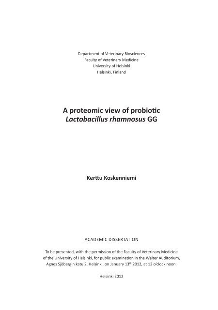
A proteomic view of probiotic Lactobacillus rhamnosus GG
A proteomic view of probiotic Lactobacillus rhamnosus GG
A proteomic view of probiotic Lactobacillus rhamnosus GG
Create successful ePaper yourself
Turn your PDF publications into a flip-book with our unique Google optimized e-Paper software.
Department <strong>of</strong> Veterinary Biosciences<br />
Faculty <strong>of</strong> Veterinary Medicine<br />
University <strong>of</strong> Helsinki<br />
Helsinki, Finland<br />
A <strong>proteomic</strong> <strong>view</strong> <strong>of</strong> probio� c<br />
<strong>Lactobacillus</strong> <strong>rhamnosus</strong> <strong>GG</strong><br />
Ker� u Koskenniemi<br />
ACADEMIC DISSERTATION<br />
To be presented, with the permission <strong>of</strong> the Faculty <strong>of</strong> Veterinary Medicine<br />
<strong>of</strong> the University <strong>of</strong> Helsinki, for public examina� on in the Walter Auditorium,<br />
Agnes Sjöbergin katu 2, Helsinki, on January 13 th 2012, at 12 o’clock noon.<br />
Helsinki 2012
Supervisor Docent Pekka Varmanen<br />
Division <strong>of</strong> Food Technology<br />
Department <strong>of</strong> Food and Environmental Sciences<br />
University <strong>of</strong> Helsinki<br />
Helsinki, Finland<br />
Re<strong>view</strong>ers Pr<strong>of</strong>essor Sirpa Kärenlampi<br />
Department <strong>of</strong> Biosciences<br />
University <strong>of</strong> Eastern Finland<br />
Kuopio, Finland<br />
Docent Jaana Mättö<br />
Finnish Red Cross Blood Service<br />
Helsinki, Finland<br />
Opponent Pr<strong>of</strong>essor Oscar Kuipers<br />
Department <strong>of</strong> Molecular Genetics<br />
Groningen Biomolecular Sciences and Biotechnology Institute<br />
University <strong>of</strong> Groningen<br />
Groningen, the Netherlands.<br />
Layout: Tinde Päivärinta<br />
Cover fi gure: A 2-D DIGE gel image (photo Kerttu Koskenniemi)<br />
ISBN 978-952-10-7403-5 (paperback)<br />
ISBN 978-952-10-7404-2 (PDF)<br />
ISSN 1799-7372<br />
http://ethesis.helsinki.fi<br />
Unigrafi a<br />
Helsinki 2012
CONTENTS<br />
Abstract<br />
List <strong>of</strong> abbreviations<br />
List <strong>of</strong> original publications<br />
1. Re<strong>view</strong> <strong>of</strong> the literature ..........................................................................................1<br />
1.1. Probiotic bacteria ..................................................................................................... 1<br />
1.1.1. Common properties <strong>of</strong> <strong>probiotic</strong> bacteria .............................................. 1<br />
1.1.1.1. Probiotic mechanisms .................................................................. 1<br />
1.1.1.2. Main properties <strong>of</strong> <strong>probiotic</strong> bacteria ......................................... 2<br />
1.1.1.3. Health-benefi cial eff ects <strong>of</strong> <strong>probiotic</strong>s ........................................ 2<br />
1.1.2. Lactobacilli .................................................................................................. 3<br />
1.1.2.1. <strong>Lactobacillus</strong> <strong>rhamnosus</strong> <strong>GG</strong> ........................................................ 3<br />
1.1.3. Bifi dobacteria and propionibacteria ........................................................ 5<br />
1.2. Proteomics ................................................................................................................. 5<br />
1.2.1. Proteomic methods ........................................................................................ 5<br />
1.3. Proteomics <strong>of</strong> potential <strong>probiotic</strong> bacteria ............................................................ 6<br />
1.3.1. Basic proteome research .......................................................................... 10<br />
1.3.1.1. Proteome maps ............................................................................ 10<br />
1.3.1.2. Comparison <strong>of</strong> growth phases ................................................... 11<br />
1.3.1.3. Comparison <strong>of</strong> diff erent strains and growth media ............... 11<br />
1.3.1.4. Development <strong>of</strong> <strong>proteomic</strong> methods ........................................ 12<br />
1.3.2. Proteomics <strong>of</strong> stress responses <strong>of</strong> potentially <strong>probiotic</strong> bacteria ........ 13<br />
1.3.2.1. Bile stress ...................................................................................... 13<br />
1.3.2.2. Acid stress .................................................................................... 22<br />
1.3.2.3. Other stresses ............................................................................... 23<br />
1.3.3. Studies <strong>of</strong> the cell envelope proteomes and secretomes <strong>of</strong><br />
potentially <strong>probiotic</strong> bacteria .................................................................. 24<br />
1.3.3.1. Cell envelope proteome .............................................................. 24<br />
1.3.3.2. Secretome ..................................................................................... 26<br />
1.3.3.3. Surfome and secretome predictions ......................................... 27<br />
1.3.4. Other <strong>proteomic</strong> studies <strong>of</strong> potentially <strong>probiotic</strong> bacteria ..................... 28<br />
2. Aims <strong>of</strong> the study ..................................................................................................30
3. Materials and methods .........................................................................................31<br />
3.1. Bacterial strain and growth conditions ............................................................... 31<br />
3.2. Proteomic methods ................................................................................................ 31<br />
3.2.1. Protein extraction ..................................................................................... 31<br />
3.2.2. Labeling <strong>of</strong> protein samples .................................................................... 31<br />
3.2.3. 2-D GE and image analysis ..................................................................... 32<br />
3.3. Protein identifi cation by MS ................................................................................. 32<br />
3.4. Transcriptomic methods ....................................................................................... 32<br />
3.4.1. Experimental design, RNA methods, cDNA synthesis, and labeling 32<br />
3.4.2. Scanning, image analysis, and data analyses ......................................... 33<br />
3.5. Analysis <strong>of</strong> the phosphoproteome ........................................................................ 33<br />
3.5.1. Protein extraction ..................................................................................... 33<br />
3.5.2. 2-D GE and image analysis ..................................................................... 33<br />
3.5.3. Phosphopeptide identifi cation ................................................................ 33<br />
4. Results and discussion ..........................................................................................34<br />
4.1. Eff ect <strong>of</strong> the growth medium on the proteome <strong>of</strong> L. <strong>rhamnosus</strong> <strong>GG</strong> .............. 34<br />
4.2. Growth phase-associated changes in L. <strong>rhamnosus</strong> <strong>GG</strong> .................................... 35<br />
4.3. Bile stress response <strong>of</strong> L. <strong>rhamnosus</strong> <strong>GG</strong> ............................................................. 37<br />
4.4. Eff ect <strong>of</strong> growth pH on L. <strong>rhamnosus</strong> <strong>GG</strong> ........................................................... 38<br />
4.5. Protein phosphorylation in L. <strong>rhamnosus</strong> <strong>GG</strong> .................................................... 39<br />
4.6. Proteome map <strong>of</strong> L. <strong>rhamnosus</strong> <strong>GG</strong> ..................................................................... 39<br />
5. Concluding remarks and future prospects ..........................................................43<br />
6. Acknowledgements ...............................................................................................45<br />
7. References .........................................................................................................46<br />
Original publications
ABSTRACT<br />
<strong>Lactobacillus</strong> <strong>rhamnosus</strong> <strong>GG</strong> is a <strong>probiotic</strong> bacterium that is known worldwide. Since its<br />
discovery in 1985, the health eff ects and biology <strong>of</strong> this health-promoting strain have<br />
been researched at an increasing rate. However, knowledge <strong>of</strong> the molecular biology<br />
responsible for these health eff ects is limited, even though research in this area has continued<br />
to grow since the publication <strong>of</strong> the whole genome sequence <strong>of</strong> L. <strong>rhamnosus</strong> <strong>GG</strong><br />
in 2009. Probiotic strains encounter various stress conditions during the production,<br />
product formulation, and passage through the gastro-intestinal tract (GIT), which may<br />
aff ect the functionality <strong>of</strong> these organisms. In this thesis, the molecular biology <strong>of</strong> L.<br />
<strong>rhamnosus</strong> <strong>GG</strong> was explored by mapping the changes in protein levels in response to<br />
diverse stress factors and environmental conditions. Th e <strong>proteomic</strong>s data were supplemented<br />
with transcriptome level mapping <strong>of</strong> gene expression.<br />
Th e harsh conditions <strong>of</strong> the GIT, which involve acidic conditions and detergent-like bile<br />
acids, are a notable challenge to the survival <strong>of</strong> <strong>probiotic</strong> bacteria. To simulate GIT conditions,<br />
L. <strong>rhamnosus</strong> <strong>GG</strong> was exposed to a sudden bile stress, and several stress response<br />
mechanisms were revealed. A noteworthy site <strong>of</strong> bile stress responses was the cell envelope,<br />
which was modifi ed in several ways to relieve the harmful eff ects <strong>of</strong> bile and to<br />
improve the adhesion properties <strong>of</strong> L. <strong>rhamnosus</strong> <strong>GG</strong> before the entrance to the gut, e.g.,<br />
debasing <strong>of</strong> the negative charge <strong>of</strong> the cell envelope and reduction in the thickness <strong>of</strong><br />
the exopolysaccharide layer. Mechanisms for recognizing bile compounds and actively<br />
removing them from the cell were activated by bile exposure, and several bile-induced<br />
changes in central metabolism were also detected. L. <strong>rhamnosus</strong> <strong>GG</strong> also responded in<br />
various ways to mild acid stress. Probiotic bacteria may face mild acid stress in dairy production<br />
because the pH is lowered by the production <strong>of</strong> acid by fermenting bacteria in<br />
fermented milk products. Th e acid stress response <strong>of</strong> L. <strong>rhamnosus</strong> <strong>GG</strong> included changes<br />
in central metabolism and in specifi c responses, such as the induction <strong>of</strong> proton-translocating<br />
ATPase, a membrane transporter used for increasing intracellular pH. Th ese<br />
results clearly showed that L. <strong>rhamnosus</strong> <strong>GG</strong> possesses a large repertoire <strong>of</strong> mechanisms<br />
for responding to stress conditions, which probably explains its good survival in GIT.<br />
Adaptation to diff erent growth conditions was studied by comparing the proteome level<br />
responses <strong>of</strong> L. <strong>rhamnosus</strong> <strong>GG</strong> to diff erent growth media and to diff erent phases <strong>of</strong><br />
growth. Th e growth phase-dependent changes were also mapped at the transcriptome<br />
level. Th e growth <strong>of</strong> L. <strong>rhamnosus</strong> <strong>GG</strong> in a rich laboratory medium, MRS (de Man –<br />
Rogosa – Sharpe), and in an industrial-type whey-based medium was compared to reveal<br />
diff erences in laboratory-grown and commercially available <strong>probiotic</strong>s. Th e rich MRS<br />
medium supported the growth <strong>of</strong> L. <strong>rhamnosus</strong> <strong>GG</strong> better than the whey medium, in<br />
which the need for purine and fatty acid biosynthesis was higher. Th e sugar composition<br />
<strong>of</strong> the glucose-containing MRS medium was also optimal for L. <strong>rhamnosus</strong> <strong>GG</strong>, while the<br />
galactose and glucose originating from hydrolyzed lactose in the whey medium created<br />
an increased need for galactose degradation pathways. Th e growth medium also potentially<br />
aff ected the cell surface properties <strong>of</strong> L. <strong>rhamnosus</strong> <strong>GG</strong> because, e.g., the abundance<br />
<strong>of</strong> exopolysaccharide biosynthetic proteins was higher in the MRS medium. Th ese results
lead us to recommend that the industrial-type media should be used in laboratory studies<br />
<strong>of</strong> L. <strong>rhamnosus</strong> <strong>GG</strong> and other <strong>probiotic</strong> bacteria to achieve a similar physiological<br />
state for the bacteria as that found in industrial products, which would thus yield more<br />
relevant information about the bacteria. Th e results could also be utilized for the development<br />
<strong>of</strong> an optimized medium for the industrial production <strong>of</strong> L. <strong>rhamnosus</strong> <strong>GG</strong>.<br />
Comparing diff erent growth phases revealed that the metabolism <strong>of</strong> L. <strong>rhamnosus</strong> <strong>GG</strong> is<br />
modifi ed markedly during shift from the exponential to the stationary phase <strong>of</strong> growth.<br />
Carbohydrate metabolism shift ed from homolactic acid fermentation and glucose consumption<br />
to mixed-acid fermentation and consumption <strong>of</strong> galactose and other alternative<br />
energy sources aft er the transition to stationary phase. Many biosynthetic activities,<br />
such as fatty acid and pyrimidine biosynthesis, were at their highest levels during<br />
the exponential phase <strong>of</strong> growth, whereas the components <strong>of</strong> the proteolytic system<br />
were expressed throughout the progression <strong>of</strong> growth, although, e.g., parallel oligopeptide<br />
transport systems responded divergently under diff erent growth phase conditions.<br />
Several factors that are known or suspected to be associated with <strong>probiotic</strong> eff ects also<br />
showed growth phase-dependent expression, but there was no clear trend in the expression<br />
pr<strong>of</strong>i les that would suggest the optimal harvesting point for the bacteria for <strong>probiotic</strong><br />
preparations. However, this extensive mapping <strong>of</strong> the growth phase-dependent gene<br />
expression pr<strong>of</strong>i les (both transcriptome and proteome level) <strong>of</strong> L. <strong>rhamnosus</strong> <strong>GG</strong> could<br />
be exploited in future studies <strong>of</strong> this important <strong>probiotic</strong> bacterium, e.g., if some new<br />
mediators <strong>of</strong> <strong>probiotic</strong> traits are identifi ed.<br />
In this thesis, a noteworthy phenomenon <strong>of</strong> protein phosphorylation was observed in L.<br />
<strong>rhamnosus</strong> <strong>GG</strong>. Phosphorylation <strong>of</strong> several proteins <strong>of</strong> L. <strong>rhamnosus</strong> <strong>GG</strong> was detected,<br />
and there were hints that the degree <strong>of</strong> phosphorylation may be dependent on the growth<br />
pH. In bacteria, protein phosphorylation has been suggested to regulate enzyme activities<br />
or to direct proteins to diff erent cellular locations, but in this study the purpose <strong>of</strong> the<br />
phosphorylation was not identifi ed. However, in this study, phosphorylation events were<br />
detected for the fi rst time in a <strong>Lactobacillus</strong> strain.
LIST OF ABBREVIATIONS<br />
2-D DIGE two-dimensional diff erence gel electrophoresis<br />
2-D GE two-dimensional gel electrophoresis<br />
ABC ATP-binding cassette<br />
CXC cation exchange chromatography<br />
DTT dithiothreitol<br />
EPS exopolysaccharide<br />
GIT gastro-intestinal tract<br />
IEF isoelectric focusing<br />
IPG immobilized pH gradient<br />
LC liquid chromatography<br />
MALDI matrix-assisted laser desorption/ionization<br />
MRS de Man – Rogosa – Sharpe<br />
MS mass spectrometry<br />
MudPIT multidimensional protein identifi cation technology<br />
ORF open reading frame<br />
pI isoelectric point<br />
PMF peptide mass fi ngerprinting<br />
SDS-PAGE sodium dodecyl sulfate polyacrylamide gel electrophoresis<br />
SILAC stable isotope labeling <strong>of</strong> amino acids in cell cultures<br />
TOF time <strong>of</strong> fl ight
LIST OF ORIGINAL PUBLICATIONS<br />
I Koskenniemi K., Koponen J., Kankainen M., Savijoki K., Tynkkynen S., de Vos<br />
W. M., Kalkkinen N., Varmanen P. (2009) Proteome analysis <strong>of</strong> <strong>Lactobacillus</strong><br />
<strong>rhamnosus</strong> <strong>GG</strong> using 2-D DIGE and mass spectrometry shows diff erential protein<br />
production in laboratory and industrial-type growth media. Journal <strong>of</strong> Proteome<br />
Research 8 (11): 4993–5007.<br />
II Laakso K., Koskenniemi K., Koponen J., Kankainen M., Surakka A., Salusjärvi<br />
T., Auvinen P., Savijoki K., Nyman T. A., Kalkkinen N., Tynkkynen S., Varmanen<br />
P. (2011) Growth phase-associated changes in the proteome and transcriptome <strong>of</strong><br />
<strong>Lactobacillus</strong> <strong>rhamnosus</strong> <strong>GG</strong> in industrial-type whey medium. Microbial Biotechnology<br />
4 (6): 746–766.<br />
III Koskenniemi K., Laakso K., Koponen J., Kankainen M., Greco D., Auvinen P.,<br />
Savijoki K., Nyman T. A., Surakka A., Salusjärvi T., de Vos W. M., Tynkkynen S.,<br />
Kalkkinen N., Varmanen P. (2011) Proteomics and transcriptomics characterization<br />
<strong>of</strong> bile stress response in <strong>probiotic</strong> <strong>Lactobacillus</strong> <strong>rhamnosus</strong> <strong>GG</strong>. Molecular &<br />
Cellular Proteomics 10 (2): M110.002741.<br />
IV Koponen J., Laakso K., Koskenniemi K., Kankainen M., Savijoki K., Nyman T.<br />
A., de Vos W. M., Tynkkynen S., Kalkkinen N., Varmanen P. (2011) Eff ect <strong>of</strong> acid<br />
stress on protein expression and phosphorylation in <strong>Lactobacillus</strong> <strong>rhamnosus</strong> <strong>GG</strong>.<br />
Journal <strong>of</strong> Proteomics doi:10.1016/j.jprot.2011.11.009.<br />
Th e original articles were reprinted with the permission <strong>of</strong> the original copyright holders.
1. REVIEW OF THE LITERATURE<br />
1.1. Probio� c bacteria<br />
Probiotic bacteria are defi ned as “live microorganisms,<br />
which, when administered in<br />
adequate amounts, confer a health benefi t on<br />
the host” (FAO/WHO, 2002). A wide range<br />
<strong>of</strong> commercial <strong>probiotic</strong> products is available,<br />
which contain diff erent <strong>probiotic</strong> strains<br />
that elicit varying health-beneficial effects.<br />
Th e majority <strong>of</strong> the <strong>probiotic</strong> bacteria in use<br />
today belong to the genera <strong>Lactobacillus</strong> and<br />
Bifidobacterium (Marco et al., 2006; Minocha,<br />
2009), but products containing strains<br />
from other genera such as Propionibacterium,<br />
Enterococcus, and Escherichia are also available<br />
(Ouwehand et al., 2002).<br />
1.1.1. Common proper� es <strong>of</strong><br />
probio� c bacteria<br />
1.1.1.1. Probio� c mechanisms<br />
Most <strong>of</strong> the proven health eff ects <strong>probiotic</strong>s<br />
elicit are provided in the gastro-intestinal tract<br />
(GIT). Th e <strong>probiotic</strong> mechanisms <strong>of</strong> action in<br />
the GIT can be roughly divided into luminal,<br />
mucosal, and submucosal eff ects (Sherman et<br />
al., 2009). Th e basic luminal eff ect (an eff ect<br />
which appears in gut lumen) <strong>of</strong> <strong>probiotic</strong>s is<br />
the improvement <strong>of</strong> intestinal microbial balance.<br />
Th e human GIT is colonized by a myriad<br />
<strong>of</strong> microbes, whose balanced composition and<br />
activity are essential for human health (Round<br />
& Mazmanian, 2009). Eating <strong>probiotic</strong>s maintains<br />
or promotes the GIT homeostasis, and<br />
<strong>probiotic</strong>s have been found to stimulate the<br />
growth <strong>of</strong> indigenous benefi cial gut microbes<br />
such as bifi dobacteria and inhibit the growth<br />
<strong>of</strong> pathogenic or opportunistic pathogenic<br />
microbes (Ohashi & Ushida, 2009; Ouwehand<br />
Re<strong>view</strong> <strong>of</strong> the Literature<br />
et al., 2002; Sherman et al., 2009). Examples<br />
<strong>of</strong> stimulatory eff ects are reported in a study<br />
by Benno and colleagues (Benno et al., 1996),<br />
who showed that the consumption <strong>of</strong> <strong>probiotic</strong><br />
<strong>Lactobacillus</strong> <strong>rhamnosus</strong> <strong>GG</strong> increased the<br />
number <strong>of</strong> fecal bifi dobacteria, and in a study<br />
by Sui et al. (Sui et al., 2002), in which ingestion<br />
<strong>of</strong> <strong>probiotic</strong> <strong>Lactobacillus</strong> acidophilus<br />
NCFM changed the colonic lactobacilli composition.<br />
Pathogenic or opportunistic pathogenic<br />
microbes are inhibited by antibacterial<br />
products such as bacteriocins and lactic acid,<br />
as shown in several in vitro studies. For example,<br />
<strong>probiotic</strong> L. <strong>rhamnosus</strong> <strong>GG</strong> inhibited the<br />
growth <strong>of</strong> pathogenic Salmonella enterica by<br />
producing lactic acid and other secreted antimicrobial<br />
molecules (Marianelli et al., 2010),<br />
and several bacteriocins produced by lactic<br />
acid bacteria have been shown to have antimicrobial<br />
activity against the gastric pathogen<br />
Helicobacter pylori (Kim et al., 2003). Probiotics<br />
can also adhere to the gut and prevent<br />
pathogens from occupying this living space<br />
(which is called colonization resistance)<br />
(Gueimonde et al., 2007; Saxelin et al., 2005;<br />
Sherman et al., 2009). Probiotic Streptococcus<br />
thermophilus ATCC 19258 and L. acidophilus<br />
ATCC 4356 have been shown to interfere with<br />
the adhesion and invasion <strong>of</strong> enteroinvasive<br />
Escherichia coli in human intestinal epithelial<br />
cells in vitro (Resta-Lenert & Barrett, 2003).<br />
Th e mucosal eff ects <strong>of</strong> <strong>probiotic</strong>s include<br />
the enhancement <strong>of</strong> host mucin production,<br />
which improves the ability <strong>of</strong> the mucus layer<br />
to act as an antibacterial shield (Mack et al.,<br />
2003; Sherman et al., 2009). Some <strong>probiotic</strong>s,<br />
such as E. coli Nissle 1917 and a few <strong>Lactobacillus</strong><br />
strains, induce antimicrobial peptide<br />
(e.g., defensin) production in the host<br />
1
Re<strong>view</strong> <strong>of</strong> the Literature<br />
and thus help the host strengthen its innate<br />
defence mechanisms (Schlee et al., 2008; Wehkamp<br />
et al., 2004). Probiotics have also been<br />
shown to enhance the integrity <strong>of</strong> the host<br />
intestinal barrier; treatment <strong>of</strong> human colonic<br />
cells with L. <strong>rhamnosus</strong> <strong>GG</strong> prevented injuries<br />
in the epithelial cell barrier that were induced<br />
by enterohemorrhagic E. coli (Johnson-Henry<br />
et al., 2008), and L. acidophilus LB protected<br />
human colonic cells from aspirin-induced<br />
damage in tight junctions (Montalto et al.,<br />
2004).<br />
Submucosal eff ects include the eff ects <strong>of</strong><br />
<strong>probiotic</strong>s on the host immune system. Probiotics<br />
have been shown to improve the intestine’s<br />
immunological barrier functions and<br />
alleviate the intestinal infl ammatory response<br />
by mechanisms that include diverse effects<br />
on immune activation, cytokine production,<br />
immunomodulation, and infl ammation (Delcenserie<br />
et al., 2008; Wells, 2011).<br />
1.1.1.2. Main proper� es <strong>of</strong> probio� c<br />
bacteria<br />
To be suitable for <strong>probiotic</strong> use, a bacterial<br />
strain should have certain characteristics.<br />
It should survive the passage through the<br />
GIT and thus be resistant to GIT conditions,<br />
including acidic pH and bile acids (Bezkorovainy,<br />
2001; Fuller, 1989). Th e safety <strong>of</strong> the<br />
strain must be evident (EFSA, 2008; FAO/<br />
WHO, 2002; Fuller, 1989), and strains <strong>of</strong><br />
human origin are usually preferred to achieve<br />
host-specifi c <strong>probiotic</strong> eff ects (Ouwehand et<br />
al., 2002). Th e ability to adhere to intestinal<br />
mucosa is a desired property <strong>of</strong> a <strong>probiotic</strong><br />
because close contact and prolonged colonization<br />
may intensify the favorable effects<br />
<strong>of</strong> <strong>probiotic</strong>s (Ouwehand et al., 2002). Good<br />
technological properties are also important<br />
for a strain to be used in a commercial <strong>probiotic</strong><br />
product; the strain should be suitable<br />
2<br />
for large-scale cultivation, remain viable and<br />
stable under storage, and should not confer<br />
an unpleasant taste on the product (Saarela et<br />
al., 2000). Naturally, a <strong>probiotic</strong> strain also has<br />
to impair benefi cial health eff ects to the host<br />
(FAO/WHO, 2002; Fuller, 1989).<br />
1.1.1.3. Health-benefi cial eff ects <strong>of</strong><br />
probio� cs<br />
Th e potential health eff ects <strong>of</strong> <strong>probiotic</strong>s have<br />
been studied for several diseases and conditions<br />
using a variety <strong>of</strong> diff erent strains, and<br />
varying results have been achieved. Th e bestproven<br />
health benefit for several <strong>probiotic</strong><br />
strains is the reduction <strong>of</strong> the risk <strong>of</strong> diarrhea<br />
(e.g., antibiotic-associated and traveler’s diarrhea)<br />
and the shortening <strong>of</strong> diarrheal episodes<br />
(Hickson et al., 2007; Minocha, 2009; Ouwehand<br />
et al., 2002; Saxelin et al., 2005; Weichselbaum,<br />
2009). A meta-analysis <strong>of</strong> 34 blinded,<br />
randomized, placebo-controlled trials studying<br />
the eff ect <strong>of</strong> diff erent <strong>probiotic</strong>s (mainly<br />
<strong>Lactobacillus</strong> strains) in the prevention <strong>of</strong><br />
acute diarrhea showed that <strong>probiotic</strong>s signifi -<br />
cantly reduced antibiotic-associated diarrhea<br />
by 52% and acute diarrhea <strong>of</strong> various causes<br />
by 34% (Sazawal et al., 2006). Other diseases<br />
<strong>of</strong> the gut may also be alleviated with <strong>probiotic</strong>s.<br />
A meta-analysis <strong>of</strong> twenty randomized,<br />
controlled, blinded trials showed that <strong>probiotic</strong><br />
use may be associated with an improvement<br />
in irritable bowel syndrome symptoms compared<br />
to placebo (McFarland & Dublin, 2008).<br />
The use <strong>of</strong> <strong>probiotic</strong>s may be related to the<br />
relief <strong>of</strong> constipation (Koebnick et al., 2003;<br />
Weichselbaum, 2009) and lactose intolerance<br />
(Vesa et al., 2000). Probiotics may increase<br />
host immune defenses and thus decrease the<br />
frequency or duration <strong>of</strong> infections like the<br />
common cold (de Vrese et al., 2005; Minocha,<br />
2009; Weichselbaum, 2009; Weizman et al.,<br />
2005). In a double-blind, randomized trial, the
ingestion <strong>of</strong> <strong>Lactobacillus</strong> gasseri PA 16/8, Bifi -<br />
dobacterium longum SP 07/3, and Bifi dobacterium<br />
bifi dum MF 20/5 shortened the duration<br />
<strong>of</strong> common cold episodes in healthy adults (de<br />
Vrese et al., 2005), and among children in daycare<br />
centers, the intake <strong>of</strong> <strong>Lactobacillus</strong> reuteri<br />
ATCC 55730 decreased the number <strong>of</strong> days<br />
with fever (Weizman et al., 2005). Probiotics<br />
have also been shown to be helpful in preventing<br />
allergic disorders (Kalliomäki et al.,<br />
2001; Minocha, 2009; Ouwehand et al., 2002;<br />
Saxelin et al., 2005); in children with allergic<br />
rhinitis, consumption <strong>of</strong> <strong>Lactobacillus</strong> casei<br />
DN-114 001-containing fermented milk lowered<br />
the annual number <strong>of</strong> rhinitis episodes<br />
(Giovannini et al., 2007), and the intake <strong>of</strong> L.<br />
casei Shirota was shown to modulate immune<br />
responses <strong>of</strong> adults suffering from seasonal<br />
allergic rhinitis (Ivory et al., 2008).<br />
To achieve the desired health eff ects <strong>of</strong><br />
a <strong>probiotic</strong>, the correct dosage is required to<br />
deliver a sufficient amount <strong>of</strong> live <strong>probiotic</strong>s<br />
to the GIT, as demonstrated by Whorwell<br />
and colleagues (Whorwell et al., 2006). In that<br />
study, a dose <strong>of</strong> 1 × 10 8 CFU/ml <strong>of</strong> Bifi dobacterium<br />
infantis 35624 signifi cantly reduced the<br />
symptoms <strong>of</strong> irritable bowel syndrome, but<br />
doses <strong>of</strong> 1 × 10 6 and 1 × 10 10 CFU/ml were not<br />
significantly different from placebo (Whorwell<br />
et al., 2006). Probiotics have to be taken<br />
regularly because <strong>probiotic</strong>s usually do not<br />
colonize the GIT permanently (Marco et al.,<br />
2006; Ohashi & Ushida, 2009; Weichselbaum,<br />
2009). It also has to be emphasized that every<br />
<strong>probiotic</strong> strain has its own specific effects,<br />
and the research results for one strain can<br />
never be directly applied to other strains. In<br />
addition, there may be diff erences in responses<br />
between individuals (Ohashi & Ushida,<br />
2009). Moreover, the form in which the <strong>probiotic</strong><br />
is ingested is also important, as has been<br />
shown with <strong>probiotic</strong> Propionibacterium and<br />
Re<strong>view</strong> <strong>of</strong> the Literature<br />
Bifi dobacterium strains, which showed higher<br />
fecal counts when they were administered as<br />
capsules or yogurt than when they were consumed<br />
in cheese (Saxelin et al., 2010).<br />
1.1.2. Lactobacilli<br />
<strong>Lactobacillus</strong> is a bacterial genus comprised<br />
<strong>of</strong> Gram-positive, rod-shaped bacteria with a<br />
low percentage <strong>of</strong> guanine and cytosine bases<br />
in their genome, and they are typically aerotolerant<br />
anaerobes. Taxonomically, the <strong>Lactobacillus</strong><br />
genus is diverse and it contains at<br />
least twelve separatable phylogenetic groups<br />
(Felis & Dellaglio, 2007). More than 150 species<br />
have been named within the <strong>Lactobacillus</strong><br />
genus, which were isolated, e.g., from human<br />
and animal GITs and mucous membranes<br />
and from plant surfaces. Several <strong>Lactobacillus</strong><br />
strains are used in the preparation <strong>of</strong> fermented<br />
dairy products and in the production<br />
<strong>of</strong> sauerkraut, pickles, and silage. Certain<br />
<strong>Lactobacillus</strong> strains have been found to have<br />
benefi cial eff ects on human health, some <strong>of</strong><br />
which are therefore used as <strong>probiotic</strong>s. One<br />
<strong>of</strong> the most important <strong>probiotic</strong> <strong>Lactobacillus</strong><br />
strain is L. <strong>rhamnosus</strong> <strong>GG</strong>, which is probably<br />
the most intensively studied <strong>probiotic</strong> bacterium<br />
worldwide. L. <strong>rhamnosus</strong> belongs to an<br />
L. casei phylogenetic group together with L.<br />
casei, <strong>Lactobacillus</strong> paracasei, and <strong>Lactobacillus</strong><br />
zeae (Felis & Dellaglio, 2007).<br />
1.1.2.1. <strong>Lactobacillus</strong> <strong>rhamnosus</strong> <strong>GG</strong><br />
Th e <strong>probiotic</strong> strain L. <strong>rhamnosus</strong> <strong>GG</strong> (ATCC<br />
53103) was originally isolated by Goldin and<br />
Gorbach in 1985 (Doron et al., 2005). Strain<br />
<strong>GG</strong> was selected from a collection <strong>of</strong> <strong>Lactobacillus</strong><br />
strains isolated from stool samples from<br />
healthy human volunteers, using the following<br />
criteria for an ideal <strong>probiotic</strong> strain: bile and<br />
acid resistance and the ability to persist in the<br />
harsh conditions <strong>of</strong> the GIT, adhere to human<br />
3
Re<strong>view</strong> <strong>of</strong> the Literature<br />
epithelial cells, and colonize the human intestine<br />
(Doron et al., 2005). Th e production <strong>of</strong><br />
antimicrobial substances such as organic acids<br />
is also desirable, and good growth characteristics<br />
are useful in large-scale commercial production<br />
(Doron et al., 2005). Th e most important<br />
characteristic for a <strong>probiotic</strong> is, naturally,<br />
benefi cial eff ects on human health. Th e fi rst<br />
report <strong>of</strong> positive health eff ects <strong>of</strong> L. <strong>rhamnosus</strong><br />
<strong>GG</strong> was published in 1987 (Gorbach et al.,<br />
1987), and more than 500 scientifi c articles<br />
on this <strong>probiotic</strong> strain have since been published.<br />
The health effects <strong>of</strong> L. <strong>rhamnosus</strong> <strong>GG</strong><br />
are based on several mechanisms. L. <strong>rhamnosus</strong><br />
<strong>GG</strong> colonizes the GIT efficiently and<br />
competes for adhesion sites and nutrients,<br />
such as monosaccharides, with pathogens. L.<br />
<strong>rhamnosus</strong> <strong>GG</strong> also modulates the microecology<br />
<strong>of</strong> the GIT, e.g., by producing short-chain<br />
fatty acids, which favor the growth <strong>of</strong> nonpathogenic<br />
organisms. In addition, L. <strong>rhamnosus</strong><br />
<strong>GG</strong> has numerous eff ects on the host<br />
immune system (Doron et al., 2005). A recent<br />
comparative genomics study <strong>of</strong> L. <strong>rhamnosus</strong><br />
<strong>GG</strong> and its close relative, a dairy strain L.<br />
<strong>rhamnosus</strong> Lc705, revealed that L. <strong>rhamnosus</strong><br />
<strong>GG</strong> carries unique pilus genes (spaCBA) in<br />
its genome (Kankainen et al., 2009). Additional<br />
studies <strong>of</strong> a mutant in which the pilus<br />
gene spaC was inactivated showed that SpaC<br />
is essential for the adherence <strong>of</strong> L. <strong>rhamnosus</strong><br />
<strong>GG</strong> to intestinal mucus in vitro, and the presence<br />
<strong>of</strong> cell surface pili was assessed to explain<br />
the ability <strong>of</strong> strain <strong>GG</strong> to persist longer in the<br />
human GIT than strain Lc705 (Kankainen et<br />
al., 2009).<br />
The best-proven health benefit <strong>of</strong> L.<br />
<strong>rhamnosus</strong> <strong>GG</strong> is lowered risk and reduced<br />
treatment days for acute diarrhea in children<br />
(Szajewska et al., 2007), as shown, e.g., in a<br />
broad study in ten European countries, where<br />
4<br />
children with acute diarrhea recovered and<br />
were discharged from the hospital faster when<br />
treated with oral rehydration solution containing<br />
L. <strong>rhamnosus</strong> <strong>GG</strong> than when treated with<br />
a corresponding placebo solution (Guandalini<br />
et al., 2000). L. <strong>rhamnosus</strong> <strong>GG</strong> can also reduce<br />
the risk for antibiotic-associated diarrhea and<br />
other intestinal side effects associated with<br />
the use <strong>of</strong> antibiotics (Cremonini et al., 2002;<br />
Doron et al., 2008). For example, the administration<br />
<strong>of</strong> L. <strong>rhamnosus</strong> <strong>GG</strong> to children<br />
receiving antibiotic therapy for respiratory<br />
infections reduced the incidence <strong>of</strong> antibioticassociated<br />
diarrhea to one-third (Arvola et al.,<br />
1999). Substantial evidence has accumulated<br />
to support the eff ect <strong>of</strong> L. <strong>rhamnosus</strong> <strong>GG</strong> in<br />
treating (Isolauri et al., 2000; Majamaa & Isolauri,<br />
1997) and preventing (Kalliomäki et<br />
al., 2001; Kalliomäki et al., 2003; Kalliomäki<br />
et al., 2007) atopic diseases in children. Th e<br />
preventive eff ect among children at high risk<br />
for atopic eczema was achieved by administering<br />
L. <strong>rhamnosus</strong> <strong>GG</strong> prenatally for 2–4 weeks<br />
and postnatally for 6 months (Kalliomäki et<br />
al., 2001), and the reduced cumulative risk for<br />
developing eczema was evident even aft er seven<br />
years (Kalliomäki et al., 2007). In a similar<br />
experiment where a mixture <strong>of</strong> L. <strong>rhamnosus</strong><br />
<strong>GG</strong>, L. acidophilus La-5, and Bifi dobacterium<br />
animalis subsp. lactis Bb-12 was given to pregnant<br />
women, the cumulative incidence <strong>of</strong><br />
atopic dermatitis in 2-year-old children was<br />
reduced, but no eff ect on atopic sensitization<br />
was observed (Dotterud et al., 2010). In addition,<br />
consumption <strong>of</strong> L. <strong>rhamnosus</strong> <strong>GG</strong> may<br />
reduce the risk and duration <strong>of</strong> respiratory<br />
tract infections in children attending day care<br />
centers (Hatakka et al., 2001; Hojsak et al.,<br />
2010), and the risk for dental caries in children<br />
has been shown to be reduced by consuming<br />
strain <strong>GG</strong> (Näse et al., 2001).
1.1.3. Bifi dobacteria and<br />
propionibacteria<br />
Two other important genera that include<br />
<strong>probiotic</strong> strains are Bifidobacterium and<br />
Propionibacterium. Th ey are, Gram-positive<br />
bacteria with a high percentage <strong>of</strong> guanine<br />
and cytosine bases in their genome, and they<br />
belong to the phylum Actinobacteria. Bifi dobacteria<br />
are important inhabitants <strong>of</strong> the GIT,<br />
and high numbers <strong>of</strong> bifi dobacteria in the GIT<br />
are considered positive indicators <strong>of</strong> health<br />
(Saulnier et al., 2009). Th e most widely studied<br />
<strong>probiotic</strong> Bifi dobacterium strain is probably<br />
B. animalis subsp. lactis Bb-12, the use<br />
<strong>of</strong> which has been shown to reduce the risk<br />
for respiratory infections in infants (Taipale<br />
et al., 2011), to have some protective effect<br />
against diarrhea in children (Chouraqui et al.,<br />
2004; Weizman et al., 2005), and to reduce the<br />
extent and severity <strong>of</strong> atopic eczema in infants<br />
(Isolauri et al., 2000). B. animalis subsp. lactis<br />
species are typically fairly stress tolerant<br />
compared with other Bifi dobacterium species,<br />
which is important for their usability in <strong>probiotic</strong><br />
preparations (Corcoran et al., 2008).<br />
Propionibacteria are commonly used as<br />
starter cultures in the dairy industry, especially<br />
in Swiss-type cheeses, and there are<br />
fewer reports on their <strong>probiotic</strong> properties<br />
than what is available for <strong>probiotic</strong> <strong>Lactobacillus</strong><br />
and Bifi dobacterium strains (Cousin et al.,<br />
2010). However, one <strong>of</strong> the most <strong>of</strong>t en studied<br />
potentially <strong>probiotic</strong> Propionibacterium strain,<br />
Propionibacterium freudenreichii subsp. shermanii<br />
JS, has been shown to have anti-infl ammatory<br />
effects during H. pylori infection in<br />
vitro (Myllyluoma et al., 2008). Furthermore,<br />
this strain has been shown to reduce serum<br />
C-reactive protein levels (CRP, a marker <strong>of</strong><br />
infl ammation) in healthy adults (Kekkonen et<br />
al., 2008).<br />
1.2. Proteomics<br />
Re<strong>view</strong> <strong>of</strong> the Literature<br />
In the research <strong>of</strong> the molecular biology <strong>of</strong><br />
<strong>probiotic</strong> bacteria, one worthy technique is<br />
<strong>proteomic</strong>s. Proteomics is a tool for studying<br />
the proteome, i.e., the set <strong>of</strong> proteins expressed<br />
under a defi ned physiological condition in an<br />
organism (or cell line or tissue). All proteins<br />
are encoded by the genome <strong>of</strong> the organism.<br />
While the genome is a relatively invariable<br />
reserve <strong>of</strong> potential functions, the proteome<br />
reveals the active functions in the organism.<br />
The proteome changes continuously, and a<br />
large amount <strong>of</strong> information on the functional<br />
responses <strong>of</strong> the organism can be obtained by<br />
studying the proteome under diff erent physiological<br />
conditions.<br />
1.2.1. Proteomic methods<br />
A traditional method for revealing proteomes<br />
is two-dimensional gel electrophoresis (2-D<br />
GE), which was developed in 1975 in parallel<br />
by O’Farrell (O’Farrell, 1975) and Klose<br />
(Klose, 1975). In 2-D GE, proteins are separated<br />
on acrylamide gels, fi rst based on their<br />
isoelectric points (pI, first dimension) and<br />
then based on their molecular weights (second<br />
dimension). Isoelectric focusing (IEF) is<br />
performed on immobilized pH gradient (IPG)<br />
strips, and the second dimension separation is<br />
performed using sodium dodecyl sulfate polyacrylamide<br />
gel electrophoresis (SDS-PAGE).<br />
Two different proteomes can be compared<br />
quantitatively by comparing their 2-D<br />
proteome maps if the proteins are stained<br />
using a quantitative dye, such as Coomassie<br />
blue or fluorescent dyes. A traditional and<br />
accurate method for quantitative <strong>proteomic</strong>s<br />
is metabolic labeling <strong>of</strong> proteins with radioactive<br />
amino acids, such as 35 S-methionine. By<br />
pulse labeling with radioactive amino acids,<br />
the newly synthesized proteins can be identi-<br />
5
Re<strong>view</strong> <strong>of</strong> the Literature<br />
fi ed and separated from the proteins that were<br />
present in the cell before the addition <strong>of</strong> the<br />
radioactive compound. Another excellent<br />
and relatively recently developed method for<br />
quantitative comparison <strong>of</strong> proteome populations<br />
is two-dimensional diff erence gel electrophoresis<br />
(2-D DIGE) (Ünlü et al., 1997).<br />
In 2-D DIGE, proteins are labeled using three<br />
diff erent fl uorescent dyes, Cy2, Cy3, and Cy5,<br />
each <strong>of</strong> which have separate adsorption and<br />
emission characteristics. Using these dyes,<br />
two diff erent protein samples can be quantitatively<br />
compared on a single gel. In addition,<br />
an internal standard can be used to improve<br />
the accuracy <strong>of</strong> the quantifi cation. Th e three<br />
proteome 2-D images are then scanned from<br />
the gel using three diff erent wavelengths. By<br />
this kind <strong>of</strong> multiplexing, the eff ect <strong>of</strong> intrinsic<br />
gel-to-gel variability <strong>of</strong> the 2-D method can be<br />
diminished.<br />
In addition to gel electrophoretic methods,<br />
bacterial proteomes can be analyzed<br />
using non-gel-based methods. Using highperformance<br />
liquid chromatography and<br />
mass spectrometric methods, it is possible to<br />
separate and identify complex peptide mixtures.<br />
Accurate protein quantifi cation can be<br />
achieved using, e.g., the isotopic labeling <strong>of</strong><br />
proteins and peptides (Monteoliva & Albar<br />
2004). Non-gel-based <strong>proteomic</strong> methods are<br />
useful in high throughput applications and<br />
in the cases where the target is to identify a<br />
maximal number <strong>of</strong> different proteins. The<br />
gel-based methods, alternatively, are useful,<br />
e.g., in getting an over<strong>view</strong> <strong>of</strong> proteome level<br />
changes or when the presence <strong>of</strong> diff erent<br />
protein is<strong>of</strong>orms, post-translational modifi cations,<br />
or protein fragments is explored.<br />
6<br />
1.3. Proteomics <strong>of</strong> poten� al<br />
probio� c bacteria<br />
Proteomics research <strong>of</strong> <strong>probiotic</strong> bacteria is<br />
a relatively recent fi eld <strong>of</strong> research, but <strong>proteomic</strong>s<br />
methods have begun to be exploited<br />
in several studies <strong>of</strong> <strong>probiotic</strong>s in the last<br />
decade. First, <strong>proteomic</strong>s has been used to<br />
obtain a proteome map or an over<strong>view</strong> <strong>of</strong> bacterial<br />
protein content. Second, adaptation to<br />
gut conditions such as low pH and bile acids<br />
has been an important research theme. Proteins<br />
localized on the cell surface have also<br />
been a subject <strong>of</strong> interest because the cell<br />
surface is the main contact point between<br />
the <strong>probiotic</strong> and the host. In addition, <strong>proteomic</strong>s<br />
has been used as a tool to answer<br />
various special questions about the molecular<br />
biology <strong>of</strong> potentially <strong>probiotic</strong> bacteria. Previous<br />
re<strong>view</strong>s have discussed the <strong>proteomic</strong>s<br />
<strong>of</strong> lactic acid bacteria and bifidobacteria in<br />
general (Champomier-Vergès et al., 2002;<br />
De Angelis & Gobbetti, 2004; Di Cagno et<br />
al., 2011; Gagnaire et al., 2009; Manso et al.,<br />
2005; Pessione et al., 2010; Sánchez et al.,<br />
2008c), but only one restricted re<strong>view</strong> focusing<br />
on <strong>proteomic</strong>s <strong>of</strong> <strong>probiotic</strong> bacteria (Aires<br />
& Butel, 2011) and two studies about secreted<br />
proteins <strong>of</strong> <strong>probiotic</strong>s (Sánchez et al., 2008a;<br />
Sánchez et al., 2010b) are available.<br />
In the following chapters, proteome studies<br />
<strong>of</strong> bacterial strains that are commercially<br />
used or scientifically proven <strong>probiotic</strong>s or<br />
potential <strong>probiotic</strong>s are re<strong>view</strong>ed. Th ese studies<br />
are also listed in Table 1.
Table 1. Proteomic studies <strong>of</strong> <strong>probiotic</strong> bacteria.<br />
Topic Separation and detection methods Identifi cation method Potentially <strong>probiotic</strong> strains Reference<br />
Basic proteome research<br />
proteome catalogue MudPIT LC-MS/MS B. infantis BI07 Vitali et al., 2005<br />
proteome catalogue no data NanoLC-MS/MS, MALDI-MS/MS, P. freudenreichii CIRM-BIA1 Falentin et al., 2010<br />
CXC-LC-MS/MS<br />
proteome catalogue, comparison <strong>of</strong> strains SDS-PAGE, LC NanoLC-MS/MS L. <strong>rhamnosus</strong> <strong>GG</strong> and Lc705 Savijoki et al., 2011<br />
proteome map 2-D GE, Coomassie staining MS B. animalis subsp. lactis Bb-12 Garrigues et al., 2005<br />
proteome map, comparison <strong>of</strong> growth phases 2-D GE, silver staining MALDI-MS/MS L. casei Zhang Wu et al., 2009<br />
proteome map, growth on diff erent sugars 2-D GE, Coomassie staining MALDI-MS, nanospray ESI-MS/MS B. longum NCC2705 Yuan et al., 2006<br />
proteome map, growth on lactitol 2-D GE, Coomassie staining; 2-D MALDI-MS/MS L. acidophilus NCFM Majumder et al., 2011<br />
DIGE<br />
development <strong>of</strong> an isotope labeling method SDS-PAGE MALDI-MS/MS B. longum NCIMB8809 Couté et al., 2007<br />
development <strong>of</strong> defi ned media for<br />
2-D GE, [<br />
radiolabeling<br />
35S]methionine labeling immunoblotting, specifi c antibodies L. brevis ATCC 8287 Savijoki et al., 2006<br />
Comparison <strong>of</strong> strains and growth phases<br />
comparison <strong>of</strong> growth phases 2-D GE, silver staining MALDI-MS L. plantarum WCFS1 Cohen et al., 2006<br />
comparison <strong>of</strong> growth phases 2-D GE, SYPRO Orange staining LC-MS/MS L. plantarum MLBPL1 Koistinen et al., 2007<br />
comparison <strong>of</strong> strains 2-D GE, Coomassie staining MALDI-MS B. longum NCC2705 Aires et al., 2010<br />
L. <strong>rhamnosus</strong> E-97800<br />
2-D GE, SYPRO Ruby staining LC-MS/MS L. plantarum MLBPL1 Plumed-Ferrer et al.,<br />
2008<br />
comparison <strong>of</strong> strains, growth on diff erent<br />
media<br />
Sánchez et al., 2007b<br />
Stress<br />
bile stress 2-D GE, Coomassie staining MALDI-MS B. animalissubsp. lactis IPLA<br />
4549<br />
Re<strong>view</strong> <strong>of</strong> the Literature<br />
bile stress 2-D GE, Coomassie staining MALDI-MS B. longum NCIMB8809 Sánchez et al., 2005<br />
bile stress 2-D GE, silver staining MALDI-MS L. casei Zhang Wu et al., 2010<br />
bile stress 2-D GE, Coomassie staining MALDI-MS L. delbrueckii subsp. lactis 200 Burns et al., 2010<br />
bile stress 2-D GE, Coomassie staining LC-MS/MS L. plantarum 299V Hamon et al., 2011<br />
bile stress 2-D GE, silver staining MALDI-MS L. reuteri ATCC 23272 Lee et al., 2008a<br />
7
Re<strong>view</strong> <strong>of</strong> the Literature<br />
Topic Separation and detection methods Identifi cation method Potentially <strong>probiotic</strong> strains Reference<br />
bile stress 2-D GE, [ 35S]methionine/cysteine N-terminal sequencing, MALDI-MS P. freundreichii SI41 Leverrier et al., 2003<br />
labeling<br />
bile and acid stress 2-D GE, [ 35S]methionine/cysteine no novel identifi cations P. freundreichii SI41 Jan et al., 2002<br />
labeling<br />
bile, acid, and heat stress 2-D GE, [ 35S]methionine/cysteine LC-MS/MS P. freundreichii SI41 Leverrier et al., 2004<br />
labeling<br />
eff ect <strong>of</strong> bile stress on cell wall proteome 2-D GE, silver staining MALDI-MS B. animalissubsp. lactis BI07 Candela et al., 2010<br />
eff ect <strong>of</strong> bile stress on cell-envelope proteome DIGE; SDS-PAGE; SILAC MALDI-MS/MS B. longum NCIMB8809 Ruiz et al., 2009a<br />
exposure to rabbit jejunum 2-D GE, Coomassie staining MALDI-MS L. fermentum I5007 Yang et al., 2007<br />
exposure to rabbit large intestine 2-D GE, Coomassie staining MALDI-MS, nanospray ESI-MS/MS B. longum NCC2705 Yuan et al., 2008<br />
acid stress 2-D GE, Coomassie staining MS B. longum NCIMB8809 Sánchez et al., 2007a<br />
acid stress 2-D GE, silver staining MALDI-MS L. casei Zhang Wu et al., 2011<br />
acid stress 2-D GE, silver staining MALDI-MS L. reuteri ATCC 23272 Lee et al., 2008b<br />
acid stress 2-D GE, silver staining MALDI-MS L. reuteri ATCC 23272 Lee and Pi, 2010<br />
acid stress 2-D GE, [ 35S]methionine/cysteine N-terminal sequencing P. freundreichii SI41 Jan et al., 2001<br />
labeling<br />
heat stress 2-D DIGE MALDI-MS L. gasseri ATCC 33323 Suokko et al., 2008<br />
heat and osmotic stress 2-D GE, [ 35 Table 1. cont.<br />
S]methionine/cysteine N-terminal sequencing L. <strong>rhamnosus</strong> HN001 Prasad et al., 2003<br />
labeling<br />
oxidative stress 2-D GE, Coomassie staining MALDI-MS B. longum subsp. longum Xiao et al., 2011<br />
BBMN68<br />
comparison <strong>of</strong> strains with diff erent stress LC LC-MS/MS B. longum NCC2705 Guillaume et al., 2009<br />
tolerance<br />
8<br />
Cell envelope proteome and secretome<br />
cell surface proteins SDS-PAGE LC-MS/MS L. plantarum 423 and WCFS1 Ramiah et al., 2008<br />
L. acidophilus ATCC 4356<br />
cell wall proteins binding to human<br />
2-D GE, Coomassie staining MALDI-MS B. lactis BI07 Candela et al., 2007<br />
plasminogen<br />
cell wall-associated proteome SDS-PAGE (and 2D- GE, silver LC-MS/MS L. salivarius UCC118 Kelly et al., 2005<br />
staining)<br />
surfome SDS-PAGE LC-MS/MS L. plantarum 299v Beck et al., 2009<br />
surfome SDS-PAGE MALDI-MS/MS L. <strong>rhamnosus</strong> <strong>GG</strong> Sánchez et al., 2009a<br />
adhesion proteins 2-D GE, Coomassie staining LC-MS/MS L. plantarum 299V Izquierdo et al., 2009<br />
comparison <strong>of</strong> diff erentially aggregative strains 2-D GE, Coomassie staining MALDI-MS, LC-MS/MS L. crispatus M247 and Mu5 Siciliano et al., 2008
Topic Separation and detection methods Identifi cation method Potentially <strong>probiotic</strong> strains Reference<br />
secretome 2-D GE, Coomassie staining MALDI-MS B. animalissubsp. lactis Bb-12 Gilad et al., 2011<br />
secretome 2-D GE, Coomassie staining MALDI-MS B. longum NCIMB8809 Sánchez et al., 2008b<br />
secretome SDS-PAGE MALDI-MS/MS L. gasseri B3 Sánchez et al., 2009b<br />
L. reuteri Protectis<br />
L. <strong>rhamnosus</strong> R-11<br />
secretome SDS-PAGE MALDI-MS/MS L. <strong>rhamnosus</strong> <strong>GG</strong> Sánchez et al., 2009d<br />
secretome, binding to fi bronectin SDS-PAGE MALDI-MS/MS L. plantarum 299v, NCIMB Sánchez et al., 2009c<br />
8826<br />
secretome, mucin degradation SDS-PAGE MALDI-MS/MS L. <strong>rhamnosus</strong> <strong>GG</strong> Sánchez et al., 2010a<br />
theoretical extracellular proteome sequence analysis no identifi cations L. reuteri ATCC 55730 Båth et al., 2005<br />
theoretical secretome sequence analysis no identifi cations L. plantarum WCFS1 Boekhorst et al., 2006<br />
theoretical secretome sequence analysis no identifi cations L. salivarius UCC118 van Pijkeren et al.,<br />
2006<br />
theoretical surfome sequence analysis no identifi cations L. acidophilus NCFM Barinov et al., 2009<br />
L. bulgaricus ATCC11842<br />
L. gasseri ATCC33323<br />
L. johnsonii NCC533<br />
Re<strong>view</strong> <strong>of</strong> the Literature<br />
Others<br />
coculture <strong>of</strong> two Bifi dobacterium strains 2-D GE, Coomassie staining MALDI-MS B. longum NCIMB8809 Ruiz et al., 2009b<br />
coculture with pathogenic E. coli 2-D DIGE MALDI-MS/MS B. longum JCM 1217 Nakanishi et al., 2011<br />
growth on diff erent sugars 2-D GE, Coomassie staining MALDI-MS/MS B. longum NCC2705 Liu et al., 2011<br />
growth on xylo-oligosaccharides 2-D DIGE MALDI-MS B. animalissubsp. lactis Bb-12 Gilad et al., 2010<br />
response to cholesterol 2-D GE, Coomassie staining MALDI-MS L. acidophilus A4 Lee et al., 2010<br />
response to cholesterol 2-D GE, Coomassie staining MALDI-MS/MS L. acidophilus ATCC 43121 Kim et al., 2008<br />
response to human intestinal mucus 2-D GE, Coomassie staining MALDI-MS B. longum NCIMB8809 Ruiz et al., 2011<br />
response to rifaximin 2-D GE, Coomassie/silver staining MALDI-MS B. infantis BI07 Vitali et al., 2007<br />
response to selenium 2-D GE, Coomassie staining MALDI-MS/MS L. reuteri Lb2 BM Lamberti et al., 2011<br />
response to tannic acid 2-D GE, Coomassie staining MALDI-MS L. plantarum WCFS1 Curiel et al., 2011<br />
Abbreviations: CXC, cation exchange chromatography; LC, liquid chromatography; MALDI, matrix-assisted laser desorption/ionization; MS, mass spectrometry;<br />
MudPIT, multidimensional protein identifi cation technology.<br />
9
Re<strong>view</strong> <strong>of</strong> the Literature<br />
1.3.1. Basic proteome research<br />
A typical way to begin <strong>proteomic</strong> research <strong>of</strong><br />
a new bacterial strain is to defi ne a proteome<br />
map. Two-dimensional proteome maps, which<br />
aim to identify all the protein spots on a 2-D<br />
gel, or protein catalogues, which list the proteins<br />
produced by the organism, have been<br />
defined for a few <strong>probiotic</strong> or potentially<br />
<strong>probiotic</strong> <strong>Lactobacillus</strong>, Bifi dobacterium, and<br />
Propionibacterium strains. In some studies,<br />
proteome mapping has been combined with<br />
a biological question, such as comparing the<br />
proteomes <strong>of</strong> one strain grown on different<br />
growth media or the proteomes <strong>of</strong> diff erent<br />
bacterial strains.<br />
1.3.1.1. Proteome maps<br />
Mapping <strong>of</strong> all the proteins on a 2-D gel is a<br />
good starting point for <strong>proteomic</strong> studies <strong>of</strong><br />
an organism because it facilitates further <strong>proteomic</strong><br />
studies. Basic 2-D proteome mapping<br />
was performed for L. casei Zhang, a potentially<br />
<strong>probiotic</strong> strain isolated from the Mongolian<br />
fermented dairy product koumiss. From<br />
2-D gels that covered the pI range <strong>of</strong> 4 to 7,<br />
131 protein spots were identifi ed, which represented<br />
several protein groups with carbohydrate<br />
metabolism proteins constituting the<br />
main group (Wu et al., 2009). Th e identifi ed<br />
proteins covered 4% <strong>of</strong> the total number <strong>of</strong><br />
predicted open reading frames (ORFs) in the<br />
genome (Wu et al., 2009), and the proteome<br />
map has since been utilized for studies on the<br />
stress responses <strong>of</strong> L. casei Zhang (Wu et al.,<br />
2010; Wu et al., 2011). In a more recent 2-D<br />
proteome mapping study, 275 unique proteins<br />
were identifi ed from a <strong>probiotic</strong> L. plantarum<br />
strain NCFM, covering up to 15% <strong>of</strong> the theoretical<br />
proteome <strong>of</strong> this strain (Majumder et<br />
al., 2011). A 2-D proteome map <strong>of</strong> a widely<br />
10<br />
used <strong>probiotic</strong> strain, Bifi dobacterium animalis<br />
subsp. lactis Bb-12, contained a restricted<br />
repertoire <strong>of</strong> diff erent proteins <strong>of</strong> basic metabolism<br />
(Garrigues et al., 2005). In B. infantis<br />
BI07, which is found in some commercial<br />
<strong>probiotic</strong> mixtures, a protein catalogue <strong>of</strong> 136<br />
proteins was constructed using a non-gelbased<br />
MudPIT approach (multidimensional<br />
protein identifi cation technology). Th e identifi<br />
ed proteins were mainly enzymes involved<br />
in energy metabolism and the biosynthesis <strong>of</strong><br />
basic building blocks or proteins required for<br />
oligosaccharide utilization and protein synthesis.<br />
Th e use <strong>of</strong> a non-gel-based approach<br />
enabled the identification <strong>of</strong> proteins with<br />
high masses and basic isoelectric points,<br />
which are usually challenging to separate by<br />
2-D GE (Vitali et al., 2005). An extensive 2-D<br />
proteome mapping <strong>of</strong> a potentially <strong>probiotic</strong><br />
strain <strong>of</strong> B. longum NCC2705 revealed several<br />
carbohydrate, amino acid, peptidoglycan, and<br />
nucleotide metabolism routes, which were<br />
active in exponentially growing bifi dobacterial<br />
cells. Th e 369 identifi ed proteins also included<br />
various stress proteins as well as proteins<br />
without any known function, and in total, the<br />
identified proteins represented 21% <strong>of</strong> the<br />
predicted 1727 ORFs in the genome (Yuan et<br />
al., 2006). Th is proteome map has presumably<br />
been utilized in subsequent <strong>proteomic</strong> studies<br />
<strong>of</strong> B. longum NCC2705 that examined the<br />
response <strong>of</strong> this bacterium to diff erent growth<br />
conditions (Liu et al., 2011; Yuan et al., 2008).<br />
A proteome catalogue <strong>of</strong> a potential <strong>probiotic</strong>,<br />
Propionibacterium freudenreichii CIRM-<br />
BIA1, was constructed using several mass<br />
spectrometric protein identifi cation methods<br />
and included 490 identifi ed proteins, which<br />
covered 20% <strong>of</strong> the predicted proteins in the<br />
genome (Falentin et al., 2010).
1.3.1.2. Comparison <strong>of</strong> growth<br />
phases<br />
Bacterial growth in laboratory or industrial<br />
cultivations consists <strong>of</strong> diff erent phases, each<br />
<strong>of</strong> which has its own features. When cells are<br />
inoculated into fresh medium, the lag phase is<br />
the period <strong>of</strong> time that the bacteria need for<br />
the synthesis <strong>of</strong> essential building blocks and<br />
enzymes before the cells are able to divide.<br />
In the exponential phase, the cells replicate<br />
rapidly, until the nutrients deplete or other<br />
stressful conditions arise and cells enter the<br />
stationary phase. In the stationary phase, the<br />
cells are no longer growing, but many functions<br />
in the cells may continue, including<br />
energy metabolism and some biosynthetic<br />
processes. Proteomics is an excellent approach<br />
for studying changes in bacterial metabolism<br />
and, e.g., stress responses during the progression<br />
<strong>of</strong> growth. In two thorough investigations,<br />
the generation <strong>of</strong> 2-D proteome maps<br />
was combined with an examination <strong>of</strong> growth<br />
phase-associated proteome changes in <strong>probiotic</strong><br />
L. plantarum strains (Cohen et al., 2006;<br />
Koistinen et al., 2007). Th e proteome <strong>of</strong> the<br />
potential <strong>probiotic</strong> L. plantarum WCFS1 was<br />
mapped at mid- and late-exponential and<br />
early- and late-stationary phases, and growth<br />
phase-dependent diff erences were detected in<br />
the abundances <strong>of</strong> 154 protein spots (Cohen<br />
et al., 2006). In a study <strong>of</strong> L. plantarum REB1,<br />
isolated from fermented feed, and the potential<br />
<strong>probiotic</strong> L. plantarum MLBPL1, isolated<br />
from white cabbage, both the growth phase<br />
(lag, early exponential, late exponential, and<br />
early stationary phases) -dependent and<br />
strain-dependent diff erences in the proteomes<br />
were compared (Koistinen et al., 2007). Th ese<br />
studies showed that the proteomes <strong>of</strong> these L.<br />
plantarum strains were highly dynamic and<br />
that each growth phase had a characteristic<br />
protein pr<strong>of</strong>i le. Th e lag phase had a distinctive<br />
Re<strong>view</strong> <strong>of</strong> the Literature<br />
protein pr<strong>of</strong>ile, and the bacteria seemed to<br />
produce at that stage a pool <strong>of</strong> building blocks<br />
and energy sources, which are needed for cell<br />
division in the exponential phase. In the early<br />
exponential phase, energy metabolism and<br />
protein synthesis became active, and later in<br />
the exponential phase, cell division and DNA<br />
metabolism proteins became more abundant,<br />
which all indicate active growth <strong>of</strong> the bacteria.<br />
In the stationary phase, proteins involved<br />
in the stress response and macromolecule biosynthesis<br />
became more abundant. However,<br />
there were strain-dependent variations in the<br />
responses at all <strong>of</strong> the growth phases (Cohen<br />
et al., 2006; Koistinen et al., 2007). In the previously<br />
described study by Wu et al. (Wu et al.,<br />
2009), proteome maps <strong>of</strong> L. casei Zhang cells<br />
grown until the exponential and stationary<br />
phases were also compared. Forty-seven protein<br />
spots showed growth phase-dependent<br />
production, and the major up-regulated proteins<br />
in the stationary phase were stress proteins<br />
and proteins involved in carbohydrate<br />
and energy metabolism, and they were suggested<br />
to be involved in the stress response<br />
mechanisms <strong>of</strong> L. casei Zhang (Wu et al.,<br />
2009).<br />
1.3.1.3. Comparison <strong>of</strong> diff erent<br />
strains and growth media<br />
Proteomics has also been used as a tool to<br />
compare different bacterial strains or the<br />
eff ect <strong>of</strong> diff erent growth media on the protein<br />
production <strong>of</strong> <strong>probiotic</strong> strains. In a study <strong>of</strong><br />
the L. plantarum strains REB1 and the potential<br />
<strong>probiotic</strong> strain MLBPL1, the <strong>proteomic</strong><br />
responses <strong>of</strong> these strains to three different<br />
growth media were compared (Plumed-Ferrer<br />
et al., 2008). The studied strains responded<br />
similarly to all the studied growth media, and<br />
carbohydrate metabolism proteins were the<br />
main protein group that exhibited media-<br />
11
Re<strong>view</strong> <strong>of</strong> the Literature<br />
dependent abundances. For example, in the<br />
rich laboratory medium MRS (de Man –<br />
Rogosa – Sharpe), proteins involved in glycolysis<br />
and pyruvate metabolism were upregulated<br />
and the abundance <strong>of</strong> the malolactic<br />
enzyme was increased in cucumber juice and<br />
liquid pig feed, indicating higher concentrations<br />
<strong>of</strong> malate and increased level <strong>of</strong> malate<br />
consumption compared to MRS medium.<br />
Stress responses and the synthesis <strong>of</strong> essential<br />
cellular components appeared to be less<br />
media-dependent. Th e most remarkable difference<br />
between the studied strains was the<br />
higher amount <strong>of</strong> growth media-associated<br />
changes in the proteome <strong>of</strong> L. plantarum<br />
REB1 (Plumed-Ferrer et al., 2008). By contrast,<br />
in the previously mentioned study <strong>of</strong> the<br />
growth phase-associated changes <strong>of</strong> the same<br />
strains, L. plantarum MLBPL1 displayed more<br />
differential protein production than strain<br />
REB1 (Koistinen et al., 2007). Whether this is<br />
an indication that these strains have diff erent<br />
capacities for adapting to varying environmental<br />
conditions remains to be studied.<br />
In a study by Aires et al. (Aires et al.,<br />
2010), three B. longum human isolates and the<br />
potentially <strong>probiotic</strong> B. longum strain NCC<br />
2705 were compared in a <strong>proteomic</strong> study.<br />
Thirty-seven proteins could be identified<br />
that were not present in all the strains. Th e<br />
strains particularly diff ered from one another<br />
in their sugar utilization preferences and cell<br />
envelope-related proteomes. Th e strain-specifi<br />
c proteins in B. longum strain NCC2705<br />
included bile salt hydrolase, which is useful<br />
for survival in the GIT, and cyclopropane fatty<br />
acid synthase, which has been linked to the<br />
strengthening <strong>of</strong> the cell membrane. Proteins<br />
involved in cell control and division and in a<br />
typical bifi dobacterial carbohydrate metabolism<br />
route, the bifidobacterial shunt, were<br />
conserved between the studied Bifidobacte-<br />
12<br />
rium strains (Aires et al., 2010). Th e growth<br />
<strong>of</strong> B. longum NCC 2705 on diff erent sugars<br />
was compared in the previously mentioned<br />
proteome mapping study by Yuan et al. (Yuan<br />
et al., 2006), and 18 diff erentially produced<br />
proteins were identifi ed when growth media<br />
containing either glucose or fructose were<br />
compared using a 2-D approach.<br />
In a recent study by Savijoki and colleagues,<br />
comprehensive proteome catalogues<br />
were determined for two L. <strong>rhamnosus</strong> strains:<br />
the <strong>probiotic</strong> strain <strong>GG</strong> and the potentially<br />
<strong>probiotic</strong> dairy starter strain Lc705 (Savijoki<br />
et al., 2011). By separating the proteins using<br />
one-dimensional gel electrophoresis and by<br />
identifying the proteins with nano-LC-MS/<br />
MS, more than 1600 proteins were identifi ed,<br />
covering nearly 60% <strong>of</strong> the predicted proteome.<br />
Using this method, surface-exposed<br />
proteins were also well covered: over 40% <strong>of</strong><br />
the predicted cell surface and secreted proteins<br />
were identified from both the strains.<br />
Comparison <strong>of</strong> the strains revealed 95 unique<br />
proteins in L. <strong>rhamnosus</strong> <strong>GG</strong> and 156 unique<br />
proteins in L. <strong>rhamnosus</strong> Lc705, which lack<br />
evolutionary counterparts in the other strain.<br />
Diff erences were found in proteins with likely<br />
roles in bi<strong>of</strong>ilm formation, phage-related<br />
functions, reshaping the bacterial cell wall, or<br />
immunomodulation (Savijoki et al., 2011).<br />
1.3.1.4. Development <strong>of</strong> <strong>proteomic</strong><br />
methods<br />
New <strong>proteomic</strong> methods have also been optimized<br />
for the study <strong>of</strong> <strong>probiotic</strong>s. In radiolabeling<br />
methods where radioactive amino<br />
acids are used in the growth media, a chemically<br />
defi ned medium is a prerequisite because<br />
the effi cient incorporation <strong>of</strong> the radioactive<br />
amino acids in the newly synthesized proteins<br />
requires that the amount <strong>of</strong> the corresponding<br />
non-labeled amino acid is minimal. However,
ecause <strong>of</strong> the complex nutrient requirements<br />
<strong>of</strong> many <strong>probiotic</strong> bacteria, including lactobacilli<br />
and bifi dobacteria, which have adapted to<br />
the nutrient-rich conditions in the GIT, the<br />
formulation <strong>of</strong> a defi ned medium is challenging<br />
and requires the inclusion <strong>of</strong> numerous<br />
amino acids, vitamins, and other components.<br />
A newly defi ned medium was designed<br />
for [ 35 S]methionine labeling <strong>of</strong> <strong>probiotic</strong> lactobacilli<br />
to create a <strong>proteomic</strong> method for<br />
the study <strong>of</strong> newly synthesized proteins. Th e<br />
convenience <strong>of</strong> this widely used metabolic<br />
labeling method for <strong>proteomic</strong> studies <strong>of</strong> several<br />
potentially <strong>probiotic</strong> <strong>Lactobacillus</strong> strains<br />
was demonstrated (Savijoki et al., 2006). For<br />
metabolic labeling <strong>of</strong> bifi dobacteria, a semidefi<br />
ned medium that included the permeate<br />
resulting from the dialysis <strong>of</strong> rich MRS medium<br />
was introduced. [ 13 C]Leucine was selected<br />
as the radioactive component in the medium<br />
because it is one <strong>of</strong> the most highly represented<br />
amino acids in B. longum proteins. This<br />
medium was applied in a SILAC (stable isotope<br />
labeling <strong>of</strong> amino acids in cell cultures)<br />
experiment with a potential <strong>probiotic</strong>, B. longum<br />
NCIMB8809 (Couté et al., 2007). SILAC<br />
is a metabolic labeling technique, as is [ 35 S]<br />
methionine labeling, but in SILAC protein<br />
quantifi cation is based on the identifi cation<br />
<strong>of</strong> peptides with diff erent carbon isotopes and<br />
thus diff erent molecular masses using mass<br />
spectrometry, while [ 35 S]methionine labeling<br />
is usually applied in 2-D GE and the quantifi<br />
cation is performed by imaging the radioactivity<br />
on the gels.<br />
1.3.2. Proteomics <strong>of</strong> stress responses<br />
<strong>of</strong> poten� ally probio� c<br />
bacteria<br />
Probiotic bacteria are exposed to several unfavorable<br />
environmental conditions during their<br />
industrial processing and ingestion by con-<br />
Re<strong>view</strong> <strong>of</strong> the Literature<br />
sumers. Industrial processes may include variable<br />
temperatures and pH values, and in the<br />
human GIT, <strong>probiotic</strong>s are fi rst exposed to the<br />
extremely low pH <strong>of</strong> the stomach, caused by<br />
hydrochloric acid, and then high concentrations<br />
<strong>of</strong> bile, which acts as a biological detergent<br />
in the small intestine. Proteomics has<br />
been used to study the responses <strong>of</strong> potential<br />
<strong>probiotic</strong>s to several stress conditions, and a<br />
number <strong>of</strong> stress-induced proteins have been<br />
identifi ed (Table 2 lists the proteins induced<br />
by bile, acid, or heat stress). Th e study <strong>of</strong> bacterial<br />
stress responses has been an important<br />
application <strong>of</strong> <strong>proteomic</strong>s, probably because<br />
stress conditions can induce rapid and marked<br />
changes in protein production that can be<br />
monitored using <strong>proteomic</strong> techniques.<br />
1.3.2.1. Bile stress<br />
Bile tolerance has <strong>of</strong>t en been used as a selection<br />
criterion for <strong>probiotic</strong> bacterial strains<br />
because the ability to tolerate the harsh conditions<br />
<strong>of</strong> the GIT is vital to <strong>probiotic</strong> bacterium<br />
survival and transient colonization <strong>of</strong> the host<br />
during passage through the GIT. Th e human<br />
liver secretes as much as a liter <strong>of</strong> bile into the<br />
small intestine each day, and thus exposure to<br />
bile is a serious challenge to <strong>probiotic</strong>s (Begley<br />
et al., 2005). Th e concentration <strong>of</strong> bile acids<br />
typically varies between 0.2 and 2% following<br />
food ingestion (H<strong>of</strong>man, 1998). Th e main<br />
function <strong>of</strong> bile is to facilitate the digestion <strong>of</strong><br />
fat by emulsifying and solubilizing lipids, but<br />
bile is also an antimicrobially active detergent<br />
(Begley et al., 2005; Coleman, 1987). When<br />
challenged with bile, bacteria are known to<br />
modify cell envelope properties such as cell<br />
membrane fatty acid composition, peptidoglycan<br />
composition, and membrane charge (Lebeer<br />
et al., 2008; Ruiz et al., 2007). Bile stress<br />
can also cause deleterious eff ects, including<br />
protein misfolding and denaturation, DNA<br />
13
Re<strong>view</strong> <strong>of</strong> the Literature<br />
Table 2. List <strong>of</strong> proteins that were induced by bile, acid, or heat stress in total proteome level studies <strong>of</strong> potentially <strong>probiotic</strong> bacteria. Proteins with<br />
annotated functions and at least 2-fold up-regulation are listed.<br />
Bile stress Acid stress Heat stress<br />
14<br />
Protein <strong>Lactobacillus</strong> Bifi dob. Propionibact. <strong>Lactobacillus</strong> Bif. Propionib. Lactob. Prop.<br />
Classifi cation Name Function 1* 2 3 4 5 6 7 8 9 10 11 12 13 14 7 9 15 16 9<br />
Stress response<br />
DnaK Chaperone protein DnaK x x x x x x x x x<br />
GroEL 60 kDa chaperone GroEL x x x x x x x x<br />
GroES 10 kDa chaperonin GroES x x x x<br />
GrpE Chaperone protein GrpE x x x<br />
HslU ATP-dependent protease x<br />
Hsp α-Small heat shock protein x<br />
HtpO Heat shock induced protein HtpO x<br />
UspA Universal stress protein UspA x x<br />
− Heat shock protein, Hsp20 family x<br />
− Repressor protein <strong>of</strong> class I heat shock genes x<br />
Clp proteins ClpB ATP-binding chain <strong>of</strong> ATP-dependent protease x x x x x<br />
ClpC ClpC x<br />
ClpL ATP-binding subunit <strong>of</strong> Clp protease x<br />
ClpP ATP-dependent Clp protease x<br />
ClpYQ ATP-dependent protease, peptidase subunit x<br />
− Protease subunit <strong>of</strong> ATP-dependent Clp protease x<br />
DNA repair RecN DNA repair protein RecN x<br />
RecR Recombinase x x x x x<br />
− DNA protection during starvation protein x<br />
− Putative ATPase involved in DNA repair x<br />
UvrB UvrABC system protein B x<br />
Oxidative stress SodA Superoxide dismutase x x x x<br />
− Th ioredoxin-dependent thiol peroxidase x<br />
pH homeostasis AtpA ATP synthase alpha chain x x<br />
AtpD ATP synthase beta chain x<br />
AtpH F 0 F 1 ATP synthase subunit delta x
Bile stress Acid stress Heat stress<br />
Protein <strong>Lactobacillus</strong> Bifi dob. Propionibact. <strong>Lactobacillus</strong> Bif. Propionib. Lactob. Prop.<br />
Classifi cation Name Function 1* 2 3 4 5 6 7 8 9 10 11 12 13 14 7 9 15 16 9<br />
Others Dps Stress-induced DNA binding protein x x<br />
− GTP-binding protein x<br />
Membrane and<br />
cell envelope<br />
Lipid metabolism AccC Acetyl-CoA carboxylase, biotin carboxylase x<br />
FabF 3-oxoacyl-(Acyl-carrier protein) synthase II x<br />
− Biotin carboxyl carrier protein x x x x<br />
− Glycerol-3-phosphate dehydrogenase x<br />
Peptidoglycan<br />
biosynthesis Ddl D-Alanine-D-alanine ligase x<br />
− Glycosyltransferase x<br />
Re<strong>view</strong> <strong>of</strong> the Literature<br />
Cell division EzrA Septation ring formation regulator ezrA x<br />
Central<br />
metabolism<br />
Glycolysis Eno Enolase x x<br />
Gap Glyceraldehyde-3-phosphate dehydrogenase x x<br />
Pfk 6-Phosph<strong>of</strong>ructokinase x<br />
Pgi Glucose-6-phosphate isomerase x x<br />
Pgm Phosphoglycerate mutase x x x x x<br />
Pyk Pyruvate kinase x<br />
Pyruvate<br />
metabolism Adh Aldehyde-alcohol dehydrogenase 2 x<br />
Pfl Formate acetyltransferase x x<br />
Pox Pyruvate oxidase x<br />
Ldh Lactate dehydrogenase x x x<br />
Galactose<br />
metabolism GalE UDP-glucose 4-epimerase x x<br />
GalM Galactose mutarotase related enzyme x<br />
GalU UDP-glucose pyrophosphorylase x<br />
LacC Tagatose-6-phosphate-kinase x<br />
LacD Tagatose 1,6-diphosphate aldolase x x<br />
15
Re<strong>view</strong> <strong>of</strong> the Literature<br />
Table 2. cont.<br />
16<br />
Bile stress Acid stress Heat stress<br />
Protein <strong>Lactobacillus</strong> Bifi dob. Propionibact. <strong>Lactobacillus</strong> Bif. Propionib. Lactob. Prop.<br />
Classifi cation Name Function 1* 2 3 4 5 6 7 8 9 10 11 12 13 14 7 9 15 16 9<br />
LacZ Beta-galactosidase x<br />
Others AglA Probable alpha-1,4-glucosidase x<br />
EstC Esterase C x<br />
GlmM Phosphomannomutase x<br />
Gnt 6-phosphogluconate dehydrogenase x<br />
NagA N-Acetylglucosamine-6-phosphate deacetylase x x<br />
NagB Glucosamine-6-phosphate isomerase x<br />
PhoE Phosphoglycerate mutase family protein x<br />
Tal Transaldolase x<br />
Tkt Transketolase x x<br />
Xfp Fructose 6-phosphate phosphoketolase x x<br />
Zwf Glucose-6-phosphate dehydrogenase x x<br />
− 2,3-Biphosphoglyceromutase x<br />
− 6-Phosphogluconate dehydrogenase x<br />
− Formyl-CoA transferase x<br />
− HPr kinase/phosphorylase x<br />
− Malate dehydrogenase x<br />
− Maltosephosphorylase x<br />
− Mannitol-1-phosphate-dehydrogenase x<br />
− NADPH-dependent aldo- or ketooxidoreductase x<br />
− Propable phosphoketolase x<br />
− Pyruvate-fl avodoxin oxidoreductase x<br />
− UTP-glucose-1-phosphate uridyltransferase x<br />
Amino acid<br />
metabolism ArgD Acetylornithine aminotransferase x<br />
AspA Aspartate ammonia-lyase x<br />
CysK Cysteine synthase x x x x<br />
DapF Diaminopimelate epimerase x<br />
GlnA Glutamine synthetase x<br />
IlvC Ketol-acid reductoisomerase x x
Bile stress Acid stress Heat stress<br />
Protein <strong>Lactobacillus</strong> Bifi dob. Propionibact. <strong>Lactobacillus</strong> Bif. Propionib. Lactob. Prop.<br />
Classifi cation Name Function 1* 2 3 4 5 6 7 8 9 10 11 12 13 14 7 9 15 16 9<br />
IlvD Dihydroxy acid dehydratase x<br />
IlvE Branched-chain amino acid aminotransferase x x<br />
LuxS S-Ribosylhomocysteinase x x<br />
MetK S-adenosylmethionine synthetase x x<br />
− 3-Phosphoshikimate-1-carboxyvinyltransferase x<br />
− Methylmalonyl-CoA mutase large subunit x<br />
− Orinithine carbamoyltransferase x<br />
− Putative agmatine deiminase x<br />
− Saccharopine dehydrogenase related protein x<br />
Nucleotide<br />
metabolism Adk Adenylate kinase x x<br />
CarB Carbamoylphosphate synthase large subunit x<br />
Fhs Formate-tetrahydr<strong>of</strong>olate ligase x<br />
GuaA GMP synthase/glutamine amidotransferase x<br />
GuaB IMP dehydrogenase x<br />
GuaC GMP reductase x<br />
PurM Phosphoribosyl-aminoimidazole synthetase x<br />
PyrD Dihydroorotate dehydrogenase x<br />
PyrG CTP synthase x x<br />
PyrH Uridylate kinase x<br />
PyrR Bifunctional protein PyrR x<br />
− Polyribonucleotide nucleotidyltransferase x x<br />
− Putative nucleoside deoxyribosyltransferase x<br />
Re<strong>view</strong> <strong>of</strong> the Literature<br />
Transcription<br />
and translation<br />
Ribosomal<br />
proteins RplJ Ribosomal protein L10 x x<br />
RplL Ribosomal protein L7/L12 x x<br />
RplY Ribosomal protein L25 x<br />
RpsA Ribosomal protein S1 x<br />
RpsB Ribosomal protein S2 x x x<br />
17
Re<strong>view</strong> <strong>of</strong> the Literature<br />
Table 2. cont.<br />
18<br />
Bile stress Acid stress Heat stress<br />
Protein <strong>Lactobacillus</strong> Bifi dob. Propionibact. <strong>Lactobacillus</strong> Bif. Propionib. Lactob. Prop.<br />
Classifi cation Name Function 1* 2 3 4 5 6 7 8 9 10 11 12 13 14 7 9 15 16 9<br />
tRNA AsnC Asparagine-tRNA ligase x<br />
CysS Cysteinyl-tRNA synthetase x<br />
GatA Glutamyl-tRNA (Gln) amidotransferase subunit A x<br />
IleS Isoleucyl-tRNA synthetase x<br />
PheS Phenyl-alanyl-tRNA syntethase alpha chain x<br />
ProS Prolyl-tRNA synthetase x<br />
− Probable tRNA-methyltransferase x<br />
Transcription NusG Probable transcription antitermination protein x<br />
RpoA DNA-directed RNA polymerase alpha chain x<br />
RpoC DNA-directed RNA polymerase beta prime chain x<br />
RpoD RNA polymerase sigma factor rpoD x<br />
− RNA polymerase subunit x<br />
Others FusA Elongation factor G x x<br />
NusA N utilization substance homolog x<br />
Tuf Elongation factor Tu x x x<br />
Prf Peptide chain release factor x<br />
− Transcription elongation factor x<br />
Other functions<br />
Proteolytic system Def Peptide deformylase x<br />
OppD Oligopeptide transport ATP-binding protein x<br />
PepP Aminopeptidase P x<br />
− ABC-type polar amino acid transport system x<br />
− Putative dipeptidyl-peptidase x<br />
− Trypsin-like serine protease x<br />
Transport CbiO Cobalt import ATP binding protein x x<br />
ATP binding protein <strong>of</strong> ABC transporter for<br />
MsiK sugars x<br />
− Multidrug resistance ABC transporter x<br />
− Sugar transport ATP binding protein x
Bile stress Acid stress Heat stress<br />
Protein <strong>Lactobacillus</strong> Bifi dob. Propionibact. <strong>Lactobacillus</strong> Bif. Propionib. Lactob. Prop.<br />
Classifi cation Name Function 1* 2 3 4 5 6 7 8 9 10 11 12 13 14 7 9 15 16 9<br />
DNA DnaA Chromosomal replication initiator protein DnaA x<br />
RepB Primase x x<br />
ScpA Segregation and condensation protein A x<br />
Ssb Single-strand binding protein x<br />
Two-component<br />
systems − Putative two-component sensor kinase x<br />
− Response regulator <strong>of</strong> two-component system x<br />
− Two-component system regulatory protein x<br />
Miscellaneous GlgP Glycogen phosphorylase x<br />
Hfl X GTPase x<br />
− Acetyltransferase, GNAT family x<br />
− Dioxygenase x<br />
− Ferrochelatase x x<br />
− Predicted hydrolase <strong>of</strong> HD superfamily x<br />
− Putative alternative sigma factor x<br />
− Putative NADH-fl avin reductase x<br />
− Sensory rhodopsin II transducer x<br />
* References: 1, Burns et al., 2010; 2, Hamon et al., 2011; 3, Lee et al., 2008a; 4, Wu et al., 2010; 5, Sánchez et al., 2005; 6, Sánchez et al., 2007b; 7, Jan et al., 2002;<br />
8, Leverrier et al., 2003; 9, Leverrier et al., 2004; 10, Lee et al., 2008b; 11, Lee & Pi, 2010; 12, Wu et al., 2011; 13, Sánchez et al., 2007a; 14, Jan et al., 2001; 15, Prasad<br />
et al., 2003; 16, Suokko et al., 2008.<br />
Re<strong>view</strong> <strong>of</strong> the Literature<br />
19
Re<strong>view</strong> <strong>of</strong> the Literature<br />
damage, the formation <strong>of</strong> secondary structure<br />
in RNA, and intracellular acidifi cation (Begley<br />
et al., 2005; Lebeer et al., 2008).<br />
Proteome level responses to bile exposure<br />
have been studied in several potentially<br />
<strong>probiotic</strong> strains from the <strong>Lactobacillus</strong>, Bifi -<br />
dobacterium, and Propionibacterium genera.<br />
In these studies, the bacteria are typically<br />
cultivated in batch cultures, which are either<br />
supplemented or not supplemented with bile<br />
acids, and the proteomes <strong>of</strong> these two types<br />
<strong>of</strong> cultures are then compared (Burns et al.,<br />
2010; Candela et al., 2010; Hamon et al., 2011;<br />
Lee et al., 2008a; Ruiz et al., 2009a; Sánchez<br />
et al., 2005; Sánchez et al., 2007b; Wu et al.,<br />
2010). In a few studies, a sudden bile stress<br />
was applied, i.e., bile was added to the culture<br />
in the middle <strong>of</strong> exponential growth, and<br />
the stress responses were monitored before<br />
and 40–60 min aft er the bile shock (Jan et al.,<br />
2002; Leverrier et al., 2003; Leverrier et al.,<br />
2004). Th e bile used in these studies is usually<br />
<strong>of</strong> bovine origin, possibly because <strong>of</strong> its<br />
high degree <strong>of</strong> usability, although porcine bile<br />
would be more similar to human bile (Begley<br />
et al., 2005). The tested bile concentrations<br />
range from 0.6 g/l (Lee et al., 2008a; Sánchez<br />
et al., 2005) to 36 g/l (Hamon et al., 2011),<br />
depending on the bile tolerance <strong>of</strong> the studied<br />
strain. In two more advanced studies, hostinduced<br />
proteome changes in the potential<br />
<strong>probiotic</strong> strains B. longum NCC2705 (Yuan<br />
et al., 2008) and L. fermentum I5007 (Yang et<br />
al., 2007) were studied in vivo by implanting<br />
the bacterial cells in rabbit large intestine and<br />
jejunum, respectively.<br />
A typical response to bile stress is an<br />
increase in the abundance <strong>of</strong> common stress<br />
response proteins, such as the chaperones<br />
GroEL, GroES, and DnaK, other heat shock<br />
proteins, and Clp family proteins (Table 2)<br />
(Burns et al., 2010; Hamon et al., 2011; Lever-<br />
20<br />
rier et al., 2003; Sánchez et al., 2005; Sánchez<br />
et al., 2007b; Wu et al., 2010). Th ese proteins<br />
are involved in the folding and degradation<br />
<strong>of</strong> misfolded and denatured proteins, and<br />
their up-regulation is a common response<br />
to various stress conditions. In some strains,<br />
bile-induced DNA damage seems to induce<br />
the production <strong>of</strong> proteins involved in DNA<br />
repair (Jan et al., 2002; Lee et al., 2008a; Leverrier<br />
et al., 2003; Sánchez et al., 2007b) (Table<br />
2), and oxidative stress caused by bile seems<br />
to be resisted by the increased production <strong>of</strong><br />
oxidative stress response proteins (Leverrier<br />
et al., 2003; Leverrier et al., 2004; Sánchez et<br />
al., 2007b) (Table 2) as well as by the increased<br />
production <strong>of</strong> oxidative stress target proteins,<br />
as suggested by Sánchez et al. (Sánchez et al.,<br />
2007b). Bile salt hydrolases are enzymes that<br />
deconjugate bile salts, and they have been suggested<br />
to be advantageous for the resistance<br />
towards the harmful eff ects <strong>of</strong> bile salts (Begley<br />
et al., 2005; Grill et al., 2000). Th e production<br />
<strong>of</strong> bile salt hydrolases is not generally<br />
regulated by bile salts (Begley et al., 2005), and<br />
bile (or in vivo intestine conditions) seems<br />
to have varying eff ects on bile salt hydrolase<br />
production in diff erent potentially <strong>probiotic</strong><br />
strains. In B. longum NCC2705, exposure to<br />
rabbit large intestine conditions induced the<br />
production <strong>of</strong> bile salt hydrolase (Yuan et al.,<br />
2008), whereas bile exposure decreased the<br />
abundance <strong>of</strong> bile salt hydrolase in L. plantarum<br />
299v (Hamon et al., 2011). However, bileresistant<br />
<strong>Lactobacillus</strong> and Bifidobacterium<br />
strains have been shown to have higher bile<br />
salt hydrolase levels than bile-sensitive strains<br />
(Hamon et al., 2011; Sánchez et al., 2007b),<br />
suggesting that this enzyme has a role in bile<br />
tolerance.<br />
In addition to common stress responses,<br />
bile stress has been shown to exert wide eff ects
on the metabolic processes <strong>of</strong> <strong>probiotic</strong> and<br />
potentially <strong>probiotic</strong> bacteria.<br />
Proteomic studies have revealed an upregulation<br />
<strong>of</strong> carbohydrate metabolism in bilestressed<br />
cultures <strong>of</strong> <strong>Lactobacillus</strong> (Burns et al.,<br />
2010; Lee et al., 2008a; Wu et al., 2010) and<br />
Bifi dobacterium strains (Sánchez et al., 2005;<br />
Sánchez et al., 2007b), and an in vivo study<br />
<strong>of</strong> potentially <strong>probiotic</strong> B. longum NCC2705<br />
showed that exposure to rabbit intestinal<br />
conditions increased the production <strong>of</strong> sugar<br />
metabolism proteins (Yuan et al., 2008). Th ese<br />
observations are an indication <strong>of</strong> the increased<br />
energy needs in bile-exposed cells that may<br />
be associated with adaptation and protection<br />
processes against bile stress. Several proteins<br />
involved in transcription and translation<br />
also exhibit bile stress-dependent production<br />
in some strains. Th e abundance <strong>of</strong> RNA<br />
polymerases, elongation factors, ribosomal<br />
proteins, and proteins involved in tRNA processes<br />
are mostly increased under bile stress<br />
(Burns et al., 2010; Lee et al., 2008a; Leverrier<br />
et al., 2003; Sánchez et al., 2005; Sánchez<br />
et al., 2007b; Wu et al., 2010) and in vivo GIT<br />
conditions (Yuan et al., 2008) (Table 2). However,<br />
some <strong>of</strong> these proteins have been shown<br />
to have additional functions beyond those<br />
associated with translation; the elongation<br />
factor Tu has been suggested to have chaperone<br />
functions in Escherichia coli (Caldas et<br />
al., 1998) and to function as an adhesin-like<br />
factor in L. johnsonii NCC533 (Granato et al.,<br />
2004), whereas some ribosomal proteins have<br />
been identifi ed in bacterial cell surface proteomes<br />
(e.g., Beck et al., 2009) and have been<br />
suggested to have immunomodulatory functions<br />
(Spence & Clark, 2000). Varying eff ects<br />
<strong>of</strong> bile stress on the production <strong>of</strong> proteins<br />
involved in nucleotide (Hamon et al., 2011;<br />
Lee et al., 2008a; Sánchez et al., 2005; Sánchez<br />
et al., 2007b; Wu et al., 2010) and amino<br />
Re<strong>view</strong> <strong>of</strong> the Literature<br />
acid metabolism (Burns et al., 2010; Hamon<br />
et al., 2011; Lee et al., 2008a; Leverrier et al.,<br />
2003; Leverrier et al., 2004; Sánchez et al.,<br />
2005; Sánchez et al., 2007b; Wu et al., 2010)<br />
and proteolysis (Burns et al., 2010; Leverrier<br />
et al., 2004; Ruiz et al., 2009a; Sánchez et al.,<br />
2007b; Wu et al., 2010; Yuan et al., 2008) have<br />
also been detected (Table 2). Explanations<br />
for these changes have included the possible<br />
increased need for nucleotides in the repair <strong>of</strong><br />
DNA injuries caused by bile (Sánchez et al.,<br />
2007b) or for hydrophobic proteins and thus<br />
hydrophobic amino acids in bile stress conditions<br />
(Sánchez et al., 2005).<br />
The cell envelope, which also includes<br />
cell surface-exposed proteins, is the prime<br />
contact point <strong>of</strong> the bacterial cell with the<br />
host. Th e eff ect <strong>of</strong> bile exposure on the cellenvelope<br />
proteome has been studied in two<br />
potentially <strong>probiotic</strong> Bifi dobacterium strains,<br />
B. longum NCIMB 8809 (Ruiz et al., 2009a)<br />
and B. animalis subsp. lactis BI07 (Candela<br />
et al., 2010). In B. longum NCIMB 8809, the<br />
main effect <strong>of</strong> bile stress was the increased<br />
abundance <strong>of</strong> two oligopeptide transport proteins,<br />
several ribosomal proteins, and enolase<br />
(Ruiz et al., 2009a). Up-regulation <strong>of</strong> cell envelope-associated<br />
enolase was also detected in B.<br />
animalis subsp. lactis BI07 when the bile stress<br />
response <strong>of</strong> the cell wall proteome, particularly<br />
certain plasminogen-binding proteins,<br />
was mapped (Candela et al., 2010). In addition<br />
to enolase, the abundance <strong>of</strong> phosphoglycerate<br />
mutase and DnaK was increased under<br />
bile stress and, as a whole, supplementation<br />
with bile salts increased plasminogen-binding<br />
activity and thus probably the adhesion capacity<br />
<strong>of</strong> B. animalis BI07 (Candela et al., 2010).<br />
Also the mapping <strong>of</strong> bile stress responses in<br />
total proteomes has revealed cell enveloperelated<br />
changes such as an altered abundance<br />
<strong>of</strong> proteins involved in lipid metabolism or<br />
21
Re<strong>view</strong> <strong>of</strong> the Literature<br />
peptidoglycan biosynthesis (Burns et al., 2010;<br />
Hamon et al., 2011; Lee et al., 2008a; Sánchez<br />
et al., 2007b; Wu et al., 2010) (proteins with<br />
increased abundance are shown in Table 2),<br />
which further emphasizes the importance<br />
<strong>of</strong> cell envelope-associated functions in bile<br />
responses. Using other methods, bile-induced<br />
cell envelope changes have been detected, e.g.,<br />
in the lipid composition <strong>of</strong> the cell membrane<br />
<strong>of</strong> L. reuteri (Taranto et al., 2003) and in the<br />
electron microscopical cell surface structures<br />
<strong>of</strong> B. animalis (Ruiz et al., 2007).<br />
1.3.2.2. Acid stress<br />
Acid tolerance is an important characteristic<br />
<strong>of</strong> <strong>probiotic</strong> bacteria. Probiotic bacteria<br />
encounter acidic conditions in food manufacturing<br />
processes such as dairy fermentations,<br />
where lactic acid and other fermentation end<br />
products lower the pH, and during the product<br />
formulation, if the bacteria are added in an<br />
acidic product. Furthermore, aft er the ingestion<br />
<strong>of</strong> a <strong>probiotic</strong> product, bacteria enter the<br />
stomach, where hydrochloric acid makes the<br />
pH extremely low. In acidic conditions, protons<br />
accumulate inside the cell, which may<br />
affect the transmembrane pH gradient that<br />
is a reserve <strong>of</strong> potential energy, the so-called<br />
proton-motive force (van de Guchte et al.,<br />
2002). Intracellular acidifi cation also reduces<br />
the activity <strong>of</strong> acid-sensitive enzymes and<br />
causes damage to other proteins and DNA<br />
(van de Guchte et al., 2002). Bacteria are able<br />
to regulate the homeostasis <strong>of</strong> intracellular pH<br />
by actively removing protons from the cell by<br />
the proton-translocating ATPase or by producing<br />
basic compounds, e.g., by the arginine<br />
deaminase pathway (De Angelis & Gobbetti,<br />
2004; van de Guchte et al., 2002).<br />
Responses to acid stress have been studied<br />
using <strong>proteomic</strong> methods in potentially<br />
<strong>probiotic</strong> strains from the <strong>Lactobacillus</strong> (Lee<br />
22<br />
et al., 2008b; Lee & Pi, 2010; Wu et al., 2011),<br />
Bifi dobacterium (Sánchez et al., 2007a), and<br />
Propionibacterium (Jan et al., 2001; Jan et al.,<br />
2002; Leverrier et al., 2004) genera. In these<br />
studies, the bacterial cultures were typically<br />
exposed to mild acid stress at pH 4–5 for<br />
30–60 minutes during the mid-exponential<br />
growth phase, but more extreme conditions,<br />
such as pH 3 for 90 min or pH 2.5 for 30 min,<br />
have also been applied (Lee & Pi, 2010; Wu et<br />
al., 2011). Despite diff erences in the acid stress<br />
conditions, the stress responses did not substantially<br />
diff er from one another.<br />
The common stress responses, i.e.,<br />
increased abundance <strong>of</strong> chaperone proteins,<br />
other heat shock proteins, and Clp family proteins,<br />
have been observed under conditions <strong>of</strong><br />
acid stress and bile stress (Jan et al., 2002; Jan<br />
et al., 2001; Lee et al., 2008b; Leverrier et al.,<br />
2004; Sánchez et al., 2007a; Wu et al., 2011)<br />
(Table 2). Th e production <strong>of</strong> proteins related<br />
to DNA repair (Jan et al., 2002; Jan et al., 2001;<br />
Lee & Pi, 2010; Sánchez et al., 2007a) and<br />
the oxidative stress response (Leverrier et al.,<br />
2004) was increased in response to acid stress<br />
(Table 2). Proteins that directly affect pH<br />
homeostasis were detected under acid stress<br />
conditions in a few studies (Lee et al., 2008b;<br />
Leverrier et al., 2004; Sánchez et al., 2007a).<br />
An increased production <strong>of</strong> proteins involved<br />
in carbohydrate metabolism was observed<br />
in potentially <strong>probiotic</strong> L. casei Zhang aft er<br />
extreme acid stress at pH 2.5, which was suggested<br />
to be a response to the high energy<br />
needs <strong>of</strong> cellular pH homeostasis maintenance<br />
(Wu et al., 2011). In a study by Sánchez et al.,<br />
the proteomes <strong>of</strong> a wild type B. longum strain<br />
NCIMB 8809 and its acid-resistant mutant<br />
8809dpH were compared in cultures grown<br />
at either pH 7 or 4.8, and they showed differences<br />
in the carbohydrate and amino acid<br />
metabolisms (Sánchez et al., 2007a). Th e acid-
esistant mutant strain was also able to maintain<br />
a higher intracellular pH in acid stress<br />
conditions (Sánchez et al., 2007a).<br />
1.3.2.3. Other stresses<br />
Probiotic bacteria face various stress conditions<br />
in addition to bile and acid stress. Th e<br />
response <strong>of</strong> potentially <strong>probiotic</strong> bacteria to<br />
heat stress (Leverrier et al., 2004; Prasad et al.,<br />
2003; Savijoki et al., 2006; Suokko et al., 2008),<br />
osmotic stress (Prasad et al., 2003), and oxidative<br />
stress (Xiao et al., 2011) have also been<br />
studied by <strong>proteomic</strong> tools. Probiotic bacteria<br />
may encounter heat stress, e.g., in industrial<br />
processes. Heat stress induces protein denaturation<br />
in bacterial cells and can also destabilize<br />
other macromolecules and aff ect membrane<br />
fl uidity and the transmembrane proton<br />
gradient (De Angelis & Gobbetti, 2004; van<br />
de Guchte et al., 2002). Protein denaturation<br />
is generally overcome by the activity <strong>of</strong> heat<br />
shock proteins such as chaperones and proteases<br />
(De Angelis & Gobbetti, 2004; van de<br />
Guchte et al., 2002). Treatment <strong>of</strong> potentially<br />
<strong>probiotic</strong> <strong>Lactobacillus</strong> and Propionibacterium<br />
strains at 42–50 °C for 20–60 min induced the<br />
production <strong>of</strong> the chaperones DnaK, GroEL,<br />
and GroES and the Clp family proteins ClpB,<br />
ClpC, ClpE, ClpL, and ClpX (Leverrier et al.,<br />
2004; Prasad et al., 2003; Savijoki et al., 2006;<br />
Suokko et al., 2008) (Table 2). In potentially<br />
<strong>probiotic</strong> P. freudenreichii SI41, production<br />
<strong>of</strong> a single-strand binding protein was<br />
increased in heat stress conditions, suggesting<br />
the need for DNA repair aft er heat shock<br />
treatment (Leverrier et al., 2004). A non-gelbased<br />
<strong>proteomic</strong> comparison <strong>of</strong> two B. longum<br />
strains that diff er in their heat shock tolerance<br />
revealed higher levels <strong>of</strong> heat shock proteins<br />
and proteins involved in protein synthesis<br />
in the heat shock-tolerant strain NCC2912,<br />
whereas the less tolerant, potentially probi-<br />
Re<strong>view</strong> <strong>of</strong> the Literature<br />
otic strain NCC2705 produced higher levels<br />
<strong>of</strong> glucose metabolism proteins (Guillaume et<br />
al., 2009).<br />
Th e response <strong>of</strong> a commercially available<br />
<strong>probiotic</strong>, L. <strong>rhamnosus</strong> HN001, to osmotic<br />
stress was studied by exposing the culture to<br />
0.6 M NaCl for 30 min (Prasad et al., 2003).<br />
Osmotic stress is caused by changes in solute<br />
concentrations in the environment and results<br />
in the movement <strong>of</strong> water from the cell to the<br />
outside or vice versa (De Angelis & Gobbetti,<br />
2004). Th e detrimental eff ects <strong>of</strong> osmotic<br />
stress can be overcome by accumulating or<br />
releasing compatible solutes (van de Guchte<br />
et al., 2002). In L. <strong>rhamnosus</strong> HN001, osmotic<br />
stress induced production <strong>of</strong> an ABC transporter,<br />
which was suggested to be involved<br />
in solute accumulation (Prasad et al., 2003).<br />
Changes were also observed in the production<br />
<strong>of</strong> glycolytic proteins, and the common<br />
stress response protein GroEL was produced<br />
at higher levels in osmotic stress conditions<br />
(Prasad et al., 2003).<br />
The oxidative stress response <strong>of</strong> an<br />
anaerobic, potentially <strong>probiotic</strong> strain <strong>of</strong> B.<br />
longum subsp. longum BBMN68 was studied<br />
by exposing the culture to 3% O 2 (Xiao et al.,<br />
2011). Probiotic bacteria are usually anaerobic<br />
or facultative anaerobic organisms that are<br />
stressed by oxygen. Under oxidative stress,<br />
reactive oxygen species cause adverse eff ects<br />
in the cell, especially in proteins, lipids, and<br />
nucleic acids, and these eff ects can be resisted,<br />
e.g., by the production <strong>of</strong> enzymes that reduce<br />
oxygen radicals (De Angelis & Gobbetti,<br />
2004). In B. longum BBMN68, oxidative stress<br />
induced the production <strong>of</strong> several proteins,<br />
e.g., alkyl hydroperoxide reductase AhpC and<br />
NTP pyrophosphohydrolase MutT, involved<br />
in the protection <strong>of</strong> proteins and nucleic acids<br />
from oxidative damage (Xiao et al., 2011).<br />
23
Re<strong>view</strong> <strong>of</strong> the Literature<br />
1.3.3. Studies <strong>of</strong> the cell envelope<br />
proteomes and secretomes<br />
<strong>of</strong> poten� ally probio� c<br />
bacteria<br />
Most <strong>probiotic</strong> bacteria are able to adhere to<br />
intestinal epithelial cells in the GIT, and this<br />
ability is considered to be important for the<br />
benefi cial eff ects <strong>of</strong> <strong>probiotic</strong>s. In addition to<br />
other factors, <strong>probiotic</strong> bacterial adherence<br />
is <strong>of</strong>ten associated with the immunological<br />
eff ects <strong>of</strong> <strong>probiotic</strong> bacteria and with the interference<br />
<strong>of</strong> the adhesion <strong>of</strong> pathogenic bacteria<br />
(Ohashi & Ushida, 2009). Bacterial adhesion<br />
is mediated mainly by cell surface-associated<br />
proteins, but other surface-associated factors,<br />
such as lipoteichoic acids and exopolysaccharides,<br />
could also be involved in adhesion<br />
(Veléz et al., 2007). In addition, in <strong>probiotic</strong> L.<br />
<strong>rhamnosus</strong> <strong>GG</strong>, cell wall-bound pili have been<br />
shown to be involved in adhesion to human<br />
intestinal mucus (Kankainen et al., 2009).<br />
Probiotic bacteria also secrete proteins<br />
to the bacterial surroundings, either<br />
by active transport through the cytoplasmic<br />
membrane or by shedding from the bacterial<br />
surface. Th ese extracellular proteins (i.e.,<br />
the secretome) may mediate <strong>probiotic</strong> eff ects<br />
in the GIT by regulating certain signaling<br />
pathways and cellular responses (Sánchez et<br />
al., 2010b). Th e cell envelope proteome and<br />
secretome <strong>of</strong> potentially <strong>probiotic</strong> lactobacilli<br />
and bifi dobacteria has been mapped in<br />
a few studies (Table 1), and protein localization<br />
and secretion have also been predicted<br />
by genome sequence analyses (Barinov et al.,<br />
2009; Båth et al., 2005; Boekhorst et al., 2006;<br />
van Pijkeren et al., 2006) (Table 1).<br />
1.3.3.1. Cell envelope proteome<br />
Th e Gram-positive cell envelope consists <strong>of</strong><br />
two main layers, the cytoplasmic membrane<br />
and peptidoglycan (or cell wall). Both <strong>of</strong> these<br />
24<br />
layers are spanned by various proteins, such as<br />
transporters. Th ere are also proteins attached<br />
on the cell surface. In cell envelope proteome<br />
studies <strong>of</strong> potentially <strong>probiotic</strong> bacteria, the<br />
cell wall protein fraction has typically been<br />
extracted by lysozyme-containing buffer<br />
(Candela et al., 2007; Candela et al., 2010;<br />
Izquierdo et al., 2009; Kelly et al., 2005; Ruiz<br />
et al., 2009a; Sánchez et al., 2009a), lithium<br />
chloride (Ramiah et al., 2008; Sánchez et al.,<br />
2009a), or some other cell surface molecule-<br />
or protein-solubilizing agent (Beck et al.,<br />
2009; Ramiah et al., 2008; Ruiz et al., 2009a;<br />
Sánchez et al., 2009a). Membrane proteins<br />
have also been studied (Ruiz et al., 2009a).<br />
In these studies, several typical cell envelope<br />
proteins were identified. The surfaceassociated<br />
proteome <strong>of</strong> <strong>probiotic</strong> L. <strong>rhamnosus</strong><br />
<strong>GG</strong> contained a cell wall-associated hydrolase,<br />
two oligopeptide transporter components,<br />
and two hypothetical proteins with predicted<br />
non-cytoplasmic localization (Sánchez et al.,<br />
2009a). In <strong>probiotic</strong> L. plantarum 299v, several<br />
proteins involved in cell envelope biogenesis<br />
were identifi ed from the cell surface-associated<br />
proteome (Beck et al., 2009). In an extensive<br />
study <strong>of</strong> the cell envelope proteome <strong>of</strong><br />
potentially <strong>probiotic</strong> B. longum biotype longum<br />
NCIMB 8809, several membrane proteins,<br />
such as ABC transporters and ATP synthase<br />
subunits, were identifi ed (Ruiz et al., 2009a).<br />
Most <strong>of</strong> the identifi ed proteins in these<br />
studies were “new surface proteins”, or proteins<br />
that have a cytoplasmic function but that<br />
also occur on the cell surface. Th ese proteins<br />
are called “moonlighting proteins”, and they<br />
may have more than one separate role in an<br />
organism (Jeff ery, 2003). One group <strong>of</strong> these<br />
new surface proteins was glycolytic proteins.<br />
Th ey function in a central sugar metabolizing<br />
route, glycolysis, inside the cell, but they have<br />
also been found to localize to the cell surface.
Glyceraldehyde-3-phosphate dehydrogenase<br />
(Gap) was found in the cell envelope proteomes<br />
<strong>of</strong> most <strong>of</strong> the studied potential <strong>probiotic</strong>s<br />
(Beck et al., 2009; Izquierdo et al., 2009;<br />
Ramiah et al., 2008; Ruiz et al., 2009a; Sánchez<br />
et al., 2009a). Gap is also surface-associated in<br />
other bacteria, such as pathogenic streptococci<br />
and staphylococci, and it has been shown to<br />
have several functions on the cell surface. Gap<br />
acts as an adhesin and binds to several human<br />
proteins, such as fi bronectin and plasmin. Gap<br />
is also involved in the acquisition <strong>of</strong> transferring-bound<br />
iron and has a role in cell-to-cell<br />
communication (Jin et al., 2005; Modun et al.,<br />
2000). Cell surface Gap has also been shown<br />
to be mandatory for the virulence <strong>of</strong> group A<br />
streptococci. In an elegant study by Jin and<br />
colleagues (Jin et al., 2011), the virulence <strong>of</strong><br />
Streptococcus strains was completely attenuated<br />
by inhibiting the exportation <strong>of</strong> Gap to<br />
the cell surface by inserting a hydrophobic tail<br />
in the Gap protein.<br />
In lactobacilli, cell surface Gap adheres to<br />
fi bronectin (Sánchez et al., 2009c) and human<br />
colonic mucin (Kinoshita et al., 2008) and<br />
activates human plasminogen (Hurmalainen<br />
et al., 2007). In addition, the association <strong>of</strong><br />
Gap with the cell surface is pH-dependent<br />
(Antikainen et al., 2007) and related to plasma<br />
membrane permeability (Saad et al., 2009).<br />
Gap has also been suggested to be involved in<br />
adhesion in L. plantarum because the highly<br />
adhesive strain WHE 92 expressed more Gap<br />
on its cell surface than the less adhesive strains<br />
299v and CECT 4185 (Izquierdo et al., 2009).<br />
All <strong>of</strong> the other glycolytic proteins (seven<br />
proteins), except phosph<strong>of</strong>ructokinase, have<br />
also been detected from the <strong>probiotic</strong> cell<br />
envelope (Beck et al., 2009; Candela et al.,<br />
2007; Izquierdo et al., 2009; Kelly et al., 2005;<br />
Ramiah et al., 2008; Ruiz et al., 2009a; Sánchez<br />
et al., 2009a), and Candela et al. have shown<br />
Re<strong>view</strong> <strong>of</strong> the Literature<br />
that enolase and phosphoglycerate mutase<br />
are putative plasminogen-binding proteins in<br />
bifi dobacteria (Candela et al., 2007). In lactobacilli,<br />
enolase binds to human fi bronectin<br />
(Castaldo et al., 2009) and enhances the activation<br />
<strong>of</strong> human plasminogen (Hurmalainen<br />
et al., 2007), and thus it seems to be involved<br />
in bacteria–host interactions.<br />
Another group <strong>of</strong> “moonlighting proteins”<br />
found on the cell surface <strong>of</strong> potentially<br />
<strong>probiotic</strong> bacteria are the elongation factors.<br />
Elongation factors are usually cytoplasmic<br />
enzymes that play a central role in protein<br />
synthesis, but in these studies, elongation factors<br />
EF-Tu (Beck et al., 2009; Izquierdo et al.,<br />
2009; Ramiah et al., 2008; Ruiz et al., 2009a),<br />
EF-Ts (Beck et al., 2009; Kelly et al., 2005; Ruiz<br />
et al., 2009a), and a few other elongation factors<br />
(Ruiz et al., 2009a) were found in the cell<br />
envelope proteomes <strong>of</strong> potentially <strong>probiotic</strong><br />
lactobacilli and bifidobacteria. In <strong>probiotic</strong><br />
L. johnsonii NCC533, EF-Tu is cell-surface<br />
exposed and binds to intestinal epithelial cells<br />
(Granato et al., 2004). In addition to acting<br />
as an adhesin-like factor, EF-Tu can induce<br />
a proinfl ammatory response (Granato et al.,<br />
2004). In a study comparing an aggregative<br />
L. crispatus strain and its non-aggregative<br />
mutant, EF-Tu was also implicated in adhesion<br />
because it was more abundant in the<br />
aggregative strain than in the mutant (Siciliano<br />
et al., 2008).<br />
Several stress-response proteins that<br />
normally function in the cytosol, such as the<br />
chaperones GroEL and DnaK, were found in<br />
the cell envelope protein fractions <strong>of</strong> a couple<br />
<strong>of</strong> potentially <strong>probiotic</strong> lactobacilli and bifi -<br />
dobacteria (Beck et al., 2009; Candela et al.,<br />
2007; Izquierdo et al., 2009; Kelly et al., 2005;<br />
Ruiz et al., 2009a). In <strong>probiotic</strong> L. johnsonii<br />
NCC533, GroEL is also present on the cell<br />
surface, as demonstrated using a whole-cell<br />
25
Re<strong>view</strong> <strong>of</strong> the Literature<br />
enzyme-linked immunosorbent assay, and it<br />
has been shown to bind to mucins and human<br />
intestinal epithelial cells (Bergonzelli et al.,<br />
2006). In addition, GroEL has immunomodulatory<br />
eff ects and mediates the aggregation <strong>of</strong><br />
the gastric pathogen H. pylori (Bergonzelli et<br />
al., 2006). In potentially <strong>probiotic</strong> B. animalis<br />
subsp. lactis BI07, the DnaK protein was verifi<br />
ed to be surface exposed by immunoelectron<br />
microscopy. The dnaK gene <strong>of</strong> B. animalis<br />
subsp. lactis BI07 was cloned and expressed<br />
in E. coli, the His-tagged DnaK was purifi ed,<br />
and using a solid-phase plasminogen-binding<br />
assay, DnaK was shown to bind to plasminogen,<br />
suggesting a function as a human plasminogen<br />
receptor (Candela et al., 2010).<br />
Ribosomal proteins are part <strong>of</strong> ribosomes,<br />
which are at the core <strong>of</strong> protein synthesis,<br />
but individual ribosomal proteins may<br />
have other functions in the cell (Wool, 1996).<br />
In a few <strong>probiotic</strong> bacteria, ribosomal proteins<br />
have been found in the cell envelope<br />
proteome. Four diff erent ribosomal proteins<br />
occurred on the cell surfaces <strong>of</strong> both <strong>probiotic</strong><br />
L. <strong>rhamnosus</strong> <strong>GG</strong> (Sánchez et al., 2009a) and<br />
L. plantarum 299v (Beck et al., 2009), whereas<br />
41 <strong>of</strong> the 52 predicted ribosomal proteins<br />
were found in the membrane and cell envelope<br />
protein fractions in potentially <strong>probiotic</strong><br />
B. longum NCIMB 8809 (Ruiz et al., 2009a).<br />
Tjalsma et al. have suggested that the bacterial<br />
cell wall is decorated with cytoplasmic proteins,<br />
such as ribosomal proteins, as a result<br />
<strong>of</strong> the lysis <strong>of</strong> a subpopulation <strong>of</strong> the cells during<br />
culture (Tjalsma et al., 2008). Th e possible<br />
functions <strong>of</strong> surface-localized ribosomal proteins<br />
remain to be clarifi ed, but currently, an<br />
immunomodulatory role has been suggested<br />
(Spence & Clark, 2000).<br />
26<br />
1.3.3.2. Secretome<br />
Th e spectrum <strong>of</strong> extracellular proteins, i.e., the<br />
secretome, has been studied in a few potentially<br />
<strong>probiotic</strong> lactobacilli (Sánchez et al.,<br />
2009b; Sánchez et al., 2009c; Sánchez et al.,<br />
2009d; Sánchez et al., 2010a) and bifi dobacteria<br />
(Gilad et al., 2011; Sánchez et al., 2008b).<br />
In <strong>probiotic</strong>s, secreted proteins have been<br />
related, e.g., to the interaction <strong>of</strong> the bacteria<br />
with the environment and certain <strong>probiotic</strong><br />
traits such as pathogen inhibition and immunomodulation<br />
(Sánchez et al., 2010b). In<br />
these secretome studies, the protein content<br />
<strong>of</strong> the supernatants <strong>of</strong> bacterial cultures was<br />
screened using SDS-PAGE or 2-D GE, and no<br />
more than ten diff erent secreted proteins were<br />
typically identifi ed. In one thorough investigation,<br />
as many as 74 diff erent extracellular proteins<br />
were identifi ed from the <strong>probiotic</strong> B. animalis<br />
subsp. lactis Bb-12 (Gilad et al., 2011).<br />
Several <strong>of</strong> the identified extracellular<br />
proteins were proteins that are known to be<br />
involved in microbe–host interactions. Some<br />
<strong>of</strong> these proteins have been shown to bind<br />
to human plasminogen and thus may be<br />
involved in colonization. In B. animalis subsp.<br />
lactis Bb-12, four potential plasminogenbinding<br />
proteins – choloylglycine hydrolase,<br />
glutamine synthetase, enolase, and DnaK<br />
– were identifi ed in the secretome (Gilad et<br />
al., 2011), while the secretomes <strong>of</strong> L <strong>rhamnosus</strong><br />
<strong>GG</strong> (Sánchez et al., 2009d; Sánchez et al.,<br />
2010a) and L. plantarum BMCM12 (Sánchez<br />
et al., 2009c) contained the glycolytic protein<br />
Gap, which has been shown to bind plasminogen<br />
and enhance its activation (Hurmalainen<br />
et al., 2007).<br />
The Gap protein also mediates adherence<br />
to host cells (Jin et al., 2005), and other<br />
adhesion-promoting proteins were also found<br />
in the <strong>probiotic</strong> secretomes. The secretome<br />
<strong>of</strong> B. animalis subsp. lactis Bb-12 included
nine proteins that are potentially involved in<br />
adhesion (Gilad et al., 2011). Th ese proteins<br />
included one collagen-binding protein, proteins<br />
involved in fi mbriae formation, and the<br />
normally cytosolic proteins GroEL and EF-Tu,<br />
which have been shown to mediate adherence<br />
when situated on the cell surface (Bergonzelli<br />
et al., 2006; Gilad et al., 2011; Granato et al.,<br />
2004). In the lactobacilli secretome studies,<br />
three unannotated proteins and Gap, which<br />
were shown to bind to mucin and fi bronectin<br />
(Sánchez et al., 2009c), and one mucus<br />
adhesion-promoting protein (Sánchez et al.,<br />
2009b) were indentifi ed.<br />
Th e third group <strong>of</strong> host-interacting proteins<br />
found in the secretomes was composed<br />
<strong>of</strong> proteins that elicit immunogenic responses.<br />
In <strong>probiotic</strong> L. <strong>rhamnosus</strong> <strong>GG</strong>, p75 and<br />
p40 proteins were secreted into the growth<br />
medium (Sánchez et al., 2009d; Sánchez et<br />
al., 2010a). In L. <strong>rhamnosus</strong> <strong>GG</strong>, p75 and p40<br />
are secreted proteins that promote intestinal<br />
homeostasis by inhibiting epithelial cell apoptosis<br />
and by promoting cell growth in human<br />
and mouse colon epithelial cells, as shown<br />
using the purifi ed p40 and p75 proteins (Yan<br />
et al., 2007). In <strong>probiotic</strong> B. animalis subsp.<br />
lactis Bb-12, eight <strong>of</strong> the identifi ed secreted<br />
proteins were predicted to be immunogenic,<br />
including proteins similar to p75 and p40<br />
(Gilad et al., 2011).<br />
Th e other main protein groups that were<br />
found in the secretomes <strong>of</strong> potential <strong>probiotic</strong>s<br />
were the solute-binding proteins and<br />
proteins involved in cell wall metabolism.<br />
Solute-binding proteins are extracellular proteins<br />
that have a key role in nutrient intake<br />
and chemotaxis, and the bifi dobacterial secretomes<br />
included solute-binding proteins <strong>of</strong><br />
ABC transporters for peptides, sugars, manganese,<br />
and phosphate (Gilad et al., 2011;<br />
Sánchez et al., 2008b). Th e cell wall-metabo-<br />
Re<strong>view</strong> <strong>of</strong> the Literature<br />
lizing proteins that were identifi ed included<br />
a glycosyltransferase and a peptidoglycan<br />
synthetase (Gilad et al., 2011; Sánchez et al.,<br />
2008b), which are involved in peptidoglycan<br />
biosynthesis, and cell wall hydrolases, including<br />
muramidase, (Gilad et al., 2011; Sánchez<br />
et al., 2008b; Sánchez et al., 2009b; Sánchez et<br />
al., 2009c; Sánchez et al., 2009d; Sánchez et al.,<br />
2010a), which catalyze the turnover and degradation<br />
<strong>of</strong> peptidoglycan (Scheff ers & Pinho,<br />
2005).<br />
1.3.3.3. Surfome and secretome<br />
predic� ons<br />
Th e composition <strong>of</strong> the cell surface-associated<br />
proteome (i.e., the surfome) and the secretome<br />
<strong>of</strong> potentially <strong>probiotic</strong> lactobacilli have<br />
also been predicted in silico from genome<br />
sequence data (Barinov et al., 2009; Båth et<br />
al., 2005; Boekhorst et al., 2006; van Pijkeren<br />
et al., 2006). In these analyses, searches were<br />
made for signal sequences characteristic for<br />
secreted proteins or lipoproteins and for<br />
domains or motifs that are involved in the<br />
attachment <strong>of</strong> proteins to the cell surface. In<br />
addition, Barinov and colleagues developed<br />
a method for estimating the potential surface<br />
exposure <strong>of</strong> membrane proteins (Barinov et<br />
al., 2009). Th ese studies identifi ed 120 to 250<br />
potential surface-exposed and secreted proteins<br />
in a selection <strong>of</strong> potentially <strong>probiotic</strong><br />
<strong>Lactobacillus</strong> strains. However, moonlighting<br />
proteins that have been found to be cellsurface<br />
exposed or secreted in several in vitro<br />
studies could not be found using these prediction<br />
methods because no signal sequences or<br />
other motifs indicating extracellular location<br />
are present in these generally intracellular<br />
proteins (Barinov et al., 2009).<br />
Various protein groups were found in the<br />
predicted cell surface-exposed and extracellular<br />
proteomes. Th e classifi ed functions includ-<br />
27
Re<strong>view</strong> <strong>of</strong> the Literature<br />
ed enzymes, transporters, regulators, phagerelated<br />
proteins, and adherence proteins (Båth<br />
et al., 2005; Boekhorst et al., 2006). When the<br />
predicted surfomes and secretomes <strong>of</strong> three<br />
<strong>Lactobacillus</strong> strains were compared, the L.<br />
bulgaricus ATCC 11842 yoghurt strain was<br />
found to dedicate a relatively important fraction<br />
<strong>of</strong> its coding capacity to secreted proteins,<br />
while the potentially <strong>probiotic</strong> GIT bacteria L.<br />
gasseri ATCC 33323 and L. johnsonii NCC533<br />
encoded a larger variety <strong>of</strong> surface-exposed<br />
proteins, which may play a role in the interaction<br />
with the host (Barinov et al., 2009).<br />
1.3.4. Other <strong>proteomic</strong> studies <strong>of</strong><br />
poten� ally probio� c bacteria<br />
Proteomic analyses <strong>of</strong> potentially <strong>probiotic</strong><br />
bacteria also include studies in which potential<br />
<strong>probiotic</strong> traits are touched. A high serum<br />
cholesterol level is a risk factor for human disease,<br />
and the ability <strong>of</strong> potentially <strong>probiotic</strong> L.<br />
acidophilus strains to reduce cholesterol levels<br />
was analyzed in two <strong>proteomic</strong> studies (Kim<br />
et al., 2008; Lee et al., 2010). In L. acidophilus<br />
A4, the cholesterol-reducing ability was found<br />
to be related to the catabolite control protein<br />
CcpA, which was shown to regulate production<br />
<strong>of</strong> several proteins (Lee et al., 2010),<br />
whereas in L. acidophilus ATCC 43121 the<br />
cholesterol-reducing component was suggested<br />
to be a secreted protein (Kim et al., 2008).<br />
Potentially <strong>probiotic</strong> Bifidobacterium<br />
and <strong>Lactobacillus</strong> strains were investigated<br />
to evaluate properties that are required for<br />
viability in the GIT. Th e interaction <strong>of</strong> potentially<br />
<strong>probiotic</strong> B. longum NCIMB 8809 with<br />
human intestinal mucus was explored using<br />
2-D GE, and the mucus was shown to induce<br />
the production <strong>of</strong> bacterial proteins that<br />
mediate interactions with mucus (Ruiz et<br />
al., 2011). B. longum NCIMB 8809 was also<br />
shown to metabolize the mucus, and the addi-<br />
28<br />
tion <strong>of</strong> mucus to the growth medium altered<br />
the carbohydrate preferences <strong>of</strong> this strain<br />
(Ruiz et al., 2011). In the GIT, bifidobacteria<br />
are able to metabolize sugars that are not<br />
absorbed or metabolized by the host. Using a<br />
2-D DIGE method, <strong>probiotic</strong> B. animalis subsp.<br />
lactis Bb-12 was proposed to metabolize<br />
non-digestible xylo-oligosaccharides using a<br />
multistep mechanism consisting <strong>of</strong> specific<br />
transport and degradation systems (Gilad et<br />
al., 2010). A synthetic sugar alcohol, lactitol,<br />
is also a carbohydrate that is not metabolized<br />
by the host and has been found to stimulate<br />
the growth <strong>of</strong> lactobacilli and bifidobacteria<br />
(Mäkeläinen et al., 2010). In a 2-D DIGE<br />
experiment, the response <strong>of</strong> <strong>probiotic</strong> L. acidophilus<br />
NCFM to lactitol was explored, and<br />
the production <strong>of</strong> several enzymes involved in<br />
the metabolism <strong>of</strong> the carbohydrate moieties<br />
<strong>of</strong> lactitol was elevated, supporting the presence<br />
<strong>of</strong> an eff ective lactitol metabolism in this<br />
<strong>probiotic</strong> bacterium (Majumder et al., 2011).<br />
Potentially <strong>probiotic</strong> B. longum NCC2705 was<br />
shown to metabolize several monosaccharides<br />
using a single primary metabolism route<br />
(called the bifi d shunt), but the end products<br />
<strong>of</strong> pyruvate metabolism changed with the<br />
fermented sugar (Liu et al., 2011), possibly<br />
reflecting differences in the redox balance<br />
under diff erent growth conditions (Miyoshi et<br />
al., 2003).<br />
Microbial interactions between pathogenic<br />
E. coli O157:H7 and potentially <strong>probiotic</strong><br />
B. longum subsp. longum JCM 1217 were<br />
studied using a broad mapping <strong>of</strong> the transcriptome-,<br />
proteome-, and metabolome-level<br />
changes in both monoculture and a co-culture<br />
<strong>of</strong> these strains (Nakanishi et al., 2011). Th e<br />
pathogen benefited from co-cultivation by<br />
receiving extracellular nutrients, such as serine<br />
and aspartate, from the B. longum strain.<br />
Th e presence <strong>of</strong> B. longum in the culture also
down-regulated the chemotaxis system <strong>of</strong> the<br />
pathogen due to the increase in the level <strong>of</strong><br />
nutrients produced by B. longum (Nakanishi<br />
et al., 2011). Th e eff ect <strong>of</strong> co-culture has also<br />
been studied in two Bifi dobacterium strains, B.<br />
breve NCIMB8807 and potentially <strong>probiotic</strong><br />
B. longum NCIMB8809 (Ruiz et al., 2009b). In<br />
this experiment, the B. longum strain seemed<br />
to benefi t from co-cultivation and up-regulate<br />
its energy metabolism, whereas B. breve was<br />
inhibited to some extent by B. longum and its<br />
stress responses were activated in the co-cultivation<br />
conditions (Ruiz et al., 2009b).<br />
In three additional studies concerning<br />
the <strong>proteomic</strong>s <strong>of</strong> potentially <strong>probiotic</strong> bacteria,<br />
the ability <strong>of</strong> selected strains to metabolize<br />
selenium (Lamberti et al., 2011) and to resist<br />
stress caused by antibiotics (Vitali et al., 2007)<br />
and tannic acids (Curiel et al., 2011) were<br />
elucidated. Selenium is an important micronutrient<br />
for humans, and selenium defi ciency<br />
causes several health problems. Potentially<br />
<strong>probiotic</strong> L. reuteri Lb2 BM was shown to<br />
metabolize selenium by utilizing the enzyme<br />
selenocysteine lyase and thus enhance the<br />
bioavailability <strong>of</strong> organic selenium for the<br />
host (Lamberti et al., 2011). The rifaximin<br />
antibiotic resistance <strong>of</strong> potentially <strong>probiotic</strong><br />
Re<strong>view</strong> <strong>of</strong> the Literature<br />
B. infantis BI07 was studied to assess the use<br />
<strong>of</strong> an antibiotic–<strong>probiotic</strong> combination for the<br />
clinical management <strong>of</strong> intestinal disorders<br />
(Vitali et al., 2007). Rifaximin induced both<br />
common and targeted stress responses in the<br />
studied strain, but no biodegradation <strong>of</strong> the<br />
antibiotic was observed, and it was therefore<br />
estimated that rifaximin and bifidobacteria<br />
could be used simultaneously (Vitali et al.,<br />
2007). Th e response <strong>of</strong> potentially <strong>probiotic</strong> L.<br />
plantarum WCFS1 to tannic acids was studied<br />
to understand how this tannin-degrading,<br />
potentially <strong>probiotic</strong> strain resists the antimicrobial<br />
eff ects <strong>of</strong> tannins, which are common<br />
components <strong>of</strong> all plant-based food (Curiel et<br />
al., 2011). Proteomic analysis suggested that<br />
L. plantarum WCFS1 responds to tannic acid<br />
challenge by conserving energy and maintaining<br />
the integrity <strong>of</strong> the cell wall. Th e harmful<br />
oxidative eff ects caused by the autooxidation<br />
<strong>of</strong> tannins were resisted by producing proteins<br />
involved in DNA repair and the inactivation<br />
<strong>of</strong> toxic radicals. Th e ability to respond to the<br />
antibacterial eff ects <strong>of</strong> tannic acid is probably<br />
benefi cial for L. plantarum WCFS1 because it<br />
may improve its competitiveness against other<br />
less resistant microbiota in the GIT (Curiel et<br />
al., 2011).<br />
29
Aims <strong>of</strong> the Study<br />
2. AIMS OF THE STUDY<br />
Th e aim <strong>of</strong> this study was to gain an over<strong>view</strong> <strong>of</strong> the <strong>proteomic</strong> responses <strong>of</strong> the <strong>probiotic</strong> bacterium<br />
L. <strong>rhamnosus</strong> <strong>GG</strong> to diff erent environmental variables and stress factors.<br />
Th e specifi c goals <strong>of</strong> the present study were:<br />
I) To analyze the proteome changes in L. <strong>rhamnosus</strong> <strong>GG</strong> caused by two diff erent types <strong>of</strong><br />
growth media.<br />
II) To investigate the growth phase-associated changes in the proteome and transcriptome <strong>of</strong> L.<br />
<strong>rhamnosus</strong> <strong>GG</strong>.<br />
III) To reveal the responses <strong>of</strong> L. <strong>rhamnosus</strong> <strong>GG</strong> to bile stress.<br />
IV) To study the eff ects <strong>of</strong> acid stress on L. <strong>rhamnosus</strong> <strong>GG</strong>.<br />
30
3. MATERIALS AND METHODS<br />
All materials and methods are described in<br />
detail in the original publications I–IV.<br />
3.1. Bacterial strain and growth<br />
condi� ons<br />
L. <strong>rhamnosus</strong> <strong>GG</strong> (ATCC 53103) was grown<br />
in MRS (studies I and III) or whey medium (I,<br />
II, and IV) as 100 ml batch cultures (I) or 750<br />
ml bioreactor cultures (II–IV); each treatment<br />
was always performed as four biological replicate<br />
cultures. Th e pH <strong>of</strong> the growth medium<br />
was maintained constant in bioreactor culture:<br />
pH 6.0 in study III, 5.8 in study II, and either<br />
5.8 or 4.8 in study IV. In study III, the culture<br />
was challenged with bile at mid-logarithmic<br />
growth phase. Cell samples were collected at<br />
several time points for proteome (I–IV) and<br />
transcriptome (II–IV) analyses.<br />
3.2. Proteomic methods<br />
3.2.1. Protein extrac� on<br />
Cells from the <strong>proteomic</strong> samples were harvested<br />
by centrifuging at +4 °C, washed twice<br />
with ice-cold 50 mM Tris-HCl (pH 8), and<br />
stored at –20 °C. Bacterial cells were lysed<br />
with glass beads in 30 mM Tris with a Fast-<br />
Prep FP 120 homogenizer (Qbiogene). The<br />
samples were then resuspended in urea buffer<br />
containing 7 M urea, 2 M thiourea, 4%<br />
3-[(3-cholamidopropyl)dimethylammonio]-<br />
1-propanesulfonate, and 30 mM Tris and<br />
incubated for 60 min at room temperature.<br />
Next, the samples were centrifuged, and the<br />
supernatant was collected and processed using<br />
a 2-D Clean-up kit (GE Healthcare). Th e protein<br />
samples were solubilized in 10–50 μl urea<br />
Materials and Methods<br />
buff er (see above). Th e protein concentrations<br />
were determined using the 2-D Quant Kit (GE<br />
Healthcare).<br />
3.2.2. Labeling <strong>of</strong> protein samples<br />
Prior to CyDye labeling, the pH <strong>of</strong> each protein<br />
sample was adjusted to pH 8.5 by the<br />
addition <strong>of</strong> 2 M Tris. Th e samples were then<br />
labeled using Cy2, Cy3, or Cy5 dye (CyDye<br />
DIGE Fluor minimal dyes, GE Healthcare)<br />
according to the Ettan 2-D DIGE protocol.<br />
Th e dyes were added in the protein samples<br />
at a ratio <strong>of</strong> 50 μg <strong>of</strong> protein per 400 pmol<br />
dye. Cy3 and Cy5 dyes were used to label the<br />
samples that were being compared, and Cy2<br />
was used for the internal pooled standard<br />
consisting <strong>of</strong> equal amounts <strong>of</strong> each sample.<br />
Th e labeling mixtures were incubated on ice<br />
in the dark for 30 min, and the reactions were<br />
quenched with 1 mM lysine followed by incubation<br />
on ice for 10 min. Th e labeled samples<br />
were pooled and separated by two-dimensional<br />
gel electrophoresis as detailed below.<br />
In study III, intact cells were also labeled<br />
with CyDyes to study the surface-exposed<br />
proteome, i.e., the surfome, <strong>of</strong> L. <strong>rhamnosus</strong><br />
<strong>GG</strong>. In the surfome experiment, the washed<br />
cell pellets were resuspended in a buff er containing<br />
50 mM Tris and 1 M urea (pH 8.5).<br />
Th e dyes were added to the cell samples at a<br />
ratio <strong>of</strong> ~10 9 cells per 200 pmol <strong>of</strong> dye. Th e<br />
reactions were incubated for 20 min on ice<br />
in the dark, aft er which they were quenched<br />
by the addition <strong>of</strong> 10 mM lysine as described<br />
above. The labeled cells were washed twice<br />
with ice-cold 50 mM Tris-HCl (pH 8) and<br />
were lysed with glass beads as described<br />
above.<br />
31
Materials and Methods<br />
3.2.3. 2-D GE and image analysis<br />
Th e labeled proteins were separated by IEF.<br />
IPG strips (24 cm, pH 3–10, nonlinear,<br />
Bio-Rad) were rehydrated in 500 μl buffer,<br />
which contained 7 M urea, 2 M thiourea, 4%<br />
3-[(3-cholamidopropyl)dimethylammonio]-<br />
1-propanesulfonate, 50 mM DTT, 2 mM tributylphosphine,<br />
and 1% Bio-Lyte pH 3–10<br />
(Bio-Rad), overnight at 20 °C using a Protean<br />
IEF Cell (Bio-Rad). Samples containing<br />
approximately 100 μg <strong>of</strong> protein in 50 mM<br />
DTT, 4 mM tributylphosphine, and 1% Bio-<br />
Lyte pH 3–10 were applied to the IPG strips<br />
via cup-loading near the acidic end <strong>of</strong> the<br />
strips. IEF was performed using a Protean<br />
IEF Cell at 20 °C for 80,000 V. Aft er IEF, the<br />
strips were equilibrated in a buff er containing<br />
50 mM Tris-HCl (pH 6.8), 6 M urea, 2% SDS,<br />
20% glycerol, and either 2% DTT (buff er A) or<br />
2.5% iodoacetamide (buff er B), fi rst in buff er<br />
A for 25 min and then in buff er B for 25 min.<br />
The strips were loaded on 12% acrylamide<br />
gels, and the gels were subjected to<br />
electrophoresis in an Ettan DALTsix Electrophoresis<br />
Unit (GE Healthcare). Th e gels were<br />
imaged for the Cy dyes Cy2, Cy3, and Cy5<br />
using an FLA-5100 laser scanner (Fujifi lm),<br />
and the images were cropped to an identical<br />
size by removing areas extraneous to the protein<br />
spots with ImageQuant TL 7.0 s<strong>of</strong>t ware<br />
(GE Healthcare). Aft er scanning, the gels were<br />
fi xed and silver-stained. Image and statistical<br />
analyses for the cropped DIGE gels were performed<br />
using DeCyder 2D 6.5 s<strong>of</strong>t ware (GE<br />
Healthcare).<br />
3.3. Protein iden� fi ca� on by MS<br />
From the silver-stained 2-D DIGE gels, protein<br />
spots <strong>of</strong> interest were excised manually<br />
and digested in-gel with trypsin, and the peptides<br />
were recovered. Th e resulting peptides<br />
were analyzed by peptide mass fingerprint-<br />
32<br />
ing (PMF) or by fragment ion analysis with<br />
LC-MS/MS. For PMF, the mass spectra were<br />
acquired using an Ultrafl ex TOF/TOF instrument<br />
(Bruker Daltonics). LC-MS/MS analysis<br />
for the tryptic peptides was performed using<br />
an Ultimate 3000 nano-LC system (Dionex)<br />
and QSTAR Elite hybrid quadrupole TOF<br />
mass spectrometer (Applied Biosystems/MDS<br />
Sciex) with nano-ESI ionization. The PMF<br />
spectra were processed with FlexAnalysis version<br />
3.0. Th e PMF and LC-MS/MS data were<br />
searched with the local Mascot version 2.2<br />
(Matrix Science) against the in-house database<br />
<strong>of</strong> the published ORF set <strong>of</strong> L. <strong>rhamnosus</strong><br />
<strong>GG</strong> using the Biotools 3.0 (Bruker Daltonics)<br />
and ProteinPilot 2.0.1 (Applied Biosystems)<br />
interface, respectively.<br />
3.4. Transcriptomic methods<br />
3.4.1. Experimental design, RNA<br />
methods, cDNA synthesis,<br />
and labeling<br />
In studies II–IV, the RNA samples from 3 or<br />
4 independent biological replicates at each<br />
time point or treatment were hybridized to<br />
microarrays. Hybridizations were performed<br />
using a balanced dye swap design. From the<br />
bacterial cultures, 1–4 ml was mixed with 2–8<br />
ml <strong>of</strong> RNAprotect Bacteria reagent (Qiagen)<br />
and treated according to the manufacturer’s<br />
instructions. The cell pellets were stored<br />
at –70 °C for subsequent RNA extraction.<br />
Th e cells were lysed, and the cell lysate was<br />
homogenized. Cell debris was removed by<br />
centrifugation, the lysate was extracted with<br />
chlor<strong>of</strong>orm, and the phases were separated by<br />
centrifugation. Th e aqueous phase was recovered<br />
and purified with an RNeasy Mini kit<br />
(Qiagen). During RNA purifi cation, DNA was<br />
removed using RNase-free DNase (Qiagen).<br />
The concentration and purity <strong>of</strong> the RNA<br />
samples were determined. For each sample, 5
μg <strong>of</strong> RNA was reverse transcribed to cDNA<br />
with the SuperScript Indirect cDNA Labeling<br />
System (Invitrogen) and fl uorescently labeled<br />
using Cy3 or Cy5 mono-reactive dyes (Amersham<br />
Biosciences). Th e labeling effi ciency was<br />
assessed with a Nano-Drop ND-1000 spectrophotometer<br />
(NanoDrop Technologies, Inc.),<br />
and the labeled cDNA samples were hybridized<br />
to microarrays following Agilent’s procedure,<br />
“Two-Color Microarray-Based Gene<br />
Expression Analysis”.<br />
3.4.2. Scanning, image analysis, and<br />
data analyses<br />
Microarrays were scanned with a GenePix<br />
4200 AL scanner (Axon Instruments). Microarray<br />
image analysis and feature detection<br />
were performed using GenePix Pro 6.0 s<strong>of</strong>t -<br />
ware, and data analysis was performed with<br />
the Bioconductor package for statistical analysis.<br />
The data were corrected for the background<br />
and normalized, and the expression<br />
ratios and their statistical signifi cances were<br />
calculated. Th e microarray data were deposited<br />
in the NCBI Gene Expression Omnibus.<br />
3.5. Analysis <strong>of</strong> the<br />
phosphoproteome<br />
3.5.1. Protein extrac� on<br />
In study IV, cell samples <strong>of</strong> L. <strong>rhamnosus</strong><br />
<strong>GG</strong> were harvested by centrifuging at +4 °C,<br />
washed twice with ice-cold 50 mM Tris-HCl<br />
(pH 8), and stored at –20 °C. Th e cells were<br />
lysed with glass beads in 30 mM Tris in the<br />
presence <strong>of</strong> four phosphatase inhibitors (5<br />
mM sodium orthovanadate, 5 mM sodium<br />
pyrophosphate, 5 mM 2-glyserolphosphate,<br />
5 mM sodium fl uoride, Sigma-Aldrich) with<br />
a FastPrep FP120 homogenizer (Qbiogene).<br />
The samples were suspended in urea buffer<br />
and processed as described in Chapter 3.2.1.<br />
Materials and Methods<br />
3.5.2. 2-D GE and image analysis<br />
The first dimension protein separation was<br />
performed on a Protean IEF Cell (Bio-Rad).<br />
Th e IPG strips (11 cm, pH 4–7, Bio-Rad) were<br />
rehydrated in 220 μL <strong>of</strong> DeStreak Rehydration<br />
solution (GE Healthcare) containing 1% Bio-<br />
Lyte pH 3–10 (Bio-Rad). Approximately 50 μg<br />
<strong>of</strong> protein from each sample, which was solubilized<br />
in rehydration buff er containing 5 mM<br />
DTT, 5 mM tributylphosphine, and 1% Bio-<br />
Lyte pH 3–10, was applied to the IPG strips<br />
via cup-loading near the acidic end <strong>of</strong> the<br />
strip. IEF was performed at 20°C for 35,000<br />
Vh. The second dimension SDS-PAGE was<br />
performed on Criterion PreCast gels (12.5%<br />
Tris-HCl gels, BioRad) in a Criterion Dodeca<br />
Cell (BioRad). Th e gels were fi rst stained with<br />
Pro-Q Diamond phosphoprotein stain (Invitrogen/Molecular<br />
Probes), and the images<br />
were acquired with an FLA-5100 laser scanner.<br />
Th is step was followed by staining with<br />
Sypro Ruby protein stain (Invitrogen/Molecular<br />
Probes) and imaging with an FLA-5100<br />
laser scanner. Subsequently, the gels were<br />
silver stained. Images <strong>of</strong> the Pro-Q Diamond-<br />
and Sypro Ruby-stained gels were matched by<br />
the defi nition <strong>of</strong> landmark pairs with Delta2D<br />
4.0 s<strong>of</strong>tware (DECODON), and spots from<br />
the Pro-Q-stained putative phosphoprotein<br />
spot strings were picked for identifi cation.<br />
3.5.3. Phosphopep� de iden� fi ca� on<br />
Database searches with the local Mascot version<br />
2.2 (Matrix Science) were performed<br />
against the in-house database <strong>of</strong> L. <strong>rhamnosus</strong><br />
<strong>GG</strong>. Phosphorylation <strong>of</strong> serine, threonine and<br />
tyrosine were included in the variable modifi -<br />
cations. To consider the phosphopeptide identifi<br />
cation accurate, a minimum ions score <strong>of</strong><br />
19 was required, and the spectra were manually<br />
inspected.<br />
33
Results and Discussion<br />
4. RESULTS AND DISCUSSION<br />
4.1. Eff ect <strong>of</strong> the growth medium<br />
on the proteome <strong>of</strong> L.<br />
<strong>rhamnosus</strong> <strong>GG</strong><br />
In study I, the <strong>proteomic</strong> responses <strong>of</strong> <strong>probiotic</strong><br />
L. <strong>rhamnosus</strong> <strong>GG</strong> to growth on two different<br />
types <strong>of</strong> media, laboratory medium<br />
MRS and an industrial-type whey medium,<br />
were compared. In laboratory studies, the<br />
rich medium MRS is most commonly used<br />
to grow lactobacilli. However, in industrial<br />
large-scale cultivations <strong>of</strong> lactobacilli, diff erent<br />
types <strong>of</strong> media, such as those containing milk<br />
components, are used (Siaterlis et al., 2009).<br />
As a consequence, <strong>probiotic</strong> cells grown in<br />
milk-based media are also frequently used in<br />
clinical studies (Alander et al., 1997; Alander<br />
et al., 1999; Goldin et al., 1992; Isolauri et al.,<br />
1991; Ling et al., 1992; Meurman et al., 1994;<br />
Siitonen et al., 1990). The growth medium<br />
might aff ect the properties <strong>of</strong> <strong>probiotic</strong> organisms;<br />
in L. <strong>rhamnosus</strong> <strong>GG</strong> and several other<br />
potentially <strong>probiotic</strong> strains, adhesion properties<br />
were shown to be medium-dependent<br />
(Ouwehand et al., 2001).<br />
Using 2-D DIGE, the proteomes <strong>of</strong> L.<br />
<strong>rhamnosus</strong> <strong>GG</strong> grown in MRS laboratory<br />
medium and hydrolyzed whey medium were<br />
compared at the exponential and stationary<br />
growth phases. Growth medium-dependent<br />
changes were observed in 157 identifi ed protein<br />
spots, which represented 100 distinct<br />
proteins. The most marked change was the<br />
increased production <strong>of</strong> purine biosynthesis<br />
proteins during the growth <strong>of</strong> L. <strong>rhamnosus</strong><br />
<strong>GG</strong> in whey medium. Whey medium does not<br />
supply L. <strong>rhamnosus</strong> <strong>GG</strong> with purines because<br />
its components are processed from milk,<br />
which is a poor source <strong>of</strong> purines (Beyer et al.,<br />
34<br />
2003). Th us, an increase in the production <strong>of</strong><br />
purine biosynthetic enzymes was expected,<br />
and the need for purine biosynthesis during<br />
growth in milk was previously reported<br />
for lactic acid bacteria (Garault et al., 2001;<br />
Kilstrup et al., 2005).<br />
Another marked growth medium-dependent<br />
change in the proteome patterns was<br />
associated with carbohydrate metabolism proteins.<br />
Th e galactose metabolism enzymes were<br />
produced in higher amounts during growth in<br />
whey medium, and interestingly, the results<br />
indicated that L. <strong>rhamnosus</strong> <strong>GG</strong> can degrade<br />
galactose using two different pathways: the<br />
Leloir pathway, which converts galactose<br />
to glucose-6-phosphate, and the tagatose-<br />
6-phosphate pathway, which metabolizes<br />
galactose to glyceraldehyde-3-phosphate and<br />
dihydroxyacetone phosphate (de Vos, 1996;<br />
Poolman, 1993). Th e increased production <strong>of</strong><br />
galactose-metabolizing enzymes was expected<br />
because the main sugar in the MRS medium<br />
is glucose, while the whey medium contains<br />
both glucose and galactose, which are derived<br />
from hydrolyzed lactose. Th e growth medium<br />
appeared to also aff ect the balance between<br />
mixed-acid and homolactic acid fermentation<br />
because the proteins required for mixed-acid<br />
fermentation were more abundant in cells<br />
growing in MRS medium. Th is eff ect may be<br />
related to possible diff erences in the redox balance<br />
<strong>of</strong> L. <strong>rhamnosus</strong> <strong>GG</strong> under the two diff erent<br />
growth media conditions (Miyoshi et al.,<br />
2003).<br />
Proteins involved in protein synthesis<br />
were produced at a higher level in MRS<br />
medium, indicating a higher protein synthesis<br />
rate in MRS medium. This result probably<br />
derives from the higher growth rate <strong>of</strong>
L. <strong>rhamnosus</strong> <strong>GG</strong> in MRS medium, and these<br />
results indicate that MRS medium fulfi lls the<br />
growth demands <strong>of</strong> L. <strong>rhamnosus</strong> <strong>GG</strong> better<br />
than the whey medium. Th e higher protein<br />
synthesis rate may have also induced the elevated<br />
levels <strong>of</strong> chaperone proteins DnaK and<br />
GroEL detected in cells grown in MRS medium<br />
because a high protein synthesis rate and<br />
cell density could necessitate the assistance <strong>of</strong><br />
chaperones for de novo protein folding.<br />
This study clearly demonstrated that<br />
the proteome <strong>of</strong> L. <strong>rhamnosus</strong> <strong>GG</strong> grown in<br />
hydrolyzed whey-based medium differed<br />
significantly from that grown in rich MRS<br />
laboratory medium. Because milk-based<br />
media are commonly used in the commercial<br />
production <strong>of</strong> lactic acid bacteria, this type<br />
<strong>of</strong> media could be recommended for use in<br />
physiological studies <strong>of</strong> <strong>probiotic</strong>s to more<br />
accurately refl ect the conditions the <strong>probiotic</strong>s<br />
are exposed to prior to consumption.<br />
4.2. Growth phase-associated<br />
changes in L. <strong>rhamnosus</strong> <strong>GG</strong><br />
In study II, gene expression changes <strong>of</strong> L.<br />
<strong>rhamnosus</strong> <strong>GG</strong> during growth from midexponential<br />
to late-stationary phase were<br />
mapped, both at the transcriptome and proteome<br />
levels. Th e cultivations were performed<br />
under strictly controlled bioreactor conditions<br />
to exclude the eff ects <strong>of</strong> other stress factors<br />
such as pH, which would drop under uncontrolled<br />
conditions because <strong>of</strong> the lactic acid<br />
produced by the cultivated bacterium. The<br />
growth medium was the same industrial-type<br />
whey medium that was used in study I.<br />
In industrial production, dairy strains,<br />
including <strong>probiotic</strong>s, are commonly grown to<br />
stationary phase to achieve maximal cell numbers<br />
and tolerance to processing before the<br />
cells are harvested. However, the cellular state<br />
Results and Discussion<br />
is not necessarily optimal during the stationary<br />
growth phase with respect to the desired<br />
<strong>probiotic</strong>-associated factors. For example,<br />
cell surface properties <strong>of</strong> L. <strong>rhamnosus</strong> <strong>GG</strong><br />
change during growth and aff ect the strain’s<br />
ability to adhere to epithelial cells (Deepika<br />
et al., 2009). Furthermore, consumption <strong>of</strong><br />
a potentially <strong>probiotic</strong> L. plantarum strain<br />
at diff erent growth phases yielded divergent<br />
transcriptional responses in human duodenal<br />
mucosa (van Baarlen et al., 2009). Th erefore,<br />
we screened the gene expression <strong>of</strong> L. <strong>rhamnosus</strong><br />
<strong>GG</strong> at diff erent growth phases to obtain<br />
an over<strong>view</strong> <strong>of</strong> the progress <strong>of</strong> the bacterial<br />
growth and to evaluate suggestions for the<br />
most favorable growth phase harvesting cells<br />
<strong>of</strong> this <strong>probiotic</strong> bacterium.<br />
Th e microarray analyses revealed growth<br />
phase-dependent transcriptional changes in<br />
636 genes, and the 2-D DIGE analysis showed<br />
altered production <strong>of</strong> 116 diff erent proteins<br />
when diff erent growth phases were compared.<br />
Th e progression <strong>of</strong> growth was evident in the<br />
carbohydrate metabolism <strong>of</strong> L. <strong>rhamnosus</strong><br />
<strong>GG</strong>. The whey medium used in this study<br />
contained glucose and galactose derived<br />
from hydrolyzed lactose. L. <strong>rhamnosus</strong> <strong>GG</strong><br />
appeared to utilize the readily fermented<br />
glucose moiety fi rst, and aft er this preferred<br />
carbon source was exhausted during the shift<br />
from exponential to stationary phase, a signifi<br />
cant increase in the expression <strong>of</strong> galactose<br />
metabolizing enzymes was detected, both at<br />
the mRNA and protein levels. Th e galactosedegrading<br />
enzymes belonged to both the<br />
Leloir and tagatose-6-phosphate pathways,<br />
confi rming the conclusion <strong>of</strong> study I that L.<br />
<strong>rhamnosus</strong> <strong>GG</strong> can utilize both <strong>of</strong> these pathways<br />
for galactose metabolism. In addition to<br />
galactose, L. <strong>rhamnosus</strong> <strong>GG</strong> appeared to start<br />
to use other alternative energy sources, such<br />
as sorbitol, dihydroxyacetone, and deoxyri-<br />
35
Results and Discussion<br />
bose-5-phosphate, at the beginning <strong>of</strong> stationary<br />
phase aft er the exhaustion <strong>of</strong> glucose.<br />
Th e shift in the energy source used was also<br />
refl ected in the fermentation end products. A<br />
shift from homolactic fermentation to mixedacid<br />
fermentation when the culture reached<br />
stationary phase was proposed because the<br />
expressions <strong>of</strong> pyruvate-formate lyase, aldehyde-alcohol<br />
dehydrogenase, and enzymes<br />
<strong>of</strong> the pyruvate oxidase pathway were upregulated<br />
during stationary phase, while the<br />
expression <strong>of</strong> lactate hydrogenase was higher<br />
during exponential growth. Th is conclusion is<br />
also supported by observations made in Lactococcus<br />
strains, which shift from homolactic to<br />
mixed-acid fermentation during limited glucose<br />
availability (Neves et al., 2005). In study<br />
I, L. <strong>rhamnosus</strong> <strong>GG</strong> was suggested to prefer<br />
mixed-acid fermentation at both the exponential-<br />
and stationary-growth phases in MRS<br />
medium, when compared to growth in whey<br />
medium. However, the diff erences were less<br />
pronounced at stationary phase. L. <strong>rhamnosus</strong><br />
<strong>GG</strong> thus possibly prefers mixed-acid fermentation<br />
during stationary growth independent<br />
<strong>of</strong> the growth medium, and in general, MRS<br />
medium supports mixed-acid fermentation<br />
better than whey medium.<br />
The progression <strong>of</strong> growth was also<br />
refl ected in changes in the expression <strong>of</strong> components<br />
<strong>of</strong> the proteolytic system. Several oligopeptide<br />
transport systems and peptidases<br />
showed growth phase-dependent expression,<br />
and a similar phenomenon was seen in studies<br />
I and IV, in which several peptidases showed<br />
growth medium- and growth pH-dependent<br />
abundances, respectively. Th is is presumably<br />
a result <strong>of</strong> the different specificities <strong>of</strong> the<br />
transport systems and peptidases towards the<br />
varying selection <strong>of</strong> peptides available in each<br />
growth condition.<br />
36<br />
Other pathways <strong>of</strong> basic metabolism also<br />
showed growth phase-dependent expression.<br />
Fatty acid metabolism and pyrimidine biosynthesis<br />
were repressed at the end <strong>of</strong> exponential<br />
growth, and these changes were apparent at<br />
both the transcriptome and proteome levels.<br />
Th e increased need for fatty acid biosynthesis<br />
during exponential growth was also apparent<br />
in study I, in which the fatty acid biosynthesis<br />
proteins were more abundant when cells were<br />
cultured in whey medium than in MRS medium<br />
at the exponential phase; this diff erence<br />
was no longer present in stationary phase.<br />
The expression <strong>of</strong> common stress<br />
response proteins was mainly elevated when<br />
the cells reached stationary phase, but two<br />
stress proteins, DnaK and HtrA, exhibited<br />
their highest levels in the exponential growth<br />
phase. Th is eff ect could be a response to the<br />
higher protein synthesis rate in the exponential<br />
phase because chaperone proteins are also<br />
required for de novo protein folding, in addition<br />
to folding and degradation <strong>of</strong> damaged<br />
proteins. Th e need for DnaK for de novo protein<br />
folding is also supported by the results <strong>of</strong><br />
studies I and IV, in which the abundance <strong>of</strong><br />
DnaK was highest under the most favorable<br />
growth conditions, that is, rich MRS medium<br />
(compared to whey medium) and growth at<br />
pH 5.8 (compared to pH 4.8).<br />
Several genes that mediate potential <strong>probiotic</strong>-associated<br />
traits in L. <strong>rhamnosus</strong> <strong>GG</strong><br />
were expressed in a growth phase-dependent<br />
manner. Cell surface-exposed pilus has been<br />
suggested to play a key role in the adhesion <strong>of</strong><br />
L. <strong>rhamnosus</strong> <strong>GG</strong> to human intestinal mucus<br />
(Kankainen et al., 2009), and the transcription<br />
<strong>of</strong> pilus-encoding genes was most active during<br />
exponential growth. However, the predicted<br />
adhesion factors inlJ and prtP were up-regulated<br />
during stationary growth. Th e expression<br />
<strong>of</strong> genes involved in exopolysaccharide
(EPS) biosynthesis and D-alanylation <strong>of</strong> lipoteichoic<br />
acids (dlt operon) largely decreased<br />
with time. EPSs are involved in the protection<br />
<strong>of</strong> L. <strong>rhamnosus</strong> <strong>GG</strong> against host defenses<br />
(Lebeer et al., 2010), but they play a negative<br />
role in adhesion and bi<strong>of</strong>i lm formation, possibly<br />
by shielding adhesion factors such as pili<br />
(Lebeer et al., 2009). D-alanylation <strong>of</strong> lipoteichoic<br />
acids has been suggested to play a role in<br />
the survival and persistence as well as the <strong>probiotic</strong><br />
eff ects <strong>of</strong> L. <strong>rhamnosus</strong> <strong>GG</strong> in the host<br />
(Claes et al., 2010; Lebeer et al., 2007). Th e<br />
transcription <strong>of</strong> a gene coding for the secreted<br />
p40 protein, which has been shown to promote<br />
in vitro intestinal epithelial cell homeostasis<br />
(Yan et al., 2007), was reduced during<br />
growth. Th e variability in the expression pr<strong>of</strong>i<br />
les <strong>of</strong> these <strong>probiotic</strong>-associated genes makes<br />
it impossible to conclude which is the best<br />
harvesting phase for L. <strong>rhamnosus</strong> <strong>GG</strong>, and in<br />
addition, there could be <strong>probiotic</strong>-associated<br />
factors in strain <strong>GG</strong> that have not yet been<br />
identifi ed. However, the results <strong>of</strong> study II can<br />
be utilized in the future when the key mediators<br />
<strong>of</strong> <strong>probiotic</strong> traits have been identifi ed.<br />
4.3. Bile stress response <strong>of</strong><br />
L. <strong>rhamnosus</strong> <strong>GG</strong><br />
In study III, a broad characterization <strong>of</strong> the<br />
bile stress response <strong>of</strong> L. <strong>rhamnosus</strong> <strong>GG</strong> was<br />
performed using <strong>proteomic</strong> and transcriptomic<br />
methods. Th e ability to resist the adverse<br />
conditions in the GIT, including gastric juices<br />
containing bile salts, is one <strong>of</strong> the vital characteristics<br />
that enable a <strong>probiotic</strong> bacterium to<br />
survive and transiently colonize the host. L.<br />
<strong>rhamnosus</strong> <strong>GG</strong> tolerates bile to some extent: it<br />
survives in MRS broth containing 0.3% ox gall<br />
for several hours but is not able to replicate<br />
under these conditions (Jacobsen et al., 1999).<br />
In study III, the response <strong>of</strong> L. <strong>rhamnosus</strong> <strong>GG</strong><br />
Results and Discussion<br />
to sudden bile stress (ox gall added to a fi nal<br />
concentration <strong>of</strong> 0.2% at exponential growth<br />
phase) was studied in constant bioreactor conditions<br />
similar to study II.<br />
Th e exposure <strong>of</strong> L. <strong>rhamnosus</strong> <strong>GG</strong> to bile<br />
stress resulted in changes in the transcription<br />
<strong>of</strong> 316 genes. Using a whole-genome DNA<br />
microarray, the changes were monitored 10,<br />
30, and 120 min aft er the addition <strong>of</strong> bile and<br />
compared to the situation right before the bile<br />
addition. Most <strong>of</strong> the changes in mRNA levels<br />
appeared within 10 min aft er bile exposure,<br />
and aft er 120 min, the transcript levels were<br />
almost restored. Th e proteome level response<br />
<strong>of</strong> L. <strong>rhamnosus</strong> <strong>GG</strong> to bile was studied using<br />
two approaches: total proteome analysis and<br />
surfome analysis, both <strong>of</strong> which were performed<br />
using 2-D DIGE. Bile stress aff ected<br />
the abundance <strong>of</strong> 23 different proteins in<br />
the total proteome when cell samples taken<br />
immediately before and 60 min aft er bile addition<br />
were compared. The surfome analysis,<br />
including the labeling <strong>of</strong> intact L. <strong>rhamnosus</strong><br />
<strong>GG</strong> cells using Cy dyes and the subsequent<br />
2-D DIGE analysis, revealed bile-induced<br />
changes in 25 surface-associated proteins.<br />
Bile stress induced several changes<br />
in the cell envelope-related functions <strong>of</strong> L.<br />
<strong>rhamnosus</strong> <strong>GG</strong>. A clear down-regulation<br />
was observed in EPS biosynthesis at both<br />
the transcriptome and proteome levels, suggesting<br />
a reduced thickness <strong>of</strong> the EPS layer<br />
in the presence <strong>of</strong> bile. Earlier studies have<br />
shown that an L. <strong>rhamnosus</strong> <strong>GG</strong> mutant with<br />
reduced EPS production capability had better<br />
adhesion capacity and bi<strong>of</strong>i lm formation<br />
than the wild-type strain, which may be due<br />
to shielding <strong>of</strong> adhesins by the thick EPS layer<br />
(Lebeer et al., 2009). Furthermore, it has been<br />
revealed that bile induces bi<strong>of</strong>i lm formation in<br />
L. <strong>rhamnosus</strong> <strong>GG</strong> (Lebeer et al., 2007). Th ese<br />
previous studies (Lebeer et al., 2007; Lebeer<br />
37
Results and Discussion<br />
et al., 2009) and the results produced in study<br />
III suggest a model in which in the absence<br />
<strong>of</strong> bile, L. <strong>rhamnosus</strong> <strong>GG</strong> cells are shielded by<br />
EPS, potentially providing protection against<br />
the harsh conditions <strong>of</strong> the stomach. The<br />
presence <strong>of</strong> bile could function as a signal <strong>of</strong><br />
gut entrance, resulting in the removal <strong>of</strong> EPS<br />
and, concomitantly, increased adherence <strong>of</strong> L.<br />
<strong>rhamnosus</strong> <strong>GG</strong> cells to the gut.<br />
Other cell envelope-related functions<br />
that were likely to be affected by bile were<br />
membrane fatty acid composition and cell<br />
envelope charge, which might help L. <strong>rhamnosus</strong><br />
<strong>GG</strong> to survive in the presence <strong>of</strong> bile.<br />
Th e active removal <strong>of</strong> bile components from<br />
L. <strong>rhamnosus</strong> <strong>GG</strong> cells could be performed by<br />
ABC transporters and the proton-translocating<br />
ATPase, which were up-regulated under<br />
bile stress conditions. Th e bile response <strong>of</strong> L.<br />
<strong>rhamnosus</strong> <strong>GG</strong> also included the up-regulation<br />
<strong>of</strong> a bile salt hydrolase and several twocomponent<br />
systems, which may be involved in<br />
detoxifi cation and sensing <strong>of</strong> bile compounds.<br />
In addition, bile induced common stress<br />
responses, including elevated expression <strong>of</strong><br />
chaperones and proteases, and aff ected some<br />
central metabolic processes, such as carbohydrate<br />
and nucleotide metabolism.<br />
The surfome analysis revealed a high<br />
number <strong>of</strong> cytoplasmic proteins, which were<br />
located on the surface <strong>of</strong> L. <strong>rhamnosus</strong> <strong>GG</strong><br />
cells and the abundance <strong>of</strong> which changed as<br />
a result <strong>of</strong> the bile shock. Th ese proteins could<br />
be so-called “moonlighting proteins”, proteins<br />
that can have diff erent functions depending<br />
on their location, as suggested previously by<br />
Jeff ery (Jeff ery, 2003). Th e altered abundance<br />
<strong>of</strong> these proteins could be related either to<br />
their functions in the bile response, e.g., as<br />
adhesins, or to their altered availability to<br />
the Cy dyes resulting from the bile-induced<br />
changes in the cell envelope EPS layer.<br />
38<br />
4.4. Eff ect <strong>of</strong> growth pH on<br />
L. <strong>rhamnosus</strong> <strong>GG</strong><br />
In study IV, the eff ect <strong>of</strong> mild acid stress on L.<br />
<strong>rhamnosus</strong> <strong>GG</strong> was studied. In industrial production,<br />
lactic acid bacteria are cultivated at<br />
constant pH, e.g., at pH 5.8, but the pH <strong>of</strong> the<br />
fi nal product in many fermented milk-based<br />
food products is decreased to around pH 4.5<br />
due to fermentation end products, such as lactic<br />
acid. Lactic acid is a weak organic acid and<br />
can easily pass the bacterial cell membrane<br />
in its protonated form at low environmental<br />
pH, thus reducing the internal pH. Intracellular<br />
acidifi cation might aff ect the transmembrane<br />
pH gradient, reduce the activity <strong>of</strong> acidsensitive<br />
enzymes, and cause damage to proteins<br />
and DNA (van de Guchte et al., 2002).<br />
In study IV, L. <strong>rhamnosus</strong> <strong>GG</strong> was grown in<br />
bioreactors in whey medium, as in study II,<br />
but the pH <strong>of</strong> the medium was adjusted either<br />
to pH 5.8, as in study II, or pH 4.8. Th e acid<br />
stress mechanisms <strong>of</strong> L. <strong>rhamnosus</strong> <strong>GG</strong> were<br />
explored by comparing the transcriptomes<br />
and proteomes under these two growth conditions.<br />
In general, pH 5.8 was clearly more<br />
favorable than pH 4.8 for the growth <strong>of</strong> L.<br />
<strong>rhamnosus</strong> <strong>GG</strong>. Th e growth rate was lower at<br />
pH 4.8, and basic biosynthetic processes, such<br />
as protein synthesis, purine and pyrimidine<br />
biosynthesis, and sugar transport, appeared<br />
to be reduced at pH 4.8. A reduction in the<br />
production <strong>of</strong> pyrimidine biosynthesis proteins<br />
was also detected in the response to bile<br />
stress in study III, and the down-regulation <strong>of</strong><br />
pyrimidine biosynthesis appears to be a common<br />
response <strong>of</strong> L. <strong>rhamnosus</strong> <strong>GG</strong> to adverse<br />
growth conditions. A specifi c response <strong>of</strong> L.<br />
<strong>rhamnosus</strong> <strong>GG</strong> to the acidic growth pH was<br />
the increased expression <strong>of</strong> proton-translocating<br />
ATPase, which is a multimeric enzyme
that exports protons from the cell with the<br />
energy provided by ATP hydrolysis and thus<br />
lowers the internal pH in bacterial cells (van<br />
de Guchte et al., 2002).<br />
4.5. Protein phosphoryla� on in<br />
L. <strong>rhamnosus</strong> <strong>GG</strong><br />
In study IV, several proteins appeared on 2-D<br />
DIGE gels as several is<strong>of</strong>orms, and these protein<br />
spots formed horizontal spot strings on<br />
the gels. Interestingly, the relative abundance<br />
<strong>of</strong> these different protein is<strong>of</strong>orms differed<br />
with the growth pH during late exponential<br />
growth. Th ese abundance diff erences seemed<br />
to follow a pattern in which the is<strong>of</strong>orms with<br />
more acidic pI values were more abundant at<br />
pH 5.8, whereas the more basic is<strong>of</strong>orms were<br />
more abundant at pH 4.8. To further study<br />
this phenomenon, the potential phosphorylation<br />
<strong>of</strong> these protein spots was examined<br />
because posttranslational modifi cations, such<br />
as phosphorylation, are known to aff ect the<br />
pI values <strong>of</strong> proteins. Th e purpose <strong>of</strong> protein<br />
phosphorylation in bacteria is not clear. Phosphorylation<br />
has been suggested to regulate<br />
enzyme activities (Macek et al., 2007) or to<br />
determine the intracellular locations <strong>of</strong> proteins<br />
(Jers et al., 2010).<br />
Using a 2-D GE approach and a fluorescent<br />
phosphoprotein-specific stain, Pro-<br />
Q Diamond, the potentially phosphorylated<br />
proteins in the L. <strong>rhamnosus</strong> <strong>GG</strong> proteome<br />
were visualized. The analysis <strong>of</strong> the Pro-Q<br />
Diamond-stained gels led to the identification<br />
<strong>of</strong> 35 diff erent potentially phosphorylated<br />
proteins. Further mass spectrometric analysis<br />
<strong>of</strong> a subset <strong>of</strong> these protein spots confi rmed<br />
the phosphorylation <strong>of</strong> 13 <strong>of</strong> the 35 proposed<br />
phosphoproteins; phosphopeptides and phosphorylation<br />
sites were identifi ed by fragment<br />
ion analyses <strong>of</strong> the protein spots.<br />
Th e identifi ed phosphoproteins included<br />
proteins involved in carbohydrate metabolism<br />
and protein synthesis. One notable group <strong>of</strong><br />
phosphorylated proteins was the glycolytic<br />
proteins. Five <strong>of</strong> the nine proteins involved in<br />
glycolysis were phosphorylated (Fba, Tpi, Gap,<br />
Pgk, and Pyk), and in addition, two proteins<br />
were potentially phosphorylated (Pgi and<br />
Eno) because they were stained with the phosphoprotein-specifi<br />
c Pro-Q Diamond stain on<br />
the 2-D gels. Pfk and Gpm were the only glycolytic<br />
proteins that were not detected in these<br />
experiments. Th e extensive phosphorylation<br />
<strong>of</strong> glycolytic enzymes was previously detected<br />
in the Gram-positive bacteria Bacillus subtilis<br />
(Eymann et al., 2007; Macek et al., 2007) and<br />
Lactococcus lactis (Soufi et al., 2008).<br />
In study II, the glycolytic proteins<br />
appeared as several is<strong>of</strong>orms on the 2-D<br />
gels, and the parallel spots showed divergent<br />
expression pr<strong>of</strong>iles during different growth<br />
phases. At the transcriptome level, no clear<br />
growth phase-dependent regulation <strong>of</strong> the<br />
expression <strong>of</strong> glycolytic enzymes was detected.<br />
Altogether, studies II and IV indicate that in L.<br />
<strong>rhamnosus</strong> <strong>GG</strong>, glycolysis is not regulated at<br />
the transcriptional or translational levels but<br />
rather at the post-translational level via phosphorylation.<br />
Th e importance <strong>of</strong> post-translational<br />
modifi cations in the regulation <strong>of</strong> glycolysis<br />
in L. lactis has also been proposed by<br />
Soufi et al. (Soufi et al., 2008).<br />
4.6. Proteome map <strong>of</strong><br />
L. <strong>rhamnosus</strong> <strong>GG</strong><br />
Results and Discussion<br />
During studies I–IV, hundreds <strong>of</strong> protein spots<br />
from the 2-D gels <strong>of</strong> the L. <strong>rhamnosus</strong> <strong>GG</strong><br />
proteomes were identifi ed. Th is accumulated<br />
data, plus an additional set <strong>of</strong> 23 newly identifi<br />
ed spots (Table 3), was used to construct a<br />
comprehensive 2-D proteome reference map<br />
39
Results and Discussion<br />
Figure 1. A 2-D proteome reference map for L. <strong>rhamnosus</strong> <strong>GG</strong>. Proteins with no annotated<br />
abbreviated name are represented by their locus tag numbers (e.g., “2098” refers to protein<br />
L<strong>GG</strong>_02098). Detailed information about protein identifi cations is presented in studies I–IV and<br />
Table 3.<br />
40
Results and Discussion<br />
41
Results and Discussion<br />
(Figure 1). The reference map contains 316<br />
unique identifi ed protein spots, which represent<br />
about 40% <strong>of</strong> the approximately 800 spots<br />
detected on the 2-D gels. Th e identifi ed spots<br />
represent 226 diff erent gene products, which<br />
is 7.7% <strong>of</strong> the 2944 predicted ORFs in the L.<br />
<strong>rhamnosus</strong> <strong>GG</strong> genome. Th e proteome map<br />
covers proteins in the molecular mass range<br />
<strong>of</strong> 10–140 kDa and pI range 3–10. Of the 2944<br />
predicted ORFs, 2028 fall between this area,<br />
Table 3. Novel protein identifi cations (not shown in studies I−IV) for the 2-D proteome reference<br />
map (Figure 1). All the identifi cations were made using MALDI-TOF as described in the Materials<br />
and Methods.<br />
Protein Th eoretical<br />
mol.<br />
Name Function Locus tag mass (kDa)/pI<br />
42<br />
and thus our identifi cations cover 11.1% <strong>of</strong> the<br />
theoretical proteome in the gel area. Although<br />
the coverage is not complete, a broad selection<br />
<strong>of</strong> proteins involved in basic metabolism<br />
and covering the main metabolic routes was<br />
identifi ed, whereas hydrophobic proteins and<br />
proteins with low abundance (e.g., membrane<br />
proteins and regulatory proteins) remained<br />
largely unresolved / unidentifi ed.<br />
Mowse<br />
score<br />
Sequence<br />
coverage<br />
(%)<br />
Dgk Deoxynucleoside kinase L<strong>GG</strong>_01222 27.2/5.0 80 27 6<br />
Frr Ribosome-recycling factor L<strong>GG</strong>_01624 20.6/5.5 82 23 5<br />
Number<br />
<strong>of</strong><br />
peptides<br />
FtsE Cell division ATP-binding protein FtsE<br />
Aspartyl/glutamyl-tRNA(Asn/Gln)<br />
L<strong>GG</strong>_00901 25.5/5.2 59 19 4<br />
GatA amidotransferase subunit A<br />
6-phosphogluconate dehydrogenase,<br />
L<strong>GG</strong>_01019 51.3/5.3 146 23 11<br />
Gnd decarboxylating<br />
2,3-bisphosphoglycerate-dependent<br />
L<strong>GG</strong>_01727 52.1/5.3 67 12 6<br />
GpmA phosphoglycerate mutase<br />
2,3-bisphosphoglycerate-dependent<br />
L<strong>GG</strong>_02138 25.9/5.3 206 55 15<br />
GpmA phosphoglycerate mutase<br />
ABC transporter, sugar transporter<br />
L<strong>GG</strong>_02138 25.9/5.3 195 44 13<br />
MalK ATPase component L<strong>GG</strong>_00954 38.4/6.5 116 24 7<br />
ManA Mannose-6-phosphate isomerase L<strong>GG</strong>_00331 36.1/6.0 79 23 6<br />
MtlD Mannitol-1-phosphate 5-dehydrogenase L<strong>GG</strong>_02909 42.6/5.2 152 32 9<br />
OadA Oxaloacetate decarboxylase, alpha chain<br />
Pyruvate dehydrogenase E1 component<br />
L<strong>GG</strong>_01912 51.8/5.4 130 19 10<br />
PdhB beta subunit L<strong>GG</strong>_01321 35.2/5.1 89 29 8<br />
Pfk A 6-phosph<strong>of</strong>ructokinase L<strong>GG</strong>_01374 34.2/5.7 169 32 12<br />
Pfk A 6-phosph<strong>of</strong>ructokinase L<strong>GG</strong>_01374 34.2/6.0 172 42 9<br />
Pta Phosphate acetyltransferase<br />
Phosphoribosylaminoimidazole<br />
L<strong>GG</strong>_00959 35.2/5.4 51 13 4<br />
PurE carboxylase, catalytic subunit<br />
Glucose-1-phosphate<br />
L<strong>GG</strong>_01814 16.8/6.0 70 29 7<br />
RmlA thymidylyltransferase L<strong>GG</strong>_01999 32.5/5.3 104 23 6<br />
RplK LSU/50S ribosomal protein L11P L<strong>GG</strong>_02289 14.9/8.9 83 45 6<br />
RpsA SSU/30S ribosomal protein S1P L<strong>GG</strong>_01389 47.1/5.1 61 9 4<br />
RpsB SSU/30S ribosomal protein S2P L<strong>GG</strong>_01628 29.6/5.0 73 27 8<br />
Tuf Elongation factor Tu (EF-TU) L<strong>GG</strong>_01342 43.5/4.8 221 51 20<br />
VraD ABC transporter, ATPase component<br />
Galactose mutarotase enzyme, lacX<br />
L<strong>GG</strong>_01986 27.2/5.7 64 19 4<br />
YqhA protein L<strong>GG</strong>_02577 32.6/5.1 79 17 7
5. CONCLUDING REMARKS AND FUTURE<br />
PROSPECTS<br />
Probiotic bacteria have been used for years<br />
as food supplements, and their consumption<br />
has been proposed to be a convenient way to<br />
promote health. Th e molecular details <strong>of</strong> the<br />
mechanisms that elicit the positive health<br />
effects <strong>of</strong> <strong>probiotic</strong>s are <strong>of</strong> great interest,<br />
and some <strong>probiotic</strong> mechanisms have been<br />
revealed. However, the general <strong>view</strong> <strong>of</strong> <strong>probiotic</strong><br />
function remains limited, and extensive<br />
eff ort is needed to achieve a better explanation<br />
<strong>of</strong> the health-promoting eff ects <strong>of</strong> <strong>probiotic</strong><br />
preparations. During industrial processing<br />
and in the GIT environment <strong>of</strong> the consumer,<br />
<strong>probiotic</strong> bacteria encounter several unfavorable<br />
environmental conditions, such as<br />
acidic pH and digestive juice. In this thesis,<br />
the intensively studied and widely used <strong>probiotic</strong><br />
L. <strong>rhamnosus</strong> <strong>GG</strong> has been examined for<br />
its adaptation to diff erent growth conditions<br />
and stressful environments using <strong>proteomic</strong><br />
methods, which are useful for mapping the<br />
changes in cellular functions in response to<br />
varying environmental conditions. In studies<br />
II–IV, the <strong>proteomic</strong> data were complemented<br />
with transcriptome data, which was achieved<br />
at the whole-genome scale and thus enabled<br />
the coverage <strong>of</strong> a wider selection <strong>of</strong> diff erent<br />
genes than <strong>proteomic</strong>s alone.<br />
In studies III and IV, the response <strong>of</strong> L.<br />
<strong>rhamnosus</strong> <strong>GG</strong> to diff erent stress conditions<br />
that <strong>probiotic</strong> bacteria encounter during their<br />
industrial production and in the GIT after<br />
ingestion, namely bile and acid stress, were<br />
explored. Sudden bile stress was shown to<br />
induce a wide stress response in L. <strong>rhamnosus</strong><br />
<strong>GG</strong>, and in particular, several cell enveloperelated<br />
functions were affected. The proteome-<br />
and transcriptome-level observations<br />
suggested that the cell envelope charge <strong>of</strong> L.<br />
Concluding Remarks and Future Prospects<br />
<strong>rhamnosus</strong> <strong>GG</strong> became more positive, the<br />
membrane became more rigid due to changes<br />
in membrane fatty acid composition, and the<br />
EPS layer became thinner in response to bile<br />
stress. The bile stress response <strong>of</strong> L. <strong>rhamnosus</strong><br />
<strong>GG</strong> also included the up-regulation <strong>of</strong><br />
specifi c mechanisms, such as two-component<br />
systems and multidrug transporters, by which<br />
this bacterium could sense and respond to bile<br />
challenge and actively remove bile compounds<br />
from the cell. In mild acid stress, the most<br />
remarkable specific stress response was the<br />
increased expression <strong>of</strong> proton-translocating<br />
ATPase, a well-known proton transport system<br />
the can be used to raise the intracellular<br />
pH. Th e acid stress conditions also reduced<br />
the growth rate <strong>of</strong> L. <strong>rhamnosus</strong> <strong>GG</strong> and<br />
consequently diminished the cellular biosynthetic<br />
activities. Altogether, L. <strong>rhamnosus</strong> <strong>GG</strong><br />
was shown to have a large repertoire <strong>of</strong> stress<br />
response mechanisms, which enables its survival<br />
in unfavorable conditions. Th is characteristic<br />
is certainly important for a <strong>probiotic</strong><br />
organism and is presumably one factor that<br />
gives a competitive advantage to L. <strong>rhamnosus</strong><br />
<strong>GG</strong>.<br />
In studies I and II, the responses <strong>of</strong><br />
L. <strong>rhamnosus</strong> <strong>GG</strong> to various growth environments<br />
were studied. Growth on different<br />
media, a rich laboratory medium and a<br />
whey-based industrial-type medium, were<br />
compared in study I by monitoring the proteome-level<br />
changes in L. <strong>rhamnosus</strong> <strong>GG</strong>. In<br />
comparing the growth <strong>of</strong> L. <strong>rhamnosus</strong> <strong>GG</strong> in<br />
diff erent media, clearly separatable responses<br />
were observed depending on the chemical<br />
compositions <strong>of</strong> the media. During growth<br />
in the whey medium, the shortage <strong>of</strong> purine<br />
bases induced the production <strong>of</strong> purine bio-<br />
43
Concluding Remarks and Future Prospects<br />
synthesis proteins and the lower concentration<br />
<strong>of</strong> fatty acids was refl ected in higher levels<br />
<strong>of</strong> fatty acid biosynthesis proteins. Diff erences<br />
in the sugar compositions <strong>of</strong> the two media –<br />
the laboratory medium MRS contains glucose,<br />
whereas the whey medium contains galactose<br />
and glucose, which originate from hydrolyzed<br />
lactose – were apparent at the proteome level<br />
because the production <strong>of</strong> galactose-degrading<br />
enzymes was clearly induced by the whey<br />
medium. Th e diff ering proteome pr<strong>of</strong>i les <strong>of</strong> L.<br />
<strong>rhamnosus</strong> <strong>GG</strong> grown in these diff erent types<br />
<strong>of</strong> media lead us to recommend the use <strong>of</strong> the<br />
industrial-type media in laboratory studies <strong>of</strong><br />
<strong>probiotic</strong>s because medical studies are also<br />
usually performed with <strong>probiotic</strong> preparations<br />
grown in this type <strong>of</strong> medium. Th ese results<br />
could also be utilized in the development <strong>of</strong><br />
an improved medium for the industrial scale<br />
production <strong>of</strong> L. <strong>rhamnosus</strong> <strong>GG</strong>. E.g., the<br />
addition <strong>of</strong> a source <strong>of</strong> purine bases and fatty<br />
acids could increase the cell yields.<br />
In study II, the progression <strong>of</strong> growth<br />
from the exponential to the stationary phase<br />
and the effects <strong>of</strong> consequent changes in<br />
growth conditions were mapped, at both the<br />
proteome and transcriptome level. Th e results<br />
obtained clearly showed that glucose is a preferred<br />
energy source for L. <strong>rhamnosus</strong> <strong>GG</strong>,<br />
and this bacterium switches to the use <strong>of</strong> other<br />
fermentable sugars, such as galactose and<br />
sorbitol, only aft er glucose is exhausted at the<br />
beginning <strong>of</strong> the stationary phase. Th e proteolytic<br />
system <strong>of</strong> L. <strong>rhamnosus</strong> <strong>GG</strong> appeared to<br />
be very fl exible, and probably depending on<br />
the prevailing selection <strong>of</strong> peptides available,<br />
specifi c peptidases and oligopeptide transport<br />
systems were expressed at each growth phase.<br />
Th e progression <strong>of</strong> growth was also refl ected<br />
in the expression <strong>of</strong> diff erent <strong>probiotic</strong>-associated<br />
factors, such as pilus and adhesion factor-encoding<br />
genes, but based on the diverg-<br />
44<br />
ing results obtained, no single growth phase<br />
could be suggested as optimal from a health<br />
benefi t perspective. Th e results <strong>of</strong> studies I and<br />
II support the adaptability <strong>of</strong> L. <strong>rhamnosus</strong><br />
<strong>GG</strong>, which copes with diff erent environmental<br />
conditions by exploiting its highly fl exible<br />
metabolism.<br />
In study IV, L. <strong>rhamnosus</strong> <strong>GG</strong> was<br />
revealed to phosphorylate proteins, which is<br />
a form <strong>of</strong> post-translational modifi cation. In<br />
some proteins, the degree <strong>of</strong> phosphorylation<br />
appeared to be dependent on growth pH. Th e<br />
protein phosphorylation phenomenon has<br />
been mapped in a few bacteria, but it is mainly<br />
an unexplored area <strong>of</strong> bacterial <strong>proteomic</strong>s.<br />
An extensive mapping <strong>of</strong> all the phosphorylated<br />
proteins <strong>of</strong> L. <strong>rhamnosus</strong> <strong>GG</strong> using specifi c<br />
enrichment methods for phosphoproteins<br />
or phosphopeptides and applying sophisticated<br />
identifi cation methods could shed light<br />
on status <strong>of</strong> protein phosphorylation in this<br />
important <strong>probiotic</strong> bacterium. Revealing the<br />
specifi c functions <strong>of</strong> protein phosphorylation<br />
would also be interesting, e.g., phosphorylation<br />
could be involved in regulating enzyme<br />
activities or determining the cellular localization<br />
<strong>of</strong> proteins.<br />
To validate the results <strong>of</strong> these studies,<br />
the changes observed at the proteome and<br />
transcriptome levels could be explored, e.g., at<br />
the level <strong>of</strong> fatty acid pr<strong>of</strong>i les or fermentation<br />
end products. The functions <strong>of</strong> some interesting<br />
genes, such as the L<strong>GG</strong>_00914 coding<br />
for a conserved hypothetical protein, which<br />
seemed to be cell surface-exposed and important<br />
for the bile stress resistance <strong>of</strong> L. <strong>rhamnosus</strong><br />
<strong>GG</strong>, could be further explored, e.g., by<br />
using knock-out mutants. In vivo studies with<br />
humans could shed light on the speculations<br />
about the optimal growth phase, where the<br />
<strong>probiotic</strong> cells should be harvested to get the<br />
best <strong>probiotic</strong> eff ect.
6. ACKNOWLEDGEMENTS<br />
Acknowledgements<br />
This work was performed at the Department <strong>of</strong> Veterinary Biosciences, Faculty <strong>of</strong> Veterinary<br />
Medicine, University <strong>of</strong> Helsinki, in 2007–2011. Th e study was funded by the Academy <strong>of</strong> Finland,<br />
University <strong>of</strong> Helsinki (Dissertation Completion Grant), and the Finnish Graduate School on Applied<br />
Bioscience (ABS), which is also acknowledged for its extensive educational facilities. Th e heads <strong>of</strong><br />
the department, pr<strong>of</strong>essors Antti Sukura and Airi Palva, are thanked for providing excellent work<br />
facilities.<br />
I am most grateful to my supervisor, docent Pekka Varmanen, for the inspiring research atmosphere,<br />
thorough guidance, and confi dence in my skills as a researcher. It has been a pleasure to work under<br />
your enthusiastic supervision.<br />
All my co-authors are greatly acknowledged for their valuable contribution. Especially I want to<br />
thank Johanna Koponen and Kati Laakso – without you these studies would never have been made.<br />
Nisse Kalkkinen is acknowledged for providing his excellent knowledge and facilities in protein<br />
chemistry. Kirsi Savijoki and Tuula Nyman are thanked for valuable ideas and the careful re<strong>view</strong><br />
<strong>of</strong> the article manuscripts. Matti Kankainen is thanked for his expertise in bioinformatics, and Petri<br />
Auvinen and Dario Greco for the design <strong>of</strong> the microarrays. Soile Tynkkynen, Kati Laakso, Tuomas<br />
Salusjärvi, and Anu Surakka are acknowledged for the fruitful collaboration with the dairy company<br />
Valio Ltd. Willem de Vos I want to thank for his incredible expertise in microbiology which he has<br />
kindly shared with us.<br />
Pr<strong>of</strong>essor Sirpa Kärenlampi and docent Jaana Mättö are thanked for re<strong>view</strong>ing this thesis and for<br />
their valuable comments.<br />
I wish to thank all my current and previous colleagues at the Department <strong>of</strong> Veterinary Biosciences<br />
for their company and help with everyday matters. I am especially grateful to my closest co-workers,<br />
Hanna, Taru, Johanna, Emilia, and Marjo, for their constant friendship and support during these<br />
years. Without you, this work would have been much tougher. Colleagues in the Pathology division<br />
are thanked for pleasant atmosphere and company. Personnel <strong>of</strong> Nisse’s lab are acknowledged for<br />
their kind help in the questions <strong>of</strong> protein chemistry.<br />
Finally, I want to thank my family and friends and Seppo for love and support.<br />
Helsinki, December 2011<br />
45
References<br />
7. REFERENCES<br />
Aires J., Anglade P., Baraige F., Zagorec M.,<br />
Champomier-Vergès M.-C., Butel M.-J.<br />
(2010) Proteomic comparison <strong>of</strong> the cytosolic<br />
proteins <strong>of</strong> three Bifi dobacterium longum human<br />
isolates and B. longum NCC2705. BMC Microbiol<br />
10, 29.<br />
Aires J. & Butel M.-J. (2011) Proteomics, human<br />
gut microbiota and <strong>probiotic</strong>s. Expert Rev Proteomics<br />
8, 279–288.<br />
Alander M., Korpela R., Saxelin M., Vilpponen-<br />
Salmela T., Mattila-Sandholm T., von Wright<br />
A. (1997) Recovery <strong>of</strong> <strong>Lactobacillus</strong> <strong>rhamnosus</strong><br />
<strong>GG</strong> from human colonic biopsies. Lett Appl<br />
Microbiol 24, 361–364.<br />
Alander M., Satokari R., Korpela R., Saxelin<br />
M., Vilpponen-Salmela T., Mattila-Sandholm<br />
T., von Wright A. (1999) Persistence <strong>of</strong> colonization<br />
<strong>of</strong> human colonic mucosa by a <strong>probiotic</strong><br />
strain, <strong>Lactobacillus</strong> <strong>rhamnosus</strong> <strong>GG</strong>, after<br />
oral consumption. Appl Environ Microbiol 65,<br />
351–354.<br />
Antikainen J., Kuparinen V., Lähteenmaki K.,<br />
Korhonen T. K. (2007) pH-dependent association<br />
<strong>of</strong> enolase and glyceraldehyde-3-phosphate<br />
dehydrogenase <strong>of</strong> <strong>Lactobacillus</strong> crispatus with<br />
the cell wall and lipoteichoic acids. J Bacteriol<br />
189, 4539–4543.<br />
Arvola T., Laiho K., Torkkeli S., Mykkänen H.,<br />
Salminen S., Maunula L., Isolauri E. (1999)<br />
Prophylactic <strong>Lactobacillus</strong> <strong>GG</strong> reduces antibiotic-associated<br />
diarrhea in children with respiratory<br />
infections: A randomized study. Pediatrics<br />
104, e64.<br />
Barinov A., Loux V., Hammani A., Nicolas P.,<br />
Langella P., Ehrlich D., Maguin E., van de<br />
Guchte M. (2009) Prediction <strong>of</strong> surface exposed<br />
proteins in Streptococcus pyogenes, with a<br />
potential application to other Gram-positive bacteria.<br />
Proteomics 9, 61–73.<br />
Båth K., Roos S., Wall T., Jonsson H. (2005)<br />
The cell surface <strong>of</strong> <strong>Lactobacillus</strong> reuteri ATCC<br />
55730 highlighted by identifi cation <strong>of</strong> 126 extracellular<br />
proteins from the genome sequence.<br />
FEMS Microbiol Lett 253, 75–82.<br />
Beck H. C., Madsen S. M., Glenting J., Petersen<br />
J., Israelsen H., Nørrelykke M. R., Antonsson<br />
M., Hansen A. M. (2009) Proteomic analysis <strong>of</strong><br />
cell surface-associated proteins from <strong>probiotic</strong><br />
<strong>Lactobacillus</strong> plantarum. FEMS Microbiol Lett<br />
297, 61–66.<br />
46<br />
Begley M., Gahan C. G. M., Hill C. (2005) The<br />
interaction between bacteria and bile. FEMS<br />
Microbiol Rev 29, 625–651.<br />
Benno Y., He F., Hosoda M., Hashimoto H.,<br />
Kojima T. U., Yamazaki K., Uno H., Mykkänen<br />
H., Salminen S. (1996) Effects <strong>of</strong> <strong>Lactobacillus</strong><br />
<strong>GG</strong> yogurt on human intestinal microecology<br />
in Japanese subjects. Nutr Today 31, 12S.<br />
Bergonzelli G. E., Granato D., Pridmore R. D.,<br />
Marvin-Guy L. F., Donnicola D., Corthésy-<br />
Theulaz I. E. (2006) GroEL <strong>of</strong> <strong>Lactobacillus</strong><br />
johnsonii La1 (NCC 533) is cell surface associated:<br />
Potential role in interactions with the host<br />
and the gastric pathogen Helicobacter pylori.<br />
Infect Immun 74, 425–434.<br />
Beyer N. H., Roepstorff P., Hammer K.,<br />
Kilstrup M. (2003) Proteome analysis <strong>of</strong> the<br />
purine stimulon from Lactococcus lactis. Proteomics<br />
3, 786–797.<br />
Bezkorovainy A. (2001) Probiotics: determinants<br />
<strong>of</strong> survival and growth in the gut. Am J Clin Nutr<br />
73, 399S–405S.<br />
Boekhorst J., Wels M., Kleerebezem M., Siezen<br />
R. J. (2006) The predicted secretome <strong>of</strong> <strong>Lactobacillus</strong><br />
plantarum WCFS1 sheds light on interactions<br />
with its environment. Microbiology 152,<br />
3175–3183.<br />
Burns P., Sánchez B., Vinderola G., Ruas-Madiedo<br />
P., Ruiz L., Margolles A., Reinheimer J.,<br />
de los Reyes-Gavilán C. G. (2010) Inside the<br />
adaptation process <strong>of</strong> <strong>Lactobacillus</strong> delbrueckii<br />
subsp. lactis to bile. Int J Food Microbiol 142,<br />
132–141.<br />
Caldas T. D., El Yaagoubi A., Richarme G.<br />
(1998) Chaperone properties <strong>of</strong> bacterial elongation<br />
factor EF-Tu. J Biol Chem 273, 11478–<br />
11482.<br />
Candela M., Bergmann S., Vici M., Vitali B.,<br />
Turroni S., Eikmanns B. J., Hammerschmidt<br />
S., Brigidi P. (2007) Binding <strong>of</strong> human plasminogen<br />
to Bifi dobacterium. J Bacteriol 189,<br />
5929–5936.<br />
Candela M., Centanni M., Fiori J., Biagi E.,<br />
Turroni S., Orrico C., Bergmann S., Hammerschmidt<br />
S., Brigidi P. (2010) DnaK from<br />
Bifidobacterium animalis subsp. lactis is a<br />
surface-exposed human plasminogen receptor<br />
upregulated in response to bile salts. Microbiology<br />
156, 1609–1618.
Castaldo C., Vastano V., Siciliano R. A., Candela<br />
M., Vici M., Muscariello L., Marasco R., Sacco<br />
M. (2009) Surface displaced alfa-enolase <strong>of</strong><br />
<strong>Lactobacillus</strong> plantarum is a fi bronectin binding<br />
protein. Microb Cell Fact 8, 14.<br />
Champomier-Vergès M.-C., Maguin E., Mistou<br />
M.-Y., Anglade P., Chich J.-F. (2002) Lactic<br />
acid bacteria and <strong>proteomic</strong>s: Current knowledge<br />
and perspectives. J Chromatogr B Analyt<br />
Technol Biomed Life Sci 771, 329–342.<br />
Chouraqui J.-P., Van Egroo L.-D., Fichot M.-C.<br />
(2004) Acidified milk formula supplemented<br />
with Bifidobacterium lactis: Impact on infant<br />
diarrhea in residential care settings. J Pediatr<br />
Gastroenterol Nutr 38, 288–292.<br />
Claes I. J. J., Lebeer S., Shen C., Verhoeven T.<br />
L. A., Dilissen E., De Hertogh G., Bullens D.<br />
M. A., Ceuppens J. L., Van Assche G., Vermeire<br />
S., Rugeerts P., Vanderleyden J., De<br />
Keersmaecker S. C. J. (2010) Impact <strong>of</strong> lipoteichoic<br />
acid modifi cation on the performance<br />
<strong>of</strong> the <strong>probiotic</strong> <strong>Lactobacillus</strong> <strong>rhamnosus</strong> <strong>GG</strong><br />
in experimental colitis. Clin Exp Immunol 162,<br />
306–314.<br />
Cohen D. P. A., Renes J., Bouwman F. G., Zoetendal<br />
E. G., Mariman E., de Vos W. M.,<br />
Vaughan E. E. (2006) Proteomic analysis <strong>of</strong> log<br />
to stationary growth phase <strong>Lactobacillus</strong> plantarum<br />
cells and a 2-DE database. Proteomics 6,<br />
6485–6493.<br />
Coleman R. (1987) Biochemistry <strong>of</strong> bile secretion.<br />
Biochem J 244, 249–261.<br />
Corcoran B. M., Stanton C., Fitzgerald G., Ross<br />
R. P. (2008) Life under stress: The <strong>probiotic</strong><br />
stress response and how it may be manipulated.<br />
Curr Pharm Des 14, 1382–1399.<br />
Cousin F. J., Mater D. D. G., Fogine B., Jan G.<br />
(2010) Dairy propionibacteria as human <strong>probiotic</strong>s:<br />
A re<strong>view</strong> <strong>of</strong> recent evidence. Dairy Sci Technol<br />
doi: 10.1051/dst/2010032.<br />
Couté Y., Hernandez C., Appel R. D., Sanchez<br />
J.-C., Margolles A. (2007) Labeling <strong>of</strong> Bifi dobacterium<br />
longum cells with 13 C-substituted leucine<br />
for quantitative <strong>proteomic</strong> analyses. Appl<br />
Environ Microbiol 73, 5653–5656.<br />
Cremonini F., Di Caro S., Covino M., Armuzzi<br />
A., Gabrielli M., Santarelli L., Nista E. C.,<br />
Cammarota G., Gasbarrini G., Gasbarrini<br />
A. (2002) Effect <strong>of</strong> different <strong>probiotic</strong> preparations<br />
on anti-Helicobacter pylori therapy-related<br />
side effects: A parallel group, triple blind, placebo-controlled<br />
study. Am J Gastroenterol 97,<br />
2744–2749.<br />
References<br />
Curiel J. A., Rodríguez H., de las Rivas B.,<br />
Anglade P., Baraige F., Zagorec M., Champomier-Vergès<br />
M., Munoz R., de Felipe F. L.<br />
(2011) Response <strong>of</strong> a <strong>Lactobacillus</strong> plantarum<br />
human isolate to tannic acid challenge assessed<br />
by <strong>proteomic</strong> analyses. Mol Nutr Food Res 55,<br />
1454–1465.<br />
De Angelis M. & Gobbetti M. (2004) Environmental<br />
stress responses in <strong>Lactobacillus</strong>: A<br />
re<strong>view</strong>. Proteomics 4, 106–122.<br />
de Vos W. M. (1996) Metabolic engineering <strong>of</strong><br />
sugar catabolism in lactic acid bacteria. Antonie<br />
van Leeuwenhoek 70, 223–242.<br />
de Vrese M., Winkler P., Rautenberg P., Harder<br />
T., Noah C., Laue C., Ott S., Hampe J., Schreiber<br />
S., Heller K., Schrezenmeir J. (2005)<br />
Effect <strong>of</strong> <strong>Lactobacillus</strong> gasseri PA 16/8, Bifi dobacterium<br />
longum SP 07/3, B. bifi dum MF 20/5<br />
on common cold episodes: A double blind, randomized,<br />
controlled trial. Clin Nutr 24, 481–491.<br />
Deepika G., Green R. J., Frazier R. A., Charalampopoulos<br />
D. (2009) Effect <strong>of</strong> growth time<br />
on the surface and adhesion properties <strong>of</strong> <strong>Lactobacillus</strong><br />
<strong>rhamnosus</strong> <strong>GG</strong>. J Appl Microbiol 107,<br />
1230–1240.<br />
Delcenserie V., Martel D., Lamoureux M., Amiot<br />
J., Boutin Y., Roy D. (2008) Immunomodulatory<br />
effects <strong>of</strong> <strong>probiotic</strong>s in the intestinal tract.<br />
Curr Issues Mol Biol 10, 37–54.<br />
Di Cagno R., De Angelis M., Calasso M., Gobbetti<br />
M. (2011) Proteomics <strong>of</strong> the bacterial<br />
cross-talk by quorum sensing. J Proteomics 74,<br />
19–34.<br />
Doron S., Snydman D. R., Gorbach S. L. (2005)<br />
<strong>Lactobacillus</strong> <strong>GG</strong>: Bacteriology and clinical<br />
applications. Gastroenterol Clin North Am 34,<br />
483–498.<br />
Doron S. I., Hibberd P. L., Gorbach S. L. (2008)<br />
Probiotics for prevention <strong>of</strong> antibiotic-associated<br />
diarrhea. J Clin Gastroenterol 42, S58–S63.<br />
Dotterud C. K., Storrø O., Johnsen R., Øien T.<br />
(2010) Probiotics in pregnant women to prevent<br />
allergic disease: a randomized, double-blind trial.<br />
Br J Dermatol 163, 611–623.<br />
EFSA (European Food Safety Authority). (2008)<br />
Opinion <strong>of</strong> the Scientific Panel on Biological<br />
Hazards on the maintenance <strong>of</strong> the list <strong>of</strong> QPS<br />
microorganisms intentionally added to food or<br />
feed. The EFSA Journal 923, 1–48.<br />
Eymann C., Becher D., Bernhardt J., Gronau<br />
K., Klutzny A., Hecker M. (2007) Dynamics <strong>of</strong><br />
protein phosphorylation on Ser/Thr/Tyr in Bacillus<br />
subtilis. Proteomics 7, 3509–3526.<br />
47
References<br />
Falentin H., Deutsch S.-M., Jan G., Loux V.,<br />
Thierry A., Parayre S., Maillard M.-B., Dherbécourt<br />
J., Cousin F. J., Jardin J., Siguier P.,<br />
Couloux A., Barbe V., Vacherie B., Wincker<br />
P., Gibrat J.-F., Gaillardin C., Lortal S. (2010)<br />
The complete genome <strong>of</strong> Propionibacterium<br />
freudenreichii CIRM-BIA1 T , a hardy Actinobacterium<br />
with food and <strong>probiotic</strong> applications.<br />
PLoS One 5, e11748.<br />
FAO/WHO. (2002) Guidelines for the evaluation<br />
<strong>of</strong> <strong>probiotic</strong>s in food. London, Ontario, Canada:<br />
Joint FAO/WHO Working Group, pp. 1–11.<br />
Felis G. E. & Dellaglio F. (2007) Taxonomy <strong>of</strong><br />
Lactobacilli and Bifidobacteria. Curr Issues<br />
Intest Microbiol 8, 44–61.<br />
Fuller R. (1989) Probiotics in man and animals. J<br />
Appl Bacteriol 66, 365–378.<br />
Gagnaire V., Jardin J., Jan G., Lortal S. (2009)<br />
Invited re<strong>view</strong>: Proteomics <strong>of</strong> milk and bacteria<br />
used in fermented dairy products: From qualitative<br />
to quantitative advances. J Dairy Sci 92,<br />
811–825.<br />
Garault P., Letort C., Juillard V., Monnet V.<br />
(2001) Branched-chain amino acids and purine<br />
biosynthesis: two pathways essential for optimal<br />
growth <strong>of</strong> Streptococcus thermophilus in milk.<br />
Lait 81, 83–90.<br />
Garrigues C., Stuer-Lauridsen B., Johansen E.<br />
(2005) Characterisation <strong>of</strong> Bifi dobacterium animalis<br />
subsp. lactis BB-12 and other <strong>probiotic</strong><br />
bacteria using genomics, transcriptomics and<br />
<strong>proteomic</strong>s. Aust J Dairy Technol 60, 84–92.<br />
Gilad O., Jacobsen S., Stuer-Lauridsen B., Pedersen<br />
M. B., Garrigues C., Svensson B. (2010)<br />
Combined transcriptome and proteome analysis<br />
<strong>of</strong> Bifi dobacterium animalis subsp. lactis BB-12<br />
grown on xylo-oligosaccharides and a model<br />
<strong>of</strong> their utilization. Appl Environ Microbiol 76,<br />
7285–7291.<br />
Gilad O., Svensson B., Viborg A. H., Stuer-Lauridsen<br />
B., Jacobsen S. (2011) The extracellular<br />
proteome <strong>of</strong> Bifidobacterium animalis subsp.<br />
lactis BB-12 reveals proteins with putative roles<br />
in <strong>probiotic</strong> effects. Proteomics 11, 2503–2514.<br />
Giovannini M., Agostoni C., Riva E., Salvini<br />
F., Ruscitto A., Zuccotti G. V., Radaelli G.,<br />
the Felicita Study Group. (2007) A randomized<br />
prospective double blind controlled trial on<br />
effects <strong>of</strong> long-term consumption <strong>of</strong> fermented<br />
milk containing <strong>Lactobacillus</strong> casei in preschool<br />
children with allergic asthma and/or rhinitis.<br />
Pediatr Res 62, 215–220.<br />
48<br />
Goldin B. R., Gorbach S. L., Saxelin M., Barakat<br />
S., Gualtieri L., Salminen S. (1992) Survival<br />
<strong>of</strong> <strong>Lactobacillus</strong> species (strain <strong>GG</strong>) in<br />
human gastrointestinal tract. Dig Dis Sci 37,<br />
121–128.<br />
Gorbach S. L., Chang T.-W., Goldin B. (1987)<br />
Successful treatment <strong>of</strong> relapsing Clostridium<br />
diffi cile colitis with <strong>Lactobacillus</strong> <strong>GG</strong>. Lancet 2,<br />
1519.<br />
Granato D., Bergonzelli G. E., Pridmore R. D.,<br />
Marvin L., Rouvet M., Corthésy-Theulaz I.<br />
E. (2004) Cell surface-associated elongation<br />
factor Tu mediates the attachment <strong>of</strong> <strong>Lactobacillus</strong><br />
johnsonii NCC533 (La1) to human intestinal<br />
cells and mucins. Infect Immun 72, 2160–2169.<br />
Grill J. P., Perrin S., Schneider F. (2000) Bile<br />
salt toxicity to some bifi dobacteria strains: Role<br />
<strong>of</strong> conjugated bile salt hydrolase and pH. Can J<br />
Microbiol 46, 878–884.<br />
Guandalini S., Pensabene L., Zikri M. A., Dias<br />
J. A., Casali L. G., Hoekstra H., Kolacek S.,<br />
Massar K., Micetic-Turk D., Papadopoulou<br />
A., de Sousa J. S., Sandhu B., Szajewska H.,<br />
Weizman Z. (2000) <strong>Lactobacillus</strong> <strong>GG</strong> administered<br />
in oral rehydration solution to children<br />
with acute diarrhea: A multicenter european trial.<br />
J Pediatr Gastroenterol Nutr 30, 54–60.<br />
Gueimonde M., Margolles A., de los Reyes-<br />
Gavilán C. G., Salminen S. (2007) Competitive<br />
exclusion <strong>of</strong> enteropathogens from human<br />
intestinal mucus by Bifi dobacterium strains with<br />
acquired resistance to bile – A preliminary study.<br />
Int J Food Microbiol 113, 228–232.<br />
Guillaume E., Berger B., Affolter M., Kussmann<br />
M. (2009) Label-free quantitative <strong>proteomic</strong>s<br />
<strong>of</strong> two Bifi dobacterium longum strains. J Proteomics<br />
72, 771–784.<br />
Hamon E., Horvatovich P., Izquierdo E., Bringel<br />
F., Marchioni E., Aoudé-Werner D., Ennahar<br />
S. (2011) Comparative <strong>proteomic</strong> analysis<br />
<strong>of</strong> <strong>Lactobacillus</strong> plantarum for the identifi cation<br />
<strong>of</strong> key proteins in bile tolerance. BMC Microbiol<br />
11, 63.<br />
Hatakka K., Savilahti E., Pönkä A., Meurman<br />
J. H., Poussa T., Näse L., Saxelin M., Korpela<br />
R. (2001) Effect <strong>of</strong> long term consumption <strong>of</strong><br />
<strong>probiotic</strong> milk on infections in children attending<br />
day care centres: double blind, randomised<br />
trial. BMJ 322, 1327–1329.
Hickson M., D’Souza A. L., Muthu N., Rogers T.<br />
R., Want S., Rajkumar C., Bulpitt C. J. (2007)<br />
Use <strong>of</strong> <strong>probiotic</strong> <strong>Lactobacillus</strong> preparation to<br />
prevent diarrhoea associated with antibiotics:<br />
randomised double blind placebo controlled<br />
trial. BMJ 335, 80.<br />
H<strong>of</strong>man A. F. (1998) Bile secretion and the<br />
enterohepatic circulation <strong>of</strong> bile acids, in Gastrointestinal<br />
and liver disease (Feldman M.,<br />
Scharschmidt B. F., and Sleisenger M. H., eds)<br />
6th Ed., pp. 937–948, W. B. Saunders, Philadelphia,<br />
PA.<br />
Hojsak I., Snovak N., Abdović S., Szajewska H.,<br />
Mišak Z., Kolaček S. (2010) <strong>Lactobacillus</strong> <strong>GG</strong><br />
in the prevention <strong>of</strong> gastrointestinal and respiratory<br />
tract infections in children who attend day<br />
care centers: A randomized, double-blind, placebo-controlled<br />
trial. Clin Nutr 29, 312–316.<br />
Hurmalainen V., Edelman S., Antikainen J.,<br />
Baumann M., Lähteenmäki K., Korhonen T.<br />
K. (2007) Extracellular proteins <strong>of</strong> <strong>Lactobacillus</strong><br />
crispatus enhance activation <strong>of</strong> human plasminogen.<br />
Microbiology 153, 1112–1122.<br />
Isolauri E., Juntunen M., Rautanen T., Sillanaukee<br />
P., Koivula T. (1991) A human <strong>Lactobacillus</strong><br />
strain (<strong>Lactobacillus</strong> casei sp strain <strong>GG</strong>) promotes<br />
recovery from acute diarrhea in children.<br />
Pediatrics 88, 90–97.<br />
Isolauri E., Arvola T., Sütas Y., Moilanen E.,<br />
Salminen S. (2000) Probiotics in the management<br />
<strong>of</strong> atopic eczema. Clin Exp Allergy 30,<br />
1604–1610.<br />
Ivory K., Chambers S. J., Pin C., Prieto E.,<br />
Arqués J. L., Nicoletti C. (2008) Oral delivery<br />
<strong>of</strong> <strong>Lactobacillus</strong> casei Shirota modifi es allergeninduced<br />
immune responses in allergic rhinitis.<br />
Clin Exp Allergy 38, 1282–1289.<br />
Izquierdo E., Horvatovich P., Marchioni E.,<br />
Aoude-Werner D., Sanz Y., Ennahar S. (2009)<br />
2-DE and MS analysis <strong>of</strong> key proteins in the<br />
adhesion <strong>of</strong> <strong>Lactobacillus</strong> plantarum, a first<br />
step toward early selection <strong>of</strong> <strong>probiotic</strong>s based<br />
on bacterial biomarkers. Electrophoresis 30,<br />
949–956.<br />
Jacobsen C. N., Rosenfeldt Nielsen V., Hayford<br />
A. E., Møller P. L., Michaelsen K. F., Pærregaard<br />
A., Sandström B., Tvede M., Jakobsen<br />
M. (1999) Screening <strong>of</strong> <strong>probiotic</strong> activities<br />
<strong>of</strong> forty-seven strains <strong>of</strong> <strong>Lactobacillus</strong> spp. by in<br />
vitro techniques and evaluation <strong>of</strong> the colonization<br />
ability <strong>of</strong> fi ve selected strains in humans.<br />
Appl Environ Microbiol 65, 4949–4956.<br />
References<br />
Jan G., Leverrier P., Pichereau V., Boyaval P.<br />
(2001) Changes in protein synthesis and morphology<br />
during acid adaptation <strong>of</strong> Propionibacterium<br />
freudenreichii. Appl Environ Microbiol<br />
67, 2029–2036.<br />
Jan G., Leverrier P., Proudy I., Roland N.<br />
(2002) Survival and benefi cial effects <strong>of</strong> propionibacteria<br />
in the human gut: in vivo and in vitro<br />
investigations. Lait 82, 131–144.<br />
Jeffery C. J. (2003) Moonlighting proteins: old<br />
proteins learning new tricks. Trends Genet 19,<br />
415–417.<br />
Jers C., Pedersen M. M., Paspaliari D. K.,<br />
Schütz W., Johnsson C., Soufi B., Macek B.,<br />
Jensen P. R., Mijakovic I. (2010) Bacillus subtilis<br />
BY-kinase PtkA controls enzyme activity<br />
and localization <strong>of</strong> its protein substrates. Mol<br />
Microbiol 77, 287–299.<br />
Jin H., Song Y. P., Boel G., Kochar J., Pancholi<br />
V. (2005) Group A streptococcal surface GAP-<br />
DH, SDH, recognizes uPAR/CD87 as its receptor<br />
on the human pharyngeal cell and mediates<br />
bacterial adherence to host cells. J Mol Biol 350,<br />
27–41.<br />
Jin H., Agarwal S., Agarwal S., Pancholi V.<br />
(2011) Surface export <strong>of</strong> GAPDH/SDH, a glycolytic<br />
enzyme, is essential for Streptococcus<br />
pyogenes virulence. MBio 2, e00068-11.<br />
Johnson-Henry K. C., Donato K. A., Shen-Tu<br />
G., Gordanpour M., Sherman P. M. (2008)<br />
<strong>Lactobacillus</strong> <strong>rhamnosus</strong> strain <strong>GG</strong> prevents<br />
enterohemorrhagic Escherichia coli O157:H7induced<br />
changes in epithelial barrier function.<br />
Infect Immun 76, 1340–1348.<br />
Kalliomäki M., Salminen S., Arvilommi H.,<br />
Kero P., Koskinen P., Isolauri E. (2001) Probiotics<br />
in primary prevention <strong>of</strong> atopic disease: a<br />
randomised placebo-controlled trial. Lancet 357,<br />
1076–1079.<br />
Kalliomäki M., Salminen S., Poussa T., Arvilommi<br />
H., Isolauri E. (2003) Probiotics and prevention<br />
<strong>of</strong> atopic disease: 4-year follow-up <strong>of</strong> a<br />
randomised placebo-controlled trial. Lancet 361,<br />
1869–1871.<br />
Kalliomäki M., Salminen S., Poussa T., Isolauri<br />
E. (2007) Probiotics during the fi rst 7 years <strong>of</strong><br />
life: A cumulative risk reduction <strong>of</strong> eczema in a<br />
randomized, placebo-controlled trial. J Allergy<br />
Clin Immunol 119, 1019–1021.<br />
49
References<br />
Kankainen M., Paulin L., Tynkkynen S., von<br />
Ossowski I., Reunanen J., Partanen P., Satokari<br />
R., Vesterlund S., Hendrickx A. P. A.,<br />
Lebeer S., De Keersmaecker S. C. J., Vanderleyden<br />
J., Hämäläinen T., Laukkanen S., Salovuori<br />
N., Ritari J., Alatalo E., Korpela R.,<br />
Mattila-Sandholm T., Lassig A., Hatakka K.,<br />
Kinnunen K. T., Karjalainen H., Saxelin M.,<br />
Laakso K., Surakka A., Palva A., Salusjärvi<br />
T., Auvinen P., de Vos W. M. (2009) Comparative<br />
genomic analysis <strong>of</strong> <strong>Lactobacillus</strong> <strong>rhamnosus</strong><br />
<strong>GG</strong> reveals pili containing a human-mucus<br />
binding protein. Proc Natl Acad Sci U S A 106,<br />
17193–17198.<br />
Kekkonen R. A., Lummela N., Karjalainen<br />
H., Latvala S., Tynkkynen S., Järvenpää S.,<br />
Kautiainen H., Julkunen I., Vapaatalo H.,<br />
Korpela R. (2008) Probiotic intervention has<br />
strain-specific anti-inflammatory effects in<br />
healthy adults. World J Gastroenterol 14, 2029–<br />
2036.<br />
Kelly P., Maguire P. B., Bennett M., Fitzgerald<br />
D. J., Edwards R. J., Thiede B., Treumann A.,<br />
Collins J. K., O’Sullivan G. C., Shanahan F.,<br />
Dunne C. (2005) Correlation <strong>of</strong> <strong>probiotic</strong> <strong>Lactobacillus</strong><br />
salivarius growth phase with its cell<br />
wall-associated proteome. FEMS Microbiol Lett<br />
252, 153–159.<br />
Kilstrup M., Hammer K., Jensen P. R., Martinussen<br />
J. (2005) Nucleotide metabolism and its<br />
control in lactic acid bacteria. FEMS Microbiol<br />
Rev 29, 555–590.<br />
Kim T.-S., Hur J.-W., Yu M.-A., Cheigh C.-I.,<br />
Kim K.-N., Hwang J.-K., Pyun Y.-R. (2003)<br />
Antagonism <strong>of</strong> Helicobacter pylori by bacteriocins<br />
<strong>of</strong> lactic acid bacteria. J Food Prot 66,<br />
3–12.<br />
Kim Y., Whang J. Y., Whang K. Y., Oh S., Kim<br />
S. H. (2008) Characterization <strong>of</strong> the cholesterolreducing<br />
activity in a cell-free supernatant <strong>of</strong><br />
<strong>Lactobacillus</strong> acidophilus ATCC 43121. Biosci<br />
Biotechnol Biochem 72, 1483–1490.<br />
Kinoshita H., Uchida H., Kawai Y., Kawasaki<br />
T., Wakahara N., Matsuo H., Watanabe M.,<br />
Kitazawa H., Ohnuma S., Miura K., Horii<br />
A., Saito T. (2008) Cell surface <strong>Lactobacillus</strong><br />
plantarum LA 318 glyceraldehyde-3-phosphate<br />
dehydrogenase (GAPDH) adheres to human<br />
colonic mucin. J Appl Microbiol 104, 1667–<br />
1674.<br />
Klose J. (1975) Protein mapping by combined isoelectric<br />
focusing and electrophoresis <strong>of</strong> mouse<br />
tissues. A novel approach to testing for induced<br />
point mutations in mammals. Humangenetik 26,<br />
231–243.<br />
50<br />
Koebnick C., Wagner I., Leitzmann P., Stern<br />
U., Zunft H. J. F. (2003) Probiotic beverage<br />
containing <strong>Lactobacillus</strong> casei Shirota improves<br />
gastrointestinal symptoms in patients with<br />
chronic constipation. Can J Gastroenterol 17,<br />
655–659.<br />
Koistinen K. M., Plumed-Ferrer C., Lehesranta<br />
S. J., Kärenlampi S. O., von Wright A. (2007)<br />
Comparison <strong>of</strong> growth-phase-dependent cytosolic<br />
proteomes <strong>of</strong> two <strong>Lactobacillus</strong> plantarum<br />
strains used in food and feed fermentations.<br />
FEMS Microbiol Lett 273, 12–21.<br />
Lamberti C., Mangiapane E., Pessione A., Mazzoli<br />
R., Giunta C., Pessione E. (2011) Proteomic<br />
characterization <strong>of</strong> a selenium-metabolizing<br />
<strong>probiotic</strong> <strong>Lactobacillus</strong> reuteri Lb2 BM<br />
for nutraceutical applications. Proteomics 11,<br />
2212–2221.<br />
Lebeer S., Verhoeven T. L. A., Vélez M. P.,<br />
Vanderleyden J., De Keersmaecker S. C. J.<br />
(2007) Impact <strong>of</strong> environmental and genetic factors<br />
on bi<strong>of</strong>i lm formation by the <strong>probiotic</strong> strain<br />
<strong>Lactobacillus</strong> <strong>rhamnosus</strong> <strong>GG</strong>. Appl Environ<br />
Microbiol 73, 6768–6775.<br />
Lebeer S., Vanderleyden J., De Keersmaecker S.<br />
C. J. (2008) Genes and molecules <strong>of</strong> lactobacilli<br />
supporting <strong>probiotic</strong> action. Microbiol Mol Biol<br />
Rev 72, 728–764.<br />
Lebeer S., Verhoeven T. L. A., Francius G.,<br />
Scho<strong>of</strong>s G., Lambrichts I., Dufrêne Y.,<br />
Vanderleyden J., De Keersmaecker S. C. J.<br />
(2009) Identifi cation <strong>of</strong> a gene cluster for the<br />
biosynthesis <strong>of</strong> a long, galactose-rich exopolysaccharide<br />
in <strong>Lactobacillus</strong> <strong>rhamnosus</strong> <strong>GG</strong> and<br />
functional analysis <strong>of</strong> the priming glycosyltransferase.<br />
Appl Environ Microbiol 75, 3554–3563.<br />
Lebeer S., Vanderleyden J., De Keersmaecker<br />
S. C. J. (2010) Host interactions <strong>of</strong> <strong>probiotic</strong><br />
bacterial surface molecules: comparison with<br />
commensals and pathogens. Nat Rev Microbiol<br />
8, 171–184.<br />
Lee K., Lee H.-G., Choi Y.-J. (2008a) Proteomic<br />
analysis <strong>of</strong> the effect <strong>of</strong> bile salts on the intestinal<br />
and <strong>probiotic</strong> bacterium <strong>Lactobacillus</strong><br />
reuteri. J Biotechnol 137, 14–19.<br />
Lee K., Lee H.-G., Pi K., Choi Y.-J. (2008b) The<br />
effect <strong>of</strong> low pH on protein expression by the<br />
<strong>probiotic</strong> bacterium <strong>Lactobacillus</strong> reuteri. Proteomics<br />
8, 1624–1630.<br />
Lee J., Kim Y., Yun H. S., Kim J. G., Oh S., Kim<br />
S. H. (2010) Genetic and <strong>proteomic</strong> analysis <strong>of</strong><br />
factors affecting serum cholesterol reduction<br />
by <strong>Lactobacillus</strong> acidophilus A4. Appl Environ<br />
Microbiol 76, 4829–4835.
Lee K. & Pi K. (2010) Effect <strong>of</strong> transient acid<br />
stress on the proteome <strong>of</strong> intestinal <strong>probiotic</strong><br />
<strong>Lactobacillus</strong> reuteri. Biochemistry (Mosc) 75,<br />
460–465.<br />
Leverrier P., Dimova D., Pichereau V., Auffray<br />
Y., Boyaval P., Jan G. (2003) Susceptibility<br />
and adaptive response to bile salts in Propionibacterium<br />
freudenreichii: Physiological and<br />
<strong>proteomic</strong> analysis. Appl Environ Microbiol 69,<br />
3809–3818.<br />
Leverrier P., Vissers J. P. C., Rouault A., Boyaval<br />
P., Jan G. (2004) Mass spectrometry <strong>proteomic</strong><br />
analysis <strong>of</strong> stress adaptation reveals both<br />
common and distinct response pathways in Propionibacterium<br />
freudenreichii. Arch Microbiol<br />
181, 215–230.<br />
Ling W. H., Hänninen O., Mykkänen H., Heikura<br />
M., Salminen S., von Wright A. (1992)<br />
Colonization and fecal enzyme activities after<br />
oral <strong>Lactobacillus</strong> <strong>GG</strong> administration in elderly<br />
nursing home residents. Ann Nutr Metab 36,<br />
162–166.<br />
Liu D., Wang S., Xu B., Guo Y., Zhao J., Liu W.,<br />
Sun Z., Shao C., Wei X., Jiang Z., Wang X.,<br />
Liu F., Wang J., Huang L., Hu D., He X., Riedel<br />
C. U., Yuan J. (2011) Proteomics analysis <strong>of</strong><br />
Bifi dobacterium longum NCC2705 growing on<br />
glucose, fructose, mannose, xylose, ribose, and<br />
galactose. Proteomics 11, 2628–2638.<br />
Macek B., Mijakovic I., Olsen J. V., Gnad F.,<br />
Kumar C., Jensen P. R., Mann M. (2007) The<br />
serine/threonine/tyrosine phosphoproteome <strong>of</strong><br />
the model bacterium Bacillus subtilis. Mol Cell<br />
Proteomics 6, 697–707.<br />
Mack D. R., Ahrne S., Hyde L., Wei S., Hollingsworth<br />
M. A. (2003) Extracellular MUC3 mucin<br />
secretion follows adherence <strong>of</strong> <strong>Lactobacillus</strong><br />
strains to intestinal epithelial cells in vitro. Gut<br />
52, 827–833.<br />
Majamaa H. & Isolauri E. (1997) Probiotics: A<br />
novel approach in the management <strong>of</strong> food allergy.<br />
J Allergy Clin Immunol 99, 179–185.<br />
Majumder A., Sultan A., Jersie-Christensen R.<br />
R., Ejby M., Schmidt B. G., Lahtinen S. J.,<br />
Jacobsen S., Svensson B. (2011) Proteome reference<br />
map <strong>of</strong> <strong>Lactobacillus</strong> acidophilus NCFM<br />
and quantitative <strong>proteomic</strong>s towards understanding<br />
the prebiotic action <strong>of</strong> lactitol. Proteomics<br />
11, 3470–3481.<br />
Mäkeläinen H., Saarinen M., Stowell J., Rautonen<br />
N., Ouwehand A. C. (2010) Xylo-oligosaccharides<br />
and lactitol promote the growth <strong>of</strong><br />
Bifi dobacterium lactis and <strong>Lactobacillus</strong> species<br />
in pure cultures. Benef Microbes 1, 139–148.<br />
References<br />
Manso M. A., Léonil J., Jan G., Gagnaire V.<br />
(2005) Application <strong>of</strong> <strong>proteomic</strong>s to the characterisation<br />
<strong>of</strong> milk and dairy products. Int Dairy J<br />
15, 845–855.<br />
Marco M. L., Pavan S., Kleerebezem M. (2006)<br />
Towards understanding molecular modes <strong>of</strong><br />
<strong>probiotic</strong> action. Curr Opin Biotechnol 17, 204–<br />
210.<br />
Marianelli C., Cifani N., Pasquali P. (2010) Evaluation<br />
<strong>of</strong> antimicrobial activity <strong>of</strong> <strong>probiotic</strong> bacteria<br />
against Salmonella enterica subsp. enterica<br />
serovar typhimurium 1344 in a common medium<br />
under different environmental conditions. Res<br />
Microbiol 161, 673–680.<br />
McFarland L. V. & Dublin S. (2008) Meta-analysis<br />
<strong>of</strong> <strong>probiotic</strong>s for the treatment <strong>of</strong> irritable<br />
bowel syndrome. World J Gastroenterol 14,<br />
2650–2661.<br />
Meurman J. H., Antila H., Salminen S. (1994)<br />
Recovery <strong>of</strong> <strong>Lactobacillus</strong> strain <strong>GG</strong> (ATCC<br />
53103) from saliva <strong>of</strong> healthy volunteers after<br />
consumption <strong>of</strong> yogurt prepared with the bacterium.<br />
Microb Ecol Health Dis 7, 295–298.<br />
Minocha A. (2009) Probiotics for preventive<br />
health. Nutr Clin Pract 24, 227–241.<br />
Miyoshi A., Rochat T., Gratadoux J.-J., Le Loir<br />
Y., Oliveira S. C., Langella P., Azevedo V.<br />
(2003) Oxidative stress in Lactococcus lactis.<br />
Genet Mol Res 2, 348–359.<br />
Modun B., Morrissey J., Williams P. (2000) The<br />
staphylococcal transferrin receptor: a glycolytic<br />
enzyme with novel functions. Trends Microbiol<br />
8, 231–237.<br />
Montalto M., Maggiano N., Ricci R., Curigliano<br />
V., Santoro L., Di Nicuolo F., Vecchio F. M.,<br />
Gasbarrini A., Gasbarrini G. (2004) <strong>Lactobacillus</strong><br />
acidophilus protects tight junctions from<br />
aspirin damage in HT-29 cells. Digestion 69,<br />
225–228.<br />
Monteoliva L. & Albar J. P. (2004) Differential<br />
<strong>proteomic</strong>s: An over<strong>view</strong> <strong>of</strong> gel and non-gel<br />
based approaches. Brief Funct Genomic Proteomic<br />
3, 220–239.<br />
Myllyluoma E., Ahonen A.-M., Korpela R.,<br />
Vapaatalo H., Kankuri E. (2008) Effects <strong>of</strong><br />
multispecies <strong>probiotic</strong> combination on Helicobacter<br />
pylori infection in vitro. Clin Vaccine<br />
Immunol 15, 1472–1482.<br />
Nakanishi Y., Fukuda S., Chikayama E., Kimura<br />
Y., Ohno H., Kikuchi J. (2011) Dynamic<br />
omics approach identifies nutrition-mediated<br />
microbial interactions. J Proteome Res 10, 824–<br />
836.<br />
51
References<br />
Näse L., Hatakka K., Savilahti E., Saxelin M.,<br />
Pönkä A., Poussa T., Korpela R., Meurman J.<br />
H. (2001) Effect <strong>of</strong> long-term consumption <strong>of</strong> a<br />
<strong>probiotic</strong> bacterium, <strong>Lactobacillus</strong> <strong>rhamnosus</strong><br />
<strong>GG</strong>, in milk on dental caries and caries risk in<br />
children. Caries Res 35, 412–420.<br />
Neves A. R., Pool W. A., Kok J., Kuipers O. P.,<br />
Santos H. (2005) Over<strong>view</strong> on sugar metabolism<br />
and its control in Lactococcus lactis – The<br />
input from in vivo NMR. FEMS Microbiol Rev<br />
29, 531–554.<br />
O’Farrell P. H. (1975) High resolution twodimensional<br />
electrophoresis <strong>of</strong> proteins. J Biol<br />
Chem 250, 4007–4021.<br />
Ohashi Y. & Ushida K. (2009) Health-benefi cial<br />
effects <strong>of</strong> <strong>probiotic</strong>s: Its mode <strong>of</strong> action. Anim<br />
Sci J 80, 361–371.<br />
Ouwehand A. C., Tuomola E. M., Tölkkö S., Salminen<br />
S. (2001) Assessment <strong>of</strong> adhesion properties<br />
<strong>of</strong> novel <strong>probiotic</strong> strains to human intestinal<br />
mucus. Int J Food Microbiol 64, 119–126.<br />
Ouwehand A. C., Salminen S., Isolauri E. (2002)<br />
Probiotics: an over<strong>view</strong> <strong>of</strong> beneficial effects.<br />
Antonie van Leeuwenhoek 82, 279–289.<br />
Pessione A., Lamberti C., Pessione E. (2010)<br />
Proteomics as a tool for studying energy metabolism<br />
in lactic acid bacteria. Mol Biosyst 6, 1419–<br />
1430.<br />
Plumed-Ferrer C., Koistinen K. M., Tolonen<br />
T. L., Lehesranta S. J., Kärenlampi S. O.,<br />
Mäkimattila E., Joutsjoki V., Virtanen V., von<br />
Wright A. (2008) Comparative study <strong>of</strong> sugar<br />
fermentation and protein expression patterns <strong>of</strong><br />
two <strong>Lactobacillus</strong> plantarum strains grown in<br />
three different media. Appl Environ Microbiol<br />
74, 5349–5358.<br />
Poolman B. (1993) Energy transduction in lactic<br />
acid bacteria. FEMS Microbiol Rev 12, 125–147.<br />
Prasad J., McJarrow P., Gopal P. (2003) Heat<br />
and osmotic stress responses <strong>of</strong> <strong>probiotic</strong> <strong>Lactobacillus</strong><br />
<strong>rhamnosus</strong> HN001 (DR20) in relation<br />
to viability after drying. Appl Environ Microbiol<br />
69, 917–925.<br />
Ramiah K., van Reenen C. A., Dicks L. M. T.<br />
(2008) Surface-bound proteins <strong>of</strong> <strong>Lactobacillus</strong><br />
plantarum 423 that contribute to adhesion <strong>of</strong><br />
Caco-2 cells and their role in competitive exclusion<br />
and displacement <strong>of</strong> Clostridium sporogenes<br />
and Enterococcus faecalis. Res Microbiol<br />
159, 470–475.<br />
Resta-Lenert S. & Barrett K. E. (2003) Live<br />
<strong>probiotic</strong>s protect intestinal epithelial cells from<br />
the effects <strong>of</strong> infection with enteroinvasive Escherichia<br />
coli (EIEC). Gut 52, 988–997.<br />
52<br />
Round J. L. & Mazmanian S. K. (2009) The gut<br />
microbiota shapes intestinal immune responses<br />
during health and disease. Nat Rev Immunol 9,<br />
313–323.<br />
Ruiz L., Sánchez B., Ruas-Madiedo P., de los<br />
Reyes-Gavilán C. G., Margolles A. (2007) Cell<br />
envelope changes in Bifi dobacterium animalis<br />
ssp. lactis as a response to bile. FEMS Microbiol<br />
Lett 274, 316–322.<br />
Ruiz L., Couté Y., Sánchez B., de los Reyes-<br />
Gavilán C. G., Sanchez J.-C., Margolles A.<br />
(2009a) The cell-envelope proteome <strong>of</strong> Bifi dobacterium<br />
longum in an in vitro bile environment.<br />
Microbiology 155, 957–967.<br />
Ruiz L., Sánchez B., de los Reyes-Gavilán C. G.,<br />
Gueimonde M., Margolles A. (2009b) Coculture<br />
<strong>of</strong> Bifi dobacterium longum and Bifi dobacterium<br />
breve alters their protein expression pr<strong>of</strong>i<br />
les and enzymatic activities. Int J Food Microbiol<br />
133, 148–153.<br />
Ruiz L., Gueimonde M., Couté Y., Salminen S.,<br />
Sanchez J.-C., de los Reyes-Gavilán C. G.,<br />
Margolles A. (2011) Evaluation <strong>of</strong> the ability<br />
<strong>of</strong> Bifi dobacterium longum to metabolize human<br />
intestinal mucus. FEMS Microbiol Lett 314,<br />
125–130.<br />
Saad N., Urdaci M., Vignoles C., Chaignepain<br />
S., Tallon R., Schmitter J. M., Bressollier P.<br />
(2009) <strong>Lactobacillus</strong> plantarum 299v surfacebound<br />
GAPDH: A new insight into enzyme<br />
cell walls location. J Microbiol Biotechnol 19,<br />
1635–1643.<br />
Saarela M., Mogensen G., Fondén R., Mättö J.,<br />
Mattila-Sandholm T. (2000) Probiotic bacteria:<br />
safety, functional and technological properties. J<br />
Biotechnol 84, 197–215.<br />
Sánchez B., Champomier-Vergès M.-C., Anglade<br />
P., Baraige F., de los Reyes-Gavilán C. G.,<br />
Margolles A., Zagorec M. (2005) Proteomic<br />
analysis <strong>of</strong> global changes in protein expression<br />
during bile salt exposure <strong>of</strong> Bifi dobacterium longum<br />
NCIMB 8809. J Bacteriol 187, 5799–5808.<br />
Sánchez B., Champomier-Vergès M.-C., Collado<br />
M. C., Anglade P., Baraige F., Sanz Y., de los<br />
Reyes-Gavilán C. G., Margolles A., Zagorec<br />
M. (2007a) Low-pH adaptation and the acid<br />
tolerance response <strong>of</strong> Bifi dobacterium longum<br />
biotype longum. Appl Environ Microbiol 73,<br />
6450–6459.<br />
Sánchez B., Champomier-Vergès M.-C., Stuer-<br />
Lauridsen B., Ruas-Madiedo P., Anglade P.,<br />
Baraige F., de los Reyes-Gavilán C. G., Johansen<br />
E., Zagorec M., Margolles A. (2007b)
Adaptation and response <strong>of</strong> Bifidobacterium<br />
animalis subsp. lactis to bile: a <strong>proteomic</strong> and<br />
physiological approach. Appl Environ Microbiol<br />
73, 6757–6767.<br />
Sánchez B., Bressollier P., Urdaci M. C. (2008a)<br />
Exported proteins in <strong>probiotic</strong> bacteria: adhesion<br />
to intestinal surfaces, host immunomodulation<br />
and molecular cross-talking with the host. FEMS<br />
Immunol Med Microbiol 54, 1–17.<br />
Sánchez B., Champomier-Vergès M.-C., Anglade<br />
P., Baraige F., de los Reyes-Gavilán C. G.,<br />
Margolles A., Zagorec M. (2008b) A preliminary<br />
analysis <strong>of</strong> Bifi dobacterium longum exported<br />
proteins by two-dimensional electrophoresis.<br />
J Mol Microbiol Biotechnol 14, 74–79.<br />
Sánchez B., Ruiz L., de los Reyes-Gavilán C.<br />
G., Margolles A. (2008c) Proteomics <strong>of</strong> stress<br />
response in Bifi dobacterium. Front Biosci 13,<br />
6905–6919.<br />
Sánchez B., Bressollier P., Chaignepain S.,<br />
Schmitter J.-M., Urdaci M. C. (2009a) Identifi<br />
cation <strong>of</strong> surface-associated proteins in the <strong>probiotic</strong><br />
bacterium <strong>Lactobacillus</strong> <strong>rhamnosus</strong> <strong>GG</strong>.<br />
Int Dairy J 19, 85–88.<br />
Sánchez B., Chaignepain S., Schmitter J.-M.,<br />
Urdaci M. C. (2009b) A method for the identifi -<br />
cation <strong>of</strong> proteins secreted by lactic acid bacteria<br />
grown in complex media. FEMS Microbiol Lett<br />
295, 226–229.<br />
Sánchez B., Schmitter J.-M., Urdaci M. C.<br />
(2009c) Identifi cation <strong>of</strong> novel proteins secreted<br />
by <strong>Lactobacillus</strong> plantarum that bind to mucin<br />
and fi bronectin. J Mol Microbiol Biotechnol 17,<br />
158–162.<br />
Sánchez B., Schmitter J.-M., Urdaci M. C.<br />
(2009d) Identifi cation <strong>of</strong> novel proteins secreted<br />
by <strong>Lactobacillus</strong> <strong>rhamnosus</strong> <strong>GG</strong> grown in de<br />
Mann–Rogosa–Sharpe broth. Lett Appl Microbiol<br />
48, 618–622.<br />
Sánchez B., Saad N., Schmitter J.-M., Bressollier<br />
P., Urdaci M. C. (2010a) Adhesive properties,<br />
extracellular protein production, and<br />
metabolism in the <strong>Lactobacillus</strong> <strong>rhamnosus</strong> <strong>GG</strong><br />
strain when grown in the presence <strong>of</strong> mucin. J<br />
Microbiol Biotechnol 20, 978–984.<br />
Sánchez B., Urdaci M. C., Margolles A. (2010b)<br />
Extracellular proteins secreted by <strong>probiotic</strong><br />
bacteria as mediators <strong>of</strong> effects that promote<br />
mucosa–bacteria interactions. Microbiology 156,<br />
3232–3242.<br />
Saulnier D. M., Kolida S., Gibson G. R. (2009)<br />
Microbiology <strong>of</strong> the human intestinal tract and<br />
approaches for its dietary modulation. Curr<br />
Pharm Des 15, 1403–1414.<br />
References<br />
Savijoki K., Suokko A., Palva A., Varmanen P.<br />
(2006) New convenient defi ned media for [ 35 S]<br />
methionine labelling and <strong>proteomic</strong> analyses <strong>of</strong><br />
<strong>probiotic</strong> lactobacilli. Lett Appl Microbiol 42,<br />
202–209.<br />
Savijoki K., Lietzén N., Kankainen M., Alatossava<br />
T., Koskenniemi K., Varmanen P.,<br />
Nyman T. A. (2011) Comparative proteome<br />
cataloging <strong>of</strong> <strong>Lactobacillus</strong> <strong>rhamnosus</strong> strains<br />
<strong>GG</strong> and Lc705. J Proteome Res 10, 3460–3473.<br />
Saxelin M., Tynkkynen S., Mattila-Sandholm T.,<br />
de Vos W. M. (2005) Probiotic and other functional<br />
microbes: from markets to mechanisms.<br />
Curr Opin Biotechnol 16, 204–211.<br />
Saxelin M., Lassig A., Karjalainen H., Tynkkynen<br />
S., Surakka A., Vapaatalo H., Järvenpää<br />
S., Korpela R., Mutanen M., Hatakka<br />
K. (2010) Persistence <strong>of</strong> <strong>probiotic</strong> strains in the<br />
gastrointestinal tract when administered as capsules,<br />
yoghurt, or cheese. Int J Food Microbiol<br />
144, 293–300.<br />
Sazawal S., Hiremath G., Dhingra U., Malik P.,<br />
Deb S., Black R. E. (2006) Effi cacy <strong>of</strong> <strong>probiotic</strong>s<br />
in prevention <strong>of</strong> acute diarrhoea: a meta-analysis<br />
<strong>of</strong> masked, randomised, placebo-controlled<br />
trials. Lancet Infect Dis 6, 374–382.<br />
Scheffers D.-J. & Pinho M. G. (2005) Bacterial<br />
cell wall synthesis: New insights from localization<br />
studies. Microbiol Mol Biol Rev 69, 585–<br />
607.<br />
Schlee M., Harder J., Köten B., Stange E. F.,<br />
Wehkamp J., Fellermann K. (2008) Probiotic<br />
lactobacilli and VSL#3 induce enterocyte<br />
β-defensin 2. Clin Exp Immunol 151, 528–535.<br />
Sherman P. M., Ossa J. C., Johnson-Henry K.<br />
(2009) Unraveling mechanisms <strong>of</strong> action <strong>of</strong> <strong>probiotic</strong>s.<br />
Nutr Clin Pract 24, 10–14.<br />
Siaterlis A., Deepika G., Charalampopoulos D.<br />
(2009) Effect <strong>of</strong> culture medium and cryoprotectants<br />
on the growth and survival <strong>of</strong> <strong>probiotic</strong> lactobacilli<br />
during freeze drying. Lett Appl Microbiol<br />
48, 295–301.<br />
Siciliano R. A., Cacace G., Mazzeo M. F., Morelli<br />
L., Elli M., Rossi M., Malorni A. (2008)<br />
Proteomic investigation <strong>of</strong> the aggregation phenomenon<br />
in <strong>Lactobacillus</strong> crispatus. Biochim<br />
Biophys Acta 1784, 335–342.<br />
Siitonen S., Vapaatalo H., Salminen S., Gordin<br />
A., Saxelin M., Wikberg R., Kirkkola A.-L.<br />
(1990) Effect <strong>of</strong> <strong>Lactobacillus</strong> <strong>GG</strong> yoghurt in<br />
prevention <strong>of</strong> antibiotic associated diarrhoea.<br />
Ann Med 22, 57–59.<br />
53
References<br />
Soufi B., Gnad F., Jensen P. R., Petranovic D.,<br />
Mann M., Mijakovic I., Macek B. (2008) The<br />
Ser/Thr/Tyr phosphoproteome <strong>of</strong> Lactococcus<br />
lactis IL1403 reveals multiply phosphorylated<br />
proteins. Proteomics 8, 3486–3493.<br />
Spence J. M. & Clark V. L. (2000) Role <strong>of</strong> ribosomal<br />
protein L12 in gonococcal invasion <strong>of</strong><br />
Hec1B cells. Infect Immun 68, 5002–5010.<br />
Sui J., Leighton S., Busta F., Brady L. (2002)<br />
16S ribosomal DNA analysis <strong>of</strong> the faecal lactobacilli<br />
composition <strong>of</strong> human subjects consuming<br />
a <strong>probiotic</strong> strain <strong>Lactobacillus</strong> acidophilus<br />
NCFM. J Appl Microbiol 93, 907–912.<br />
Suokko A., Poutanen M., Savijoki K., Kalkkinen<br />
N., Varmanen P. (2008) ClpL is essential for<br />
induction <strong>of</strong> thermotolerance and is potentially<br />
part <strong>of</strong> the HrcA regulon in <strong>Lactobacillus</strong> gasseri.<br />
Proteomics 8, 1029–1041.<br />
Szajewska H., Skórka A., Ruszczyński M.,<br />
Gieruszczak-Białek D. (2007) Meta-analysis:<br />
<strong>Lactobacillus</strong> <strong>GG</strong> for treating acute diarrhoea in<br />
children. Aliment Pharmacol Ther 25, 871–881.<br />
Taipale T., Pienihäkkinen K., Isolauri E., Larsen<br />
C., Brockmann E., Alanen P., Jokela J.,<br />
Söderling E. (2011) Bifi dobacterium animalis<br />
subsp. lactis BB-12 in reducing the risk <strong>of</strong> infections<br />
in infancy. Br J Nutr 105, 409–416.<br />
Taranto M. P., Fernandez Murga M. L., Lorca<br />
G., deValdez G. F. (2003) Bile salts and cholesterol<br />
induce changes in the lipid cell membrane<br />
<strong>of</strong> <strong>Lactobacillus</strong> reuteri. J Appl Microbiol 95,<br />
86–91.<br />
Tjalsma H., Lambooy L., Hermans P. W., Swinkels<br />
D. W. (2008) Shedding & shaving: Disclosure<br />
<strong>of</strong> <strong>proteomic</strong> expressions on a bacterial<br />
face. Proteomics 8, 1415–1428.<br />
Ünlü M., Morgan M. E., Minden J. S. (1997)<br />
Difference gel electrophoresis: A single gel<br />
method for detecting changes in protein extracts.<br />
Electrophoresis 18, 2071–2077.<br />
van Baarlen P., Troost F. J., van Hemert S.,<br />
van der Meer C., de Vos W. M., de Groot P.<br />
J., Hooiveld G. J. E. J., Brummer R.-J. M.,<br />
Kleerebezem M. (2009) Differential NF-κB<br />
pathways induction by <strong>Lactobacillus</strong> plantarum<br />
in the duodenum <strong>of</strong> healthy humans correlating<br />
with immune tolerance. Proc Natl Acad Sci U S<br />
A 106, 2371–2376.<br />
van de Guchte M., Serror P., Chervaux C.,<br />
Smokvina T., Ehrlich S. D., Maguin E. (2002)<br />
Stress responses in lactic acid bacteria. Antonie<br />
van Leeuwenhoek 82, 187–216.<br />
54<br />
van Pijkeren J.-P., Canchaya C., Ryan K. A.,<br />
Li Y., Claesson M. J., Sheil B., Steidler L.,<br />
O’Mahony L., Fitzgerald G. F., van Sinderen<br />
D., O’Toole P. W. (2006) Comparative and<br />
functional analysis <strong>of</strong> sortase-dependent proteins<br />
in the predicted secretome <strong>of</strong> <strong>Lactobacillus</strong><br />
salivarius UCC118. Appl Environ Microbiol 72,<br />
4143–4153.<br />
Veléz M. P., De Keersmaecker S. C. J., Vanderleyden<br />
J. (2007) Adherence factors <strong>of</strong> <strong>Lactobacillus</strong><br />
in the human gastrointestinal tract. FEMS<br />
Microbiol Lett 276, 140–148.<br />
Vesa T. H., Marteau P., Korpela R. (2000) Lactose<br />
intolerance. J Am Coll Nutr 19, 165S–175S.<br />
Vitali B., Wasinger V., Brigidi P., Guilhaus M.<br />
(2005) A <strong>proteomic</strong> <strong>view</strong> <strong>of</strong> Bifidobacterium<br />
infantis generated by multi-dimensional chromatography<br />
coupled with tandem mass spectrometry.<br />
Proteomics 5, 1859–1867.<br />
Vitali B., Turroni S., Dal Piaz F., Candela M.,<br />
Wasinger V., Brigidi P. (2007) Genetic and <strong>proteomic</strong><br />
characterization <strong>of</strong> rifaximin resistance<br />
in Bifi dobacterium infantis BI07. Res Microbiol<br />
158, 355–362.<br />
Wehkamp J., Harder J., Wehkamp K., Wehkamp-von<br />
Meissner B., Schlee M., Enders<br />
C., Sonnenborn U., Nuding S., Bengmark S.,<br />
Fellermann K., Schröder J. M., Stange E. F.<br />
(2004) NF-κB- and AP-1-mediated induction<br />
<strong>of</strong> human beta defensin-2 in intestinal epithelial<br />
cells by Escherichia coli Nissle 1917: a novel<br />
effect <strong>of</strong> a <strong>probiotic</strong> bacterium. Infect Immun 72,<br />
5750–5758.<br />
Weichselbaum E. (2009) Probiotics and health: a<br />
re<strong>view</strong> <strong>of</strong> the evidence. Nutr Bull 34, 340–373.<br />
Weizman Z., Asli G., Alsheikh A. (2005) Effect<br />
<strong>of</strong> a <strong>probiotic</strong> infant formula on infections in<br />
child care centers: Comparison <strong>of</strong> two <strong>probiotic</strong><br />
agents. Pediatrics 115, 5–9.<br />
Wells J. M. (2011) Immunomodulatory mechanisms<br />
<strong>of</strong> lactobacilli. Microb Cell Fact 10 (Suppl<br />
1), S17<br />
Whorwell P. J., Altringer L., Morel J., Bond<br />
Y., Charbonneau D., O’Mahony L., Kiely B.,<br />
Shanahan F., Quigley E. M. M. (2006) Effi cacy<br />
<strong>of</strong> an encapsulated <strong>probiotic</strong> Bifi dobacterium<br />
infantis 35624 in women with irritable bowel<br />
syndrome. Am J Gastroenterol 101, 1581–1590.<br />
Wool I. G. (1996) Extraribosomal functions <strong>of</strong><br />
ribosomal proteins. Trends Biochem Sci 21,<br />
164–165.
Wu R., Wang W., Yu D., Zhang W., Li Y., Sun<br />
Z., Wu J., Meng H., Zhang H. (2009) Proteomics<br />
analysis <strong>of</strong> <strong>Lactobacillus</strong> casei Zhang,<br />
a new <strong>probiotic</strong> bacterium isolated from traditional<br />
home-made koumiss in inner Mongolia <strong>of</strong><br />
China. Mol Cell Proteomics 8, 2321–2338.<br />
Wu R., Sun Z., Wu J., Meng H., Zhang H.<br />
(2010) Effect <strong>of</strong> bile salts stress on protein synthesis<br />
<strong>of</strong> <strong>Lactobacillus</strong> casei Zhang revealed by<br />
2-dimensional gel electrophoresis. J Dairy Sci<br />
93, 3858–3868.<br />
Wu R., Zhang W., Sun T., Wu J., Yue X., Meng<br />
H., Zhang H. (2011) Proteomic analysis <strong>of</strong><br />
responses <strong>of</strong> a new <strong>probiotic</strong> bacterium <strong>Lactobacillus</strong><br />
casei Zhang to low acid stress. Int J Food<br />
Microbiol 147, 181–187.<br />
Xiao M., Xu P., Zhao J., Wang Z., Zuo F., Zhang<br />
J., Ren F., Li P., Chen S., Ma H. (2011) Oxidative<br />
stress-related responses <strong>of</strong> Bifi dobacterium<br />
longum subsp. longum BBMN68 at the <strong>proteomic</strong><br />
level after exposure to oxygen. Microbiology<br />
157, 1573–1588.<br />
References<br />
Yan F., Cao H., Cover T. L., Whitehead R.,<br />
Washington M. K., Polk D. B. (2007) Soluble<br />
proteins produced by <strong>probiotic</strong> bacteria regulate<br />
intestinal epithelial cell survival and growth.<br />
Gastroenterology 132, 562–575.<br />
Yang F., Wang J., Li X., Ying T., Qiao S., Li D.,<br />
Wu G. (2007) 2-DE and MS analysis <strong>of</strong> interactions<br />
between <strong>Lactobacillus</strong> fermentum I5007<br />
and intestinal epithelial cells. Electrophoresis<br />
28, 4330–4339.<br />
Yuan J., Zhu L., Liu X., Li T., Zhang Y., Ying T.,<br />
Wang B., Wang J., Dong H., Feng E., Li Q.,<br />
Wang J., Wang H., Wei K., Zhang X., Huang<br />
C., Huang P., Huang L., Zeng M., Wang H.<br />
(2006) A proteome reference map and <strong>proteomic</strong><br />
analysis <strong>of</strong> Bifi dobacterium longum NCC2705.<br />
Mol Cell Proteomics 5, 1105–1118.<br />
Yuan J., Wang B., Sun Z., Bo X., Yuan X., He<br />
X., Zhao H., Du X., Wang F., Jiang Z., Zhang<br />
L., Jia L., Wang Y., Wei K., Wang J., Zhang<br />
X., Sun Y., Huang L., Zeng M. (2008) Analysis<br />
<strong>of</strong> host-inducing proteome changes in Bifi dobacterium<br />
longum NCC2705 grown in vivo. J Proteome<br />
Res 7, 375–385.<br />
55


