PeloNews Feb/March2019

Create successful ePaper yourself
Turn your PDF publications into a flip-book with our unique Google optimized e-Paper software.
PELONEWS<br />
WITH FOCUS ON 3D INNVOATIONS<br />
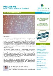
PELOBiotech Newsletter March 2019<br />
“Vita brevis, ars longa.“<br />
(Seize the day.)<br />
Dear Scientist,<br />
Dear Scientist,<br />
The UK is due to leave the European Union on Friday 29th March 2019,<br />
after the UK voted to leave in the 2016 referendum.<br />
Understandably you will be considering the changes it may have on<br />
your supply deliveries. PELOBiotech GmbH as your trusted distributor<br />
has prepared for all potential Brexit outcomes and is committed to<br />
supporting you during Brexit and to manage the supply chain<br />
risks in these challenging conditions.<br />
We have made extensive preparations to ensure continuity of service<br />
during and post-Brexit. Our core capability is clarifying and optimising<br />
global supply chains across international borders and managing regulatory<br />
complexity. Any new regulatory requirements will be integrated into<br />
our “ready to send” process so we can minimizedthe risk of customs<br />
delays at the border due to paperwork discrepancies. We will continue<br />
to keep you updated as appropriate. In the meantime, to discuss how<br />
we can support your Brexit preparations, please contact us anytime.<br />
Cheers. Best regards,<br />
Christiane Büchsel<br />
Editor in Chief PELONews<br />
Hippokrates, Founder of Medicine<br />
as Science-<br />
Content<br />
PELOAcademy: Learn<br />
from the best<br />
Thank you alll<br />
Meet us at…<br />
Paper Altert<br />
Featured Fields<br />
&Products<br />
<br />
<br />
<br />
<br />
<br />
3D<br />
BBB<br />
Cancer<br />
Chimerism Assay<br />
Microfluidic Chambers<br />
You like what you see? Sign<br />
up for PELONews now at<br />
www.pelobiotech.com or just<br />
fill out the following form<br />
here.
PELONews<br />
PELOAcademy<br />
Didn´t have time on these<br />
lIVE events? No worries.<br />
Sign up and we<br />
send you the recording<br />
asap. You are very welcome.<br />
Want to stay tuned in<br />
and informed? Then<br />
sign up for our regular<br />
Webinar Invitation Reminder.<br />
Just check the<br />
box with the infos you<br />
like to recieive and sign<br />
up here.<br />
From Bedside to Bench: Translating<br />
Clinical Tumour Treatments into 3D Culture<br />
'Translational research' is mainly seen as the effort to develop basic research<br />
into clinical routine, this webinar shows how 3D culture systems<br />
may serve as a comprehensive tool to reproduce patient treatment protocols<br />
in your laboratory.<br />
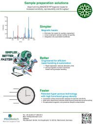
From a lab perspective, the clinical situation looks amazingly simple:<br />
Both patients and doctors do not bother much about biological endpoints<br />
as apoptosis or DNA damage of cancer cells, but focus on general ones:<br />
Does the tumour shrink? And finally: Is the tumour cured?<br />
Applying such 'simple' endpoints on 3D tumour cell cultures can make a<br />
lot of sense:<br />
<br />
Volume response as a readout summarises all anti-tumour effects<br />
into one<br />
Sign up here<br />
Watch now here<br />
<br />
<br />
<br />
<br />
<br />
<br />
In long-term cultures, each single micro-tumour may be assigned to<br />
be either 'cured' or 'resistant'<br />
The concept of clonal evolution requires time and large 'cohorts" of<br />
micro-tumours. Size-based screening is an efficient method to detect<br />
specific mutations and other rare events. In this webinar, you will<br />
learn:<br />
How to design and apply complex treatment schedules to microtumours,<br />
as chemoradiation, fractionated radiotherapy and hyperthermia<br />
treatment •<br />
How to handle cancer cell types that normally do not grow as spheroids<br />
How optical scanning and paraffin sections are easily used to assess<br />
treatment response<br />
How to detect and isolate micro-tumours that did not respond to treatment<br />
Watch now this great vivid lecture by Dr. Thomsen.
Advanced in-vitro Models for Tumor Models<br />
presented by Tommaso Sbrana, CEO IVtech, Italy<br />
A standard 2D static in-vitro models is not representative of the reality.<br />
This is well known and it is more than accepted if the goal is the estimation of an in-vitro model to mimic<br />
a tumor environment. The lack of crosstalk between tissues as well as the static cell culture conditions<br />
are limitations in the correlation between the standard 2D in-vitro model and the scenario that we desire<br />
to reproduce. It is required an update of the technology used to set up an in-vitro model, in order to be<br />
able to recreat the pato-physiological stimuli, increasing its predictivity of the human scanario. Therefore<br />
IVTech offers an enabling technology, represented by advanced cell culture chambers, to permit the study<br />
of a 3D dynamic in-vitro model, characterized by a crosstalk between different tissues.<br />
Using these products, it is now possible to add the IVth dimension (the dynamicity) and the<br />
Vth dimension (the crosstalk between different tissues) to a 3D in-vitro model. The focus is shifted from<br />
a single tissue to a pathway evaluated in dynamic conditions. The use of this chambers in oncology for<br />
instance allows to develop advanced in-vitro models to mimic the tumor environment, test the effects of<br />
an antitumor compound as well as evaluate the interactions between healthy and disease tissues.<br />
Therefore it is possible to:<br />
<br />
recreat the tumor micro-environment and its main characteristics<br />
(i.e. hipoxia )<br />
tissues, in order to study the uptake mechanism<br />
<br />
investigate the tumor cell resistance to a drug exposure, as it is<br />
sometimes observed in human (i.e. brest cancer applications)<br />
<br />
Watch now here<br />
evaluate the interactions between floating metastasis and healthy<br />
study the new frontiers of tumor treatments represented by immunotherapy, combining tumor and limphatic<br />
models. Watch now this fantatstic webinar by Tommaso.Sbrana.
PELONews<br />
Let‘s talk<br />
Optimised<br />
Cancer<br />
Treatments<br />
3D Models<br />
Symposium Munich:<br />
Wed, 27 March 2019<br />
Meet high end experts in the field<br />
of 3D cancer models and treatments.<br />
Learn what they are doing<br />
and how they have optimized<br />
their treatments.<br />
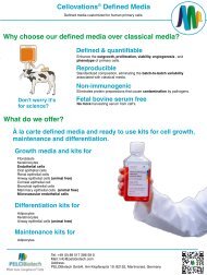
3D CoSeedis: a novel and innovative<br />
3D microwell array in the<br />
analysis of adhesion independent<br />
micro-organoids<br />
3D CoSeedis consists of a unique<br />
conical agarose matrix array that<br />
allows the formation of spheroidal<br />
and non-spheroidal cell aggregates<br />
in a highly reproducible and consistent<br />
manner. Furthermore, 3D<br />
CoSeedis allows the supporting<br />
growth of feeder cells in the formation<br />
of 3D cell constructs that are<br />
not in direct contact with the test<br />
cells themselves. Validated protocols<br />
and workflows have been developed<br />
to analyse fully formed 3D<br />
aggregates.<br />
In summary, the modular and standardised<br />
3D CoSeedis systems<br />
broaden the number of cell types<br />
accessible to examination in 3D<br />
substantially and present a versatile,<br />
robust and reliable platform for<br />
3D cell culture studies.<br />
Sign up now at<br />
info@pelobiotech.com and let<br />
Katja and Christiane know that you<br />
will be part of this groundbreaking<br />
Symposium.
New Products<br />
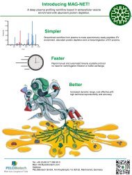
SynVivo 3D Tissue/Organ on Chip Models<br />
SynVivo is a cell-based microfluidic platform that provides a morphologically and biologically realistic microenvironment<br />
allowing real-time study of cellular behavior, drug delivery and drug discovery. Watch video showing<br />
SynVivo in action.<br />
SynVivo has developed 3D tissue models that recreate complex in vivo microvasculature including scale, morphology,<br />
hemodynamic shear stress and cellular interactions with side-by-side architecture enabling real-time<br />
visualization. 3D tissue models include SynBBB (blood brain barrier) SynTumor<br />
(Cancer), SynRAM (Inflammation) and SynTox (Toxicology). The platform can be adapted to replicate the<br />
unique features of any desired organ and tissue for your specific applications.<br />
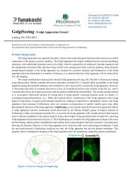
SynBBB in vitro blood brain barrier (BBB) model allows visualization of cellular interactions and barrier functionality<br />
in real time. Evaluate and quantitate permeability of therapeutics and small molecules across the endothelium<br />
of the BBB in real-time. Unlike BBB models that are membrane-based and arranged in top to bottom<br />
architecture (i.e., Transwell), small molecule transport can be visualized, accessed and quantified in real-time<br />
using the SynBBB model due to its side-by-side architecture.<br />
Human or rat species in co-culture with endothelial cells and astrocytes or tri-culture to include pericytes can<br />
be used to assess the passage of molecules through the BBB and the effect of molecules on the BBB physiology<br />
(normal or diseased). Various types of molecules can be assessed such as nanoparticles, small<br />
molecules, peptides or large molecules including antibodies. Cellular interactions including vascular - immune<br />
cell interactions can be studied in this model.<br />
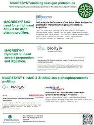
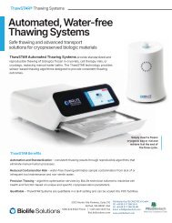
Apical chamber (outer blue channels) is for culture of<br />
vascular cells while the basolateral chamber (central<br />
red chamber) is for culture of brain tissue cells<br />
(astrocytes, pericytes) which can be mixed or separated<br />
in different compartments. Engineered porous architecture<br />
enables communication between the vascular<br />
and tissue cells.<br />
F<br />
The SynBBB model is available as kits or microfluidic chips.<br />
Publications using the SynBBB model<br />
(1) A Microfluidic model of human brain (uHuB) for assessment of blood brain barrier.<br />
Tyler D. Brown, Maksymillian Nowak, Alexandra V. Bayles, Balabhaskar Prabhakarpandian, Pankaj Karande, Joerg Lahann,<br />
Matthew E. Helgeson and Samir Mitragotri. BioEngineering and Translational Medicine 2019; 1-13.<br />
(2) Protein kinase C-delta inhibition protects blood brain barrier from sepsis-induced vascular damage.Yuan Tang, Fariborz<br />
Sorush, Shuang Sun, Elisbetta Liverani, Jordan Langston, Qingliang Yang, Laurie Kilpatrick and Mohammad Kiani. Journal of<br />
Neuroinflammation 15:309 (2018)<br />
(3) Trastuzumab Distribution in an In-Vivo and In-Vitro Model of Brain Metastases of Breast Cancer Tori B. Terrell-Hall, Mohamed<br />
Ismail Nounou, Fatema El-Amrawy, Jessica I.G. Griffith and Paul R. Lockman Oncotarget. 2017; 8:83734-83744<br />
Learn more<br />
(4) Permeability across a novel microfluidic blood‐tumor barrier model. Tori B. Terrell‐Hall, Amanda G. Ammer, Jessica I.<br />
G. Griffith and Paul R. Lockman Fluids and Barriers of the CNS (2017) 14:3<br />
(5) A Novel Dynamic Neonatal Blood-Brain Barrier on a Chip. S. Deosarkar, B. Prabhakarpandian, B. Wang, J.B. Sheffield,<br />
B. Krynska, M. Kiani. PLOS ONE, 2015
PELONews<br />
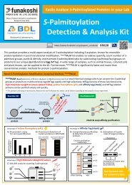
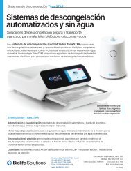
SynBBB exhibits significant tight junction formation and shows excellent correlation<br />
with in vivo permeability<br />
Other validated tissue models
Chimerism analysis: Non-T & Non-B Genomic Detection Kits<br />
· Q: Is my current sorting method compatible with these kits?<br />
A: Yes! The Non-T Genomic and Non-B Genomic Detection Kits determine cell purity at the nucleic<br />
acid level. It is therefore insensitive to the method used to sort the cells.<br />
Q: Do I need to introduce a new technique and procedures in the lab to use these kits?<br />
A:These kits are designed to be used along the traditional chimerism analysis workflow. All there is to<br />
do is add the DNA sample to the reaction tube. Then the reaction can be run at the same time than the<br />
chimerism PCR, because the reaction parameters are the same. The PCR products are analyzed on a<br />
sequencer in the same way than the chimerism sample. Chimerism labs already have all the expertise<br />
and equipment to run the Non-T Genomic and Non-B Genomic Detection Kits.<br />
Q: I don't have a lot of DNA sample to start with, I need it all for the chimerism analysis and<br />
cannot waste any. Will I have enough sample to check for purity?<br />
A:The Non-T Genomic and Non-B Genomic Detection Kits can be run with as little as 0.5 ng of genomic<br />
DNA. This corresponds to the amount of DNA present in approximately 200 cells. The lowest DNA<br />
concentration that can be detected with classical UV methods is about 2 ng/µL. So even with samples<br />
with very low, non detectable DNA content, 1 µL of sample is usually sufficient to run the Genomic Detection<br />
Kit.<br />
Learn more here<br />
Q: I validated my cell sorting method and had good purities. Why would I need to check the<br />
purity of the sorted cells every time?<br />
A:The quality of the cell sorting is very sample-dependant. The first source of viability is of course the<br />
patient. Shortly after hematopoietic stem cell transplant, it is common for patients to have a very low<br />
blood cell count, as well as a very low T cell count (often less than 5% of total leukocytes, whereas<br />
normal levels are 25-30%). The risk of T Cells isolated from these samples to be contaminated by monocytes<br />
or granulocytes is very high. In addition, blood samples sometimes reach the lab several days<br />
after collection, and can be exposed to extreme temperatures during transportation. These parameters<br />
can vary between samples, are out of the lab's control, and can have a very negative impact on cell<br />
sorting quality. Assessing the purity of each isolated cell subset is an essential<br />
quality control step, and is required for ASHI or EFI accreditation.<br />
Learn more<br />
Q: Isn't flow cytometry a more accurate method of assessing purity?<br />
A: Actually, flow cytometry is not as accurate as one might think, because measured purity can vary<br />
considerably depending on the way the data is analyzed. On most samples, changing the way the cells are<br />
gated can lead to purities readings anywhere between 80% and 100%. In contrast, calculating purity is<br />
straightforward with the Non-T Genomic and Non-B Genomic Detection Kits, and there is no interpretation<br />
or ambiguities during the analysis.<br />
In addition, chimerism tests are based on DNA analysis, and so are the Non-T Genomic and Non-B Genomic<br />
Detection Kits. All the DNA present in the sample will be taken into account. In contrast, flow cytometry<br />
analysis excludes "debris" and events that have a small size. These may contain DNA fragments or even<br />
entire nuclei. This DNA, excluded from the flow cytometry data, might have an impact on the chimerism<br />
analysis results.
PELONews<br />
PELONews: Refer a friend<br />
We are convinced that we convince you with our<br />
expertise and excellent service. So if you are<br />
already a PELO-Fan, please recommend us to a<br />
colleague and friend – you will get a nice thankyou<br />
personalized reward. More details? Call us<br />
now.<br />
Thank you all: Here‘s a quote from our Customer-survey: 100% are very satisfied<br />
or satiesfied with PELOBiotech!! You are wonderful and we are happy to<br />
serve you..<br />
Paper Alert Our partner ReSyn has publisehd a new paper.<br />
„Immune genes are primed for robust transcription by proximal long noncoding<br />
RNAs located in nuclear compartments.“<br />
by Stoyan Stoychev and his scientific partners. Nature Geneticsvolume<br />
51, pages138–150 (2019) |<br />
Accumulation of trimethylation of histone H3 at lysine 4 (H3K4me3) on immune<br />
-related gene promoters underlies robust transcription during trained immunity.<br />
However, the molecular basis for this remains unknown. Here we show threedimensional<br />
chromatin topology enables immune genes to engage in chromosomal<br />
contacts with a subset of long noncoding RNAs (lncRNAs) we have defined<br />
as immune gene–priming lncRNAs (IPLs). We show that the prototypical<br />
IPL, UMLILO, acts in cis to direct the WD repeat-containing protein 5 (WDR5)–<br />
mixed lineage leukemia protein 1 (MLL1) complex across the chemokine promoters,<br />
facilitating their H3K4me3 epigenetic priming. This mechanism is<br />
shared amongst several trained immune genes. Training mediated by β-glucan<br />
epigenetically reprograms immune genes by upregulating IPLs in manner dependent<br />
on nuclear factor of activated T cells. The murine chemokine topologically<br />
associating domain lacks an IPL, and the Cxclgenes are not trained. Strikingly,<br />
the insertion of UMLILO into the chemokine topologically associating<br />
domain in mouse macrophages resulted in training of Cxcl genes. This provides<br />
strong evidence that lncRNA-mediated regulation is central to the establishment<br />
of trained immunity.<br />
Want to meet Stoyan and talk about<br />
what ReSyn can do for you:<br />
EUPA 24-28th March, Potsdam.<br />
Meet also our sales expert Katja<br />
Keystone Symposium, 7-11th<br />
April, Stockholm, Sweden.<br />
We like your feedback – tell us what you love, don’t like so much and what you would<br />
like to get, please. Just reply Please update your subscription anytime, as we like to<br />
comply to the new GDPR guidelines.<br />
Save these<br />
dates<br />
Let`s talk:<br />
Optimised Cancer<br />
Treatments with 3D<br />
Models<br />
March 27, 2019<br />
10 AM ct – 1PM<br />
Sky Lounge G2B<br />
Club im IZB<br />
IGLD: 07.- 09.<br />
March 2019, Frankfurt<br />
Elrig: 07. March<br />
2019, Darmstadt<br />
ADF: 13.- 16.March<br />
2019, Munich<br />
EUPA 24-28th<br />
March, 2019, Potsdam<br />
How to<br />
reach us<br />
If you need any<br />
further assistance or<br />
if you like what you<br />
see, tell us:<br />
PELOBiotech GmbH<br />
Klopferspitz 19<br />
82152 Planegg |<br />
Germany<br />
Tel.: +49 89 517286<br />
59 0 | info@pelobiotech.com<br />
|<br />
www.pelobiotech.com<br />
Managing Directors:<br />
Dr. Peter Frost, Dr<br />
Lothar Steeb