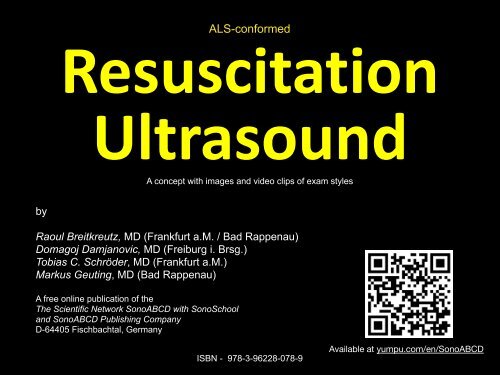
Resuscitation Ultrasound
This is the full version of ALS-conformed "Resuscitation Ultrasound" methods / protocols for practitioners. Almost all have been suggested within the ERC resuscitation guidelines of 2015 or later with POCUS in ERC 2021. Our clips should put a new complexion on those exam styles. The collection can be downloaded and may support your teaching activity. ... can be shared, kept, as you like...;-))
This is the full version of ALS-conformed "Resuscitation Ultrasound" methods / protocols for practitioners. Almost all have been suggested within the ERC resuscitation guidelines of 2015 or later with POCUS in ERC 2021.
Our clips should put a new complexion on those exam styles. The collection can be downloaded and may support your teaching activity. ... can be shared, kept, as you like...;-))
Create successful ePaper yourself
Turn your PDF publications into a flip-book with our unique Google optimized e-Paper software.
ALS-conformed<br />
<strong>Resuscitation</strong><br />
<strong>Ultrasound</strong><br />
A concept with images and video clips of exam styles<br />
by<br />
Raoul Breitkreutz, MD (Frankfurt a.M. / Bad Rappenau)<br />
Domagoj Damjanovic, MD (Freiburg i. Brsg.)<br />
Tobias C. Schröder, MD (Frankfurt a.M.)<br />
Markus Geuting, MD (Bad Rappenau)<br />
A free online publication of the<br />
The Scientific Network SonoABCD with SonoSchool<br />
and SonoABCD Publishing Company<br />
D-64405 Fischbachtal, Germany<br />
ISBN - 978-3-96228-078-9<br />
Available at yumpu.com/en/SonoABCD
ABCD
Sonoscopy of trachea in emergencies<br />
Tracheal and Esophageal tube placement<br />
- Simple pattern recognition -<br />
Normal: Single (airway) tract<br />
Double (airway) tract sign*<br />
Edited by Raoul Breitkreutz (2020)<br />
Scientific Network SonoABCD<br />
The fine art of Sonoscopy.<br />
No ventilation required.<br />
Observe: both lateral regions of airway tract<br />
*Zechner P, Breitkreutz R <strong>Resuscitation</strong> 2011<br />
Chou et al. <strong>Resuscitation</strong> 2011
Airway <strong>Ultrasound</strong> Exam<br />
Main Questions<br />
In emergency always use post-intubation check of trachea only.<br />
Edited by Raoul Breitkreutz (2020)<br />
#Q1. Esophageal misplacement? Yes or No?<br />
Only if ruled out - goto 2.<br />
#Q2. Main stem intubation? Yes or No?<br />
If ruled in - slightly withdraw and re-check.<br />
Scientific Network SonoABCD<br />
The fine art of Sonoscopy.<br />
Available at www.yumpu.com/en/SonoABCD
Distinct clinical emergency scenarios define<br />
two widely different tracheal ultrasound exam methods<br />
Exam style #1: Post-intubation check<br />
(e.g. CPR at point-of-care or on arrival at shock room)<br />
- immediate, urgent action -<br />
Edited by Raoul Breitkreutz (2020)<br />
On-off evaluation<br />
Scientific Network SonoABCD<br />
The fine art of Sonoscopy.
Team H.K. Stanis, M.D., M. Negele, Bad Rappenau, Germany (2019)<br />
Airway <strong>Ultrasound</strong> Exam<br />
#Q1 - Esophageal misplacement? Yes or No?<br />
Scientific Network SonoABCD<br />
The fine art of Sonoscopy.<br />
Edited by Raoul Breitkreutz (2020)<br />
In emergency always use post-intubation check of trachea only.
Team H.K. Stanis, M.D., M. Negele, Bad Rappenau, Germany (2019)<br />
Airway <strong>Ultrasound</strong> Exam<br />
#Q1 - Esophageal misplacement? Yes or No?<br />
Finding 1a: No, normal!<br />
Scientific Network SonoABCD<br />
The fine art of Sonoscopy.<br />
No ventilation required.<br />
Edited by Raoul Breitkreutz (2020)
Sonoscopy of trachea post-intubation<br />
Check tracheal tube placement<br />
Exam style #1 - Simple pattern recognition<br />
Edited by Raoul Breitkreutz (2020)<br />
Single (airway) tract<br />
Scientific Network SonoABCD<br />
The fine art of Sonoscopy.<br />
Observe: both lateral regions of airway tract<br />
No ventilation required.
Team H.K. Stanis, M.D., M. Negele, Bad Rappenau, Germany (2019)<br />
Airway <strong>Ultrasound</strong> Exam<br />
#Q1 - Esophageal misplacement? Yes or No?<br />
Finding 1b: Yes! Immediate correction!<br />
Scientific Network SonoABCD<br />
The fine art of Sonoscopy.<br />
No ventilation required.<br />
Edited by Raoul Breitkreutz (2020)
Sonoscopy of trachea post-intubation<br />
Tracheal and Esophageal tube placement<br />
Exam style #1 - Simple pattern recognition<br />
Normal: Single (airway) tract<br />
Double (airway) tract sign*<br />
Edited by Raoul Breitkreutz (2020)<br />
Scientific Network SonoABCD<br />
The fine art of Sonoscopy.<br />
No ventilation required.<br />
Observe: both lateral regions of airway tract<br />
*Zechner P, Breitkreutz R <strong>Resuscitation</strong> 2011<br />
Chou et al. <strong>Resuscitation</strong> 2011
Distinct clinical emergency scenarios define<br />
two widely different tracheal ultrasound exam methods<br />
Exam style #2: Visualization<br />
during entire intubation process<br />
(e.g. RSI, teaching) - planned action -<br />
Continuous evaluation<br />
Edited by Raoul Breitkreutz (2020)<br />
Scientific Network SonoABCD<br />
The fine art of Sonoscopy.
Distinct clinical emergency scenarios define<br />
two widely different tracheal ultrasound exam methods<br />
Exam style #2: Visualization<br />
during entire intubation process<br />
(e.g. RSI, teaching) - planned action -<br />
Continuous evaluation<br />
Scientific Network SonoABCD<br />
The fine art of Sonoscopy.<br />
Edited by Raoul Breitkreutz (2020)<br />
Nota bene:<br />
1st trial —> esophagus.<br />
Upon instruction of clinician supervisor,<br />
observing by sonography,<br />
—> uninterrupted 2nd trial.<br />
With support of M. Nickel, MD and M. Geuting, MD, Bad Rappenau
Sonoscopy of trachea during intubation<br />
Tracheal and Esophageal tube placement<br />
Exam style #2 - Simple pattern recognition<br />
Normal: Single (airway) tract<br />
- unspectacular observation -<br />
Double (airway) tract sign*<br />
prominent monitoring observation<br />
(2 attempts and withdrawals)<br />
Edited by Raoul Breitkreutz (2020)<br />
Scientific Network SonoABCD<br />
The fine art of Sonoscopy.<br />
No ventilation required.<br />
Observe: both lateral regions of airway tract<br />
*Zechner P, Breitkreutz R <strong>Resuscitation</strong> 2011<br />
Chou et al. <strong>Resuscitation</strong> 2011
Comparison of<br />
Clinician sonographer<br />
Probe movement <br />
at frontal neck<br />
Supervision<br />
Immediate correction?<br />
Action of sonographer?<br />
Exam styles<br />
#1 #2<br />
post-intubation<br />
active,<br />
exploration of region<br />
yes, onto and away<br />
dynamic<br />
no add role <br />
or not necessary<br />
no,<br />
check only<br />
during entire<br />
intubation process<br />
passive<br />
no, still /static <br />
waiting for action<br />
active option for role,<br />
e.g. in teaching<br />
yes,<br />
option to correction <br />
while indwelling ETT<br />
Image as is can change markedly<br />
Time (secs)
Team K. Stanis, M.D., M. Negele, Bad Rappenau, Germany (2019)<br />
Airway <strong>Ultrasound</strong> Exam<br />
#Q2 - Main stem intubation?<br />
Extension of trachea check (exam style 1) and addition of the evaluation of lung motion<br />
Scientific Network SonoABCD<br />
The fine art of Sonoscopy.<br />
In emergency, after tracheal check, start lung sliding evaluation<br />
at left hemithorax. In this scenario, there was no emergency.<br />
Edited by Raoul Breitkreutz (2020)
Team K. Stanis, M.D., M. Negele, Bad Rappenau, Germany (2019)<br />
Airway <strong>Ultrasound</strong> Exam<br />
#Q2 - Main stem intubation?<br />
Finding 2a: No, normal!<br />
Scientific Network SonoABCD<br />
The fine art of Sonoscopy.<br />
Edited by Raoul Breitkreutz (2020)<br />
In emergency, after tracheal check, start lung sliding evaluation at left hemithorax.
Sonoscopy of lung post-intubation<br />
Check for lung pulse / lung sliding<br />
Simple pattern recognition<br />
Edited by Raoul Breitkreutz (2020)<br />
Scientific Network SonoABCD<br />
The fine art of Sonoscopy.<br />
Observe:<br />
Lung Sliding. No full ventilation cycle required.
Sonoscopy of lung post-intubation<br />
Check for lung pulse / lung sliding<br />
Simple pattern recognition<br />
Edited by Raoul Breitkreutz (2020)<br />
Scientific Network SonoABCD<br />
The fine art of Sonoscopy.<br />
Observe: Lung Sliding. No full ventilation cycle required.
Team Markus Geuting, M.D., M. Negele, Bad Rappenau, Germany (2019)<br />
Airway <strong>Ultrasound</strong> Exam<br />
#Q2 - Main stem intubation?<br />
Exam style #1<br />
Scientific Network SonoABCD<br />
The fine art of Sonoscopy.<br />
Edited by Raoul Breitkreutz (2020)<br />
In emergency, after tracheal check, start lung sliding evaluation at left hemithorax.
Team Markus Geuting, M.D., M. Negele, Bad Rappenau, Germany (2019)<br />
Airway <strong>Ultrasound</strong> Exam<br />
#Q2 - Main stem intubation?<br />
Finding 2b: Yes, correction required!<br />
Scientific Network SonoABCD<br />
The fine art of Sonoscopy.<br />
Edited by Raoul Breitkreutz (2020)<br />
In emergency, after tracheal check, start lung sliding evaluation at left hemithorax.
Team Markus Geuting, M.D., M. Negele, Bad Rappenau, Germany (2019)<br />
Sonoscopy of lung post-intubation<br />
Check for lung pulse / lung sliding<br />
Simple pattern recognition<br />
Scientific Network SonoABCD<br />
The fine art of Sonoscopy.<br />
Observe: Lung pulse.<br />
Action: SlIghtly withdraw tube and re-check. No full ventilation cycle required.<br />
Edited by Raoul Breitkreutz (2020)
Team Markus Geuting, M.D., M. Negele, Bad Rappenau, Germany (2019)<br />
Sonoscopy of lung post-intubation<br />
Check for lung pulse / lung sliding<br />
Simple pattern recognition<br />
Scientific Network SonoABCD<br />
The fine art of Sonoscopy.<br />
Observe: Lung pulse or lung sliding.<br />
Action: Slightly withdraw tube and re-check. No full ventilation cycle required.<br />
Edited by Raoul Breitkreutz (2020)
Airway <strong>Ultrasound</strong> Exam<br />
Limitations of the check for main stem intubation<br />
Interprete sonographic signs per hemithorax<br />
In presence of (known or unknown) pneumothorax<br />
Trachea ultrasound is applicable to check for normal or esophageal intubation. <br />
Lung ultrasound: Mainstem intubation cannot be diagnosed. The air in the pleural<br />
space, resulting in sonographic signs of pneumothorax (no lung sliding, no lung<br />
pulse, no B-lines) overlay signs of one-sided intubation. Pneumothorax still could<br />
be ruled in.<br />
Edited by Raoul Breitkreutz (2020)<br />
In presence of (known or unknown) cardiac standstill<br />
Trachea ultrasound is applicable to check for normal or esophageal intubation. <br />
Lung ultrasound: A firm statement regarding mainstem intubation is not possible<br />
for the finding will be similar to sonographic findings as in pneumothorax (such as<br />
no lung sliding and no lung pulse). Pneumothorax still could be ruled out (if lung<br />
sliding or B-lines are present).<br />
In presence of both pneumothorax and cardiac standstill<br />
Trachea ultrasound is applicable to check for normal or esophageal intubation. <br />
Lung ultrasound: Neither mainstem intubation nor pneumothorax can be firmly<br />
diagnosed.<br />
Scientific Network SonoABCD<br />
The fine art of Sonoscopy.
Airway <strong>Ultrasound</strong> Exam<br />
Finding 3: no lung sliding, no lung pulse<br />
Edited by Raoul Breitkreutz (2020)<br />
Scientific Network SonoABCD<br />
The fine art of Sonoscopy.<br />
Consider pneumothorax, but only when heart beats!
Sonoscopy of lung post-intubation<br />
Check for lung pulse / lung sliding<br />
Simple pattern recognition<br />
Edited by Raoul Breitkreutz (2020)<br />
Scientific Network SonoABCD<br />
The fine art of Sonoscopy.<br />
Observe: Lung movements during ventilation.<br />
Think: If none, consider pneumothorax, but only if cardiac motion has been confirmed.<br />
Action: Decide side, consider or perform immediate needle puncture.
Additional information<br />
Esophageal misplacement?<br />
Comparing methods<br />
<strong>Ultrasound</strong> and Capnometry<br />
Scientific Network SonoABCD<br />
The fine art of Sonoscopy.<br />
3 2<br />
Use B-Mode<br />
Regarding the question <br />
of esophageal misplacement, in <br />
post-intubation check of trachea, <br />
ultrasound is faster and<br />
does not require ventilation trials.<br />
Capnometry is slower and <br />
insecure for this purpose<br />
because of slow drop, requirement of ventilation trials and not providing zero line results<br />
Zechner PM, Breitkreutz R<br />
<strong>Resuscitation</strong> (2011)
Additional information<br />
Airway <strong>Ultrasound</strong> Exam<br />
Main questions<br />
Edited by Raoul Breitkreutz (2020)<br />
#Q1. Esophageal<br />
misplacement? Yes or No?<br />
Esophageal misplacement?<br />
Re-Check after re-intubation<br />
Immediate action?<br />
Yes<br />
No, if<br />
normal<br />
Expected frequency<br />
ruling in<br />
ruling out<br />
#Q2. Main stem<br />
Main stem intubation?<br />
intubation? Yes or No?<br />
Re-Check after withdrawal<br />
Yes<br />
No, if<br />
normal<br />
Scientific Network SonoABCD<br />
The fine art of Sonoscopy.<br />
Available at yumpu.com/en/SonoABCD
Additional information<br />
Airway <strong>Ultrasound</strong> Exam 1,2<br />
Method<br />
Edited by Raoul Breitkreutz (2020)<br />
Method: Sonoscopy<br />
Sonoscopy is the art of stethoscope-like use<br />
of ultrasound.<br />
Regarding Airway and Lung <strong>Ultrasound</strong> as part<br />
of „<strong>Resuscitation</strong> <strong>Ultrasound</strong>“ in the clinical<br />
context of Peri-<strong>Resuscitation</strong>: Check for trachea<br />
post-intubation, and, in case of tracheal<br />
placement, for lung sliding and -pulse.<br />
Scientific Network SonoABCD<br />
The fine art of Sonoscopy.
Additional information<br />
Types of airway ultrasound exams within a defined clinical context<br />
Types of airway ultrasound exams<br />
within a defined clinical context<br />
Action / ultrasound<br />
exam type<br />
Aim, emphasis on<br />
Approx. time for<br />
exam (seconds)<br />
Time pressure?<br />
/ remark<br />
Pre-check, e.g. for<br />
prep of dLT, difficult<br />
laryngoscopy,<br />
identify cricoid<br />
membrane<br />
planned evaluation<br />
of upper airway<br />
related<br />
sonoanatomy<br />
> 60<br />
no time pressure<br />
applicable, because of<br />
measurements<br />
Trachea exam only<br />
after any emergency<br />
intubation, double<br />
tract or single tract<br />
before ventilation<br />
< 10, when<br />
evaluating postintubation<br />
only<br />
yes, integration into a<br />
(fast driving) process,<br />
similar to one by one item<br />
in a RSI<br />
Airway <strong>Ultrasound</strong><br />
Exam<br />
Airway <strong>Ultrasound</strong> as<br />
part of <strong>Resuscitation</strong><br />
<strong>Ultrasound</strong><br />
routine check for<br />
trachea plus lung<br />
sliding or lung pulse<br />
or diaphragm or<br />
parts of it<br />
check of treatable<br />
conditions<br />
< 120 no, variety of indications*<br />
A;
Airway <strong>Ultrasound</strong> Exam 1,2<br />
References<br />
Slovis TL, Poland RL. Radiology. 1986; 160:262–3.<br />
Endotracheal tubes in neonates: (Its) sonographic positioning.<br />
Raphael DT, Conard FU. <strong>Ultrasound</strong> confirmation of endotracheal tube placement.<br />
J Clin <strong>Ultrasound</strong>. 1987; 15:459–62.<br />
Ma G et al. J Emerg Med 1999 (SAEM meeting Abstract 515), Acad Emerg Med. 1999; 6:515. full paper<br />
published in 2007 (Trachea, while and post-intubation, principle, cadavers)<br />
Drescher MJ et al.. Acad Emerg Med 2000;7:722–5.<br />
(First detailed description of sonograms in esophageal intubation, while and post-intubation, principle,<br />
cadavers)<br />
Weaver B et al. Acad Emerg Med. 2006;13(3):239-44 <br />
(Lung sliding to confirm trachea placement, post-intubation, principle, cadavers)<br />
Ma G et al. J Emerg Med 2007;32:405 (Trachea, while and post-intubation, principle, cadavers)<br />
Werner SL et al. Ann Em Med 2007;49:75–80 (Trachea, while intubation, principle)<br />
Chou HC et al <strong>Resuscitation</strong> 2011;82:1279–84 (Trachea, while intubation, double tract sign)<br />
1Zechner P, Breitkreutz R <strong>Resuscitation</strong> 82 (2011) 1259–61 (First description of the <br />
„Airway ultrasound protocol“, combining ALS-conformed trachea and lung evaluation) <br />
2Breitkreutz R et al. <strong>Resuscitation</strong> 83 (2012) 273–274 (add of Lung sliding, Lung pulse)<br />
Edited by Raoul Breitkreutz (2020)<br />
Adi O et al. Crit <strong>Ultrasound</strong> J 2013; 5:7 (Trachea, Post-intubation)<br />
Abbasi et al. Eur J Emerg Med 2015;22(1):10–6 (Trachea, Post-intubation)<br />
Soar J et al. <strong>Resuscitation</strong> 95 (2015) 100-142 (ERC guideline 2015)<br />
Chou EH et al. / <strong>Resuscitation</strong> 90 (2015) 97–103 (Metaanalysis)<br />
Scientific Network SonoABCD<br />
The fine art of Sonoscopy.
Additional exam styles<br />
Sonoscopy of lung post-intervention<br />
Ruling out pneumothorax<br />
Simple pattern recognition<br />
Scientific Network SonoABCD<br />
The fine art of Sonoscopy.<br />
Edited by Raoul Breitkreutz (2020)<br />
Observe: Lung movements during ventilation.<br />
Think: If lung sliding or lung pulse are present, you can rule out pneumothorax.<br />
Action: Perform planned repeated exam two-staged in clinical time course.
Additional exam styles<br />
Sonoscopy of lung: Screening for pleural effusion<br />
Simple pattern recognition<br />
Edited by Raoul Breitkreutz (2020)<br />
Scientific Network SonoABCD<br />
The fine art of Sonoscopy.<br />
Observe: Lung sliding, diaphragm<br />
Think: separation of diaphragm and lung?<br />
Action: evaluate clinical symptoms for puncture decision and if urgent
Additional exam styles<br />
Sonoscopy tests of double lumen intubation<br />
Simple pattern recognition<br />
Edited by Raoul Breitkreutz (2020)<br />
by Tobias C. Schröder, Frankfurt a.M.<br />
Scientific Network SonoABCD<br />
The fine art of Sonoscopy.<br />
Observe: Lung movements during ventilation.<br />
Think: Lung sliding or lung pulse if not clamped or clamped.<br />
Action: Visualisation, clamping, visualisation and conclusion
Additional exam styles<br />
Sonoscopy test of congestion - B-lines<br />
Simple pattern recognition<br />
Edited by Raoul Breitkreutz (2020)<br />
Scientific Network SonoABCD<br />
The fine art of Sonoscopy.<br />
Observe: B-lines, multiple B-lines, effusion.<br />
Think: multiple B-lines? bilateral? white lung?<br />
Action: reporting, oxygen, induction of therapy
Review article<br />
nt, especially in its specificity value. Different settings, operperience,<br />
Ultrasonography and timing of the for ultrasound confirmation can cause of a endotracheal small but was tube used placement:<br />
in ten studies. 9,11,12,14–20 The sensitivity and spe<br />
mon ultrasound technique to detect esophageal intubatio<br />
nificant A influence on the diagnostic accuracy. Ultrasonogran<br />
be a useful tool for confirmation of tracheal intubation. real-time sonographic imaging during intubation has higher<br />
are both high in cadaveric models, ORs, and EDs. In ge<br />
systematic review and meta-analysis <br />
er, Eric the use H. Chou of ultrasonography a,1 , Eitan Dickmanor a , any Po-Yang method Tsouas b , Mark the sole Tessarotivity a , Yang-Ming for detection Tsai c , of esophageal Additional intubation information<br />
than post-intu<br />
Matthew Huei-Ming Ma d , Chien-Chang Lee c,d,∗ , John Marshall a<br />
a Department of Emergency Medicine, Maimonides Medical Center, Brooklyn, NY, USA<br />
b College of Medicine, National Yang-Ming University, Taipei, Taiwan<br />
c Department of Emergency Medicine, National Taiwan University Hospital Yunlin Branch, Douliou, Taiwan<br />
d Department of Emergency Medicine, National Taiwan University Hospital, Taipei, Taiwan<br />
<strong>Resuscitation</strong> 90 (2015) 97–103<br />
a r t i c l e i n f o<br />
Article history:<br />
Received 19 November 2014<br />
Received in revised form 11 February 2015<br />
Accepted 12 February 2015<br />
Keywords:<br />
Intubation<br />
Airway management<br />
Ultrasonography<br />
<strong>Resuscitation</strong><br />
Meta-analysis<br />
Critical care<br />
a b s t r a c t<br />
Objective: This study aimed to undertake a systematic review and meta-analysis to summarize evidence<br />
on the diagnostic value of ultrasonography for the assessment of endotracheal tube placement in adult<br />
patients.<br />
Methods: The major databases, PubMed, EMBASE, and the Cochrane Library, were searched for studies<br />
published from inception to June 2014. We selected studies that used ultrasonography to confirm endotracheal<br />
tube placement. The search was limited to human studies, and had no publication date or country<br />
restrictions. Exclusion criteria included case reports, comments, reviews, guidelines and animal studies.<br />
Two reviewers extracted and verified the data independently. We summarized test performance characteristics<br />
with the use of forest plots, hierarchical summary receiver operating characteristic (HSROC)<br />
curves, and bivariate random effect models. Meta-regression analysis was performed to explore the<br />
source of heterogeneity. The methodological quality of individual studies was evaluated using the Quality<br />
Assessment of Diagnostic Accuracy Studies (QUADAS) tool.<br />
Results: A total of 12 eligible studies involving adult patients and cadaveric models were identified<br />
from 1488 references. For detection of esophageal intubation, the pooled sensitivity was 0.93 (95%CI:<br />
0.86–0.96) and the specificity was 0.97 (95%CI: 0.95–0.98). The area under the summary ROC curve was<br />
0.97 (95%CI: 0.95–0.98). The positive and negative likelihood ratios were 26.98 (95%CI: 19.32–37.66) and<br />
0.08 (95%CI: 0.04–0.15), respectively.<br />
Conclusions: Current evidence supports that ultrasonography has high diagnostic value for identifying<br />
esophageal intubation. With optimal sensitivity and specificity, ultrasonography can be a valuable adjunct<br />
in this aspect of airway assessment, especially in situations where capnography may be unreliable.<br />
© 2015 Elsevier Ireland Ltd. All rights reserved.<br />
1. Introduction<br />
Tracheal intubation serves as definite airway control when<br />
resuscitating critically ill patients. Confirmation of proper tube<br />
placement should be completed in all patients at the time of initial<br />
intubation. Unrecognized misplacement of the endotracheal tube<br />
may lead to avoidable morbidity including neurological damage,<br />
and death, with a reported incidence of 6–16%. 1,2 Thus, immediate<br />
post-intubation airway assessment is an essential clinical skill<br />
for every physician in emergency medicine (EM), anesthesia and<br />
critical care medicine. There are multiple options for confirming<br />
tracheal intubation and all methods have unique limitations. 3–5<br />
According to the 2010 American Heart Association (AHA) guideline,
Breitkreutz R et al. (2012)<br />
Additional<br />
information<br />
Scientific Network SonoABCD<br />
The fine art of Sonoscopy.
Sonoscopy of lung post-intubation<br />
Check for lung pulse / lung sliding<br />
Additional information<br />
Consider obtaining findings. Don´t try diagnoses.<br />
Edited by Raoul Breitkreutz (2020)<br />
Decide C-AB or A-B-C or else upon clinical context.<br />
Don´t get distracted by insecure interpretation of a finding.<br />
Team leaders should decide CPR process independently.<br />
B-Mode provides overview and is fast. Avoid M-Mode for lung<br />
evaluation in emergencies because of lesser overview and time consuming<br />
interaction with the ultrasound machine.<br />
Consider this method as secondary option whenever available. With<br />
ultrasound or capnometry you cannot intubate. However, with<br />
bronchoscopy you could.<br />
Train tracheal quick check, lung sliding or pulse and subcostal<br />
window. On yourself or in any health environment. These are very simple<br />
exams<br />
Scientific Network SonoABCD<br />
The fine art of Sonoscopy.
ABCD
30% VF only<br />
>60% PEA / asystole
The problem is….<br />
High-quality CPR!<br />
Identify treatable<br />
conditions!<br />
How to integrate ALS-conformed ultrasound in (existing) interruptions?
The solution is<br />
1) With training<br />
ALS-conformed ultrasound is feasible.<br />
2) It has a potential to identify<br />
- tamponade<br />
- acute right heart pressure overload<br />
- hypovolemia.<br />
3) Minimize interruptions!
2015
2015
2015<br />
„Consider“ ≙ VAS 5!
2016<br />
1. Cardiac activity on ultrasound was most<br />
associated with survival following cardiac<br />
arrest.<br />
2. <strong>Ultrasound</strong> during cardiac arrest identifies<br />
interventions outside of the standard ACLS<br />
algorithm.
2018
Is <strong>Resuscitation</strong> <strong>Ultrasound</strong> = „Emergency TTE“?<br />
Main emphasis on<br />
Findings<br />
Probe positions<br />
Further regions<br />
Method of evaluation<br />
<strong>Resuscitation</strong> <strong>Ultrasound</strong><br />
resume chest compressions, driving force is<br />
ALS, identify treatable conditions, image at<br />
a glimpse only<br />
Pathologies (3) and „kinetic activity“<br />
2: cardiac, cava (sweep in subcostal area)<br />
A: airway - trachea only, post-intubation for<br />
observing single or double tract<br />
B: lung (sliding, pulse) main stem intubat.<br />
Screening, „Scanning“, simple gross<br />
pathologies, binary answer<br />
Emergency TTE<br />
function, acquisition of<br />
various views<br />
Pathologies (>10) and<br />
functional analysis<br />
5+: parasternal LAX,<br />
SAX, apical, subcostal,<br />
IVC LAX…<br />
-<br />
Diagnostic accuracy,<br />
quantitative information<br />
Duration 400 PoCUS<br />
longer time spans<br />
full specialized TTE<br />
training<br />
Concept with<br />
DEGUM / ÖGUM / SGUM
Scientific Network SonoABCD<br />
The fine art of Sonoscopy.
low-cost, available at<br />
www.SonoABCD-Verlag.org
Sweep subcostal-LAX - IVC
During CPR<br />
subcostal window<br />
Edited by Raoul Breitkreutz (2020)<br />
Raoul Breitkreutz, Scientific Network Scientific Network SonoABCD<br />
The fine art of Sonoscopy.
During CPR: EMD<br />
subcostal window<br />
interruption<br />
pause of chest compressions<br />
evaluation<br />
Edited by Raoul Breitkreutz (2020)<br />
Scientific Network SonoABCD<br />
The fine art of Sonoscopy.
asystole, after<br />
cessation no flow<br />
subcostal window<br />
Edited by Raoul Breitkreutz (2020)<br />
Scientific Network SonoABCD<br />
The fine art of Sonoscopy.
True - EMD<br />
parasternal, LAX<br />
Courtesy of H. Steiger, Darmstadt<br />
Scientific Network SonoABCD<br />
The fine art of Sonoscopy.
Pseudo - EMD<br />
parasternal, LAX<br />
Courtesy of H. Steiger, Darmstadt<br />
Scientific Network SonoABCD<br />
The fine art of Sonoscopy.
Tamponade<br />
subcostal window<br />
Edited by Raoul Breitkreutz (2020)<br />
Scientific Network SonoABCD<br />
The fine art of Sonoscopy.
Tamponade<br />
subcostal window<br />
parasternal, SAX<br />
same patient<br />
by Tobias C. Schröder, Frankfurt a.M.<br />
Field examples, pocket-sized devices<br />
Edited by Raoul Breitkreutz (2020)<br />
Scientific Network SonoABCD<br />
The fine art of Sonoscopy.
Signs of acute right heart pressure overload<br />
subcostal 4-chamber view<br />
Edited by Raoul Breitkreutz (2020)<br />
Scientific Network SonoABCD<br />
The fine art of Sonoscopy.
Signs of acute right heart<br />
pressure overload<br />
parasternal window, SAX<br />
Edited by Raoul Breitkreutz (2020)<br />
Scientific Network SonoABCD<br />
The fine art of Sonoscopy.
two patients<br />
Field examples, pocket-sized devices<br />
by Tobias C. Schröder, Frankfurt a.M.<br />
Signs of acute right heart<br />
pressure overload<br />
parasternal window, SAX<br />
immediately after ROSC<br />
Edited by Raoul Breitkreutz (2020)<br />
Scientific Network SonoABCD<br />
The fine art of Sonoscopy.
Signs of hypovolemia<br />
IVC, SAX, transhepatic view<br />
Edited by Raoul Breitkreutz (2020)<br />
Scientific Network SonoABCD<br />
The fine art of Sonoscopy.
Field example, pocket-sized device<br />
after ROSC<br />
by Tobias C. Schröder, Frankfurt a.M.<br />
Signs of congestion<br />
IVC, SAX, transhepatic view<br />
Edited by Raoul Breitkreutz (2020)
IVC , eye-balling, B-Mode<br />
Edited by Raoul Breitkreutz (2020)<br />
„eye-balling“ evaluation<br />
✓1. diameter (end-expiratory) < 1,5 cm?<br />
✓2. pulsation visible?<br />
ev. 3. respiratory variability? 0-50%<br />
- (in CPR no sniff test to assess “collapsibility”)<br />
Scientific Network SonoABCD<br />
The fine art of Sonoscopy.
IVC, B-Mode, LAX, “eye-balling”<br />
Edited by Raoul Breitkreutz (2020)<br />
Scientific Network SonoABCD<br />
The fine art of Sonoscopy.
VCI, LAX - Pitfall false Diameter (fD)!<br />
- n o horizontal diameter<br />
- vertical diameter: median or not?<br />
Edited by Raoul Breitkreutz (2020)<br />
fD<br />
Scientific Network SonoABCD<br />
The fine art of Sonoscopy.<br />
after Scheiermann P et al. Ultraschall in der A&I, Kapitel 7.2., DÄV (2007)
(very) low EF - eyeballing<br />
sweep of IVC and<br />
subcostal 4-chamber view<br />
1st exam after ROSC<br />
Edited by Raoul Breitkreutz (2020)<br />
Scientific Network SonoABCD<br />
The fine art of Sonoscopy.
Therapy: Puncture/Aspiration, Lysis, Volume, Catecholamines<br />
ER / ICU / shockroom - system has to be prepared - What about yours?<br />
Decide!<br />
If diagnostics - prepare also for therapy!<br />
Edited by Raoul Breitkreutz (2020)<br />
http://leitlinien.dgk.org/<br />
e.g.. 1:10<br />
(100 µg/ml)<br />
Scientific Network SonoABCD<br />
The fine art of Sonoscopy.
Immediate<br />
(specialist / cardiologist was informed but immediate action was required)<br />
Emergency: First aspiration / withdrawal of fluid to unload pressure!<br />
Edited by Raoul Breitkreutz (2020)<br />
Scientific Network SonoABCD<br />
The fine art of Sonoscopy.
Urgent<br />
(specialist / cardiologist was informed but not available)<br />
Scientific Network SonoABCD<br />
The fine art of Sonoscopy.<br />
Edited by Raoul Breitkreutz (2020)
Scientific Network SonoABCD<br />
The fine art of Sonoscopy.<br />
Emergencies: main goal if tamponade<br />
unload pressure = puncture + aspiration/withdrawal<br />
Catheter insertion takes time. Can be done later!<br />
puncture / withdrawal<br />
Edited by Raoul Breitkreutz (2020)<br />
no primary catheter insertion
ALS-conformed<br />
<strong>Resuscitation</strong><br />
<strong>Ultrasound</strong><br />
A concept with images and video clips of exam styles<br />
by<br />
Raoul Breitkreutz, MD (Frankfurt a.M. / Bad Rappenau)<br />
Domagoj Damjanovic, MD (Freiburg i. Brsg.)<br />
Tobias C. Schröder, MD (Frankfurt a.M.)<br />
Markus Geuting, MD (Bad Rappenau)<br />
A free online publication of the<br />
The Scientific Network SonoABCD with SonoSchool<br />
and SonoABCD Publishing Company<br />
D-64405 Fischbachtal, Germany<br />
ISBN - 978-3-96228-078-9<br />
Available at yumpu.com/en/SonoABCD
Acknowledgement<br />
This work is dedicated to WINFOCUS, a non-profit organization.<br />
Ideas are results of mutual scientific exchange with world leading experts<br />
in the field of focused ultrasound now more known as „point-of-care ultrasound“.<br />
We gathered at several annual congresses and consensus conferences<br />
and listened and learned and talked with empathy and enthusiasm.<br />
WINFOCUS´ vision is to spread the ideas<br />
of the new methods of sonoscopy to the one world.


