Active IQ Level 3 Diploma in Personal Training (sample manual)
- No tags were found...

You also want an ePaper? Increase the reach of your titles
YUMPU automatically turns print PDFs into web optimized ePapers that Google loves.
Manual<br />
<strong>Level</strong> 3 <strong>Diploma</strong> <strong>in</strong><br />
<strong>Personal</strong> Tra<strong>in</strong><strong>in</strong>g<br />
Version A<strong>IQ</strong>004228
Unit 1<br />
Section 1: The cardiovascular system<br />
Heart valves<br />
Heart valves are formed from tough connective tissue and are made up of cusps, or flaps, that cover the entrance<br />
or exit to a vessel or chamber. They open and close passively, either sucked <strong>in</strong>to place or blown open depend<strong>in</strong>g on<br />
the differential pressure <strong>in</strong> each chamber or vessel.<br />
• Semilunar valves lie between the ventricles and arteries and prevent backflow of blood from the chamber<br />
to the vessel. The aortic semilunar valve separates the left ventricle and the aorta, and the pulmonary<br />
semilunar valve separates the right ventricle and pulmonary artery.<br />
• Atrioventricular (AV) valves lie between the atria and ventricles and prevent backflow of blood from the<br />
upper to lower chambers. The left AV valve is also known as the bicuspid valve (two cusps) or the mitral valve.<br />
The right AV valve is also known as the tricuspid valve (three cusps).<br />
Figure 1.1 The valves of the heart<br />
Contraction of the heart<br />
SA node<br />
The stimulation starts <strong>in</strong> the s<strong>in</strong>oatrial<br />
(SA) node.<br />
The heart is stimulated to contract by a complex series of <strong>in</strong>tegrated<br />
systems. The heart’s pacemaker – the s<strong>in</strong>oatrial (SA) node – <strong>in</strong>itiates the<br />
cardiac muscle contraction. The SA node is located <strong>in</strong> the wall of the right<br />
atrium (see Figure 1.2). The heart muscle is stimulated to contract about<br />
72 times per m<strong>in</strong>ute.<br />
Atria contract<br />
The <strong>in</strong>terconnected cardiac muscle<br />
fibres pass the impulse across the atria.<br />
AV node<br />
The atrioventricular (AV) node<br />
is stimulated and allows the full<br />
contraction of the atria before<br />
stimulat<strong>in</strong>g the ventricular muscle to<br />
contract.<br />
Ventricles contract<br />
The AV node stimulates the ventricular<br />
muscles to contract.<br />
Figure 1.2 The contraction of the heart<br />
8<br />
Copyright © 2017 <strong>Active</strong> <strong>IQ</strong> Ltd. Not for resale
Section 1: The cardiovascular system<br />
Unit 1<br />
Circulation<br />
Pulmonary<br />
arteries<br />
Lungs<br />
Pulmonary<br />
ve<strong>in</strong>s<br />
Right<br />
ventricle<br />
Right<br />
atrium<br />
Venae<br />
cavae<br />
Atherosclerosis<br />
The right side of the heart receives blood from the upper and lower body<br />
via the superior vena cava and <strong>in</strong>ferior vena cava. The blood is saturated <strong>in</strong><br />
carbon dioxide (CO 2<br />
) and is referred to as deoxygenated. The deoxygenated<br />
blood is ejected to the lungs by the right ventricle via the pulmonary artery.<br />
In the pulmonary capillaries, CO 2<br />
diffuses <strong>in</strong>to the lungs to be expired, while<br />
oxygen (O 2<br />
) enters the blood (gaseous exchange). The oxygenated blood<br />
travels to the left atrium of the heart via the merg<strong>in</strong>g venules and ve<strong>in</strong>s that<br />
f<strong>in</strong>ally become the pulmonary ve<strong>in</strong>. The left ventricle then ejects the blood<br />
and O 2<br />
via the aorta to the tissues of the body. Once the oxygenated blood<br />
reaches the capillaries, gaseous exchange occurs – the oxygen diffuses <strong>in</strong>to<br />
the tissues and CO 2<br />
diffuses <strong>in</strong>to the bloodstream.<br />
Body<br />
Aorta<br />
Left<br />
atrium<br />
Left<br />
ventricle<br />
In a healthy blood vessel, blood flows smoothly to reach its target tissue or organ and supply it with the nutrients<br />
and oxygen it requires for optimal function. Vascular disease is the narrow<strong>in</strong>g of the blood vessels, and it is one<br />
of the ma<strong>in</strong> causes of death <strong>in</strong> the developed world. It is triggered by <strong>in</strong>flammation with<strong>in</strong> blood vessels and<br />
the subsequent accumulation of m<strong>in</strong>eral, prote<strong>in</strong> and fat deposits. This creates a build-up of plaques on vessel<br />
walls which can ultimately lead to a blockage that can severely restrict, or completely prevent, blood flow. As a<br />
consequence of this, tissues and organs can be starved of vital nutrients and oxygen.<br />
Vascular disease is most commonly caused by the harden<strong>in</strong>g of arteries – a condition called atherosclerosis (see<br />
Figure 1.3). Atherosclerosis is <strong>in</strong>itiated by the <strong>in</strong>flammatory response to vessel damage that creates plaques us<strong>in</strong>g<br />
cholesterol, prote<strong>in</strong> and m<strong>in</strong>eral deposits <strong>in</strong> an attempt to heal the area.<br />
As plaques build up, the artery wall becomes thicker, harder and less elastic. The artery narrows as a consequence<br />
of the build-up and cannot effectively stretch to accommodate blood.<br />
A lack of blood flow as a result of atherosclerosis can cause target tissue death unless those tissues are supplied<br />
by blood from alternative arteries.<br />
1 – Inflammation<br />
Known as the ‘fatty streak’ stage because this is how<br />
the condition first becomes visible.<br />
Damage (e.g. caused by smok<strong>in</strong>g or hypertension)<br />
<strong>in</strong>itiates an <strong>in</strong>flammatory response.<br />
Applied anatomy and physiology for exercise, health and fitness<br />
2 – Narrow<strong>in</strong>g<br />
The body tries to repair the damaged area us<strong>in</strong>g<br />
cholesterol, prote<strong>in</strong>s and m<strong>in</strong>erals.<br />
These substances build up, creat<strong>in</strong>g a ‘plaque’ that<br />
thickens and hardens the artery walls.<br />
The artery is narrowed by the plaque build-up.<br />
Figure 1.3 Atherosclerosis<br />
3 – Blockage<br />
The plaques build up so much that they rupture and<br />
release cholesterol and connective tissue <strong>in</strong>to the<br />
artery.<br />
The body’s protective mechanism creates a blood<br />
clot around the rupture which further scars, hardens<br />
and can block the entire artery. These are called<br />
‘complicated lesions’.<br />
Copyright © 2017 <strong>Active</strong> <strong>IQ</strong> Ltd. Not for resale 9
Section 2: The musculoskeletal system<br />
Unit 1<br />
Sarcoplasmic reticulum<br />
Myofibril<br />
Endomysium<br />
surround<strong>in</strong>g the<br />
muscle fibre<br />
Myos<strong>in</strong><br />
Epimysium<br />
Act<strong>in</strong><br />
KEY<br />
POINTS<br />
• Each muscle is covered <strong>in</strong> a connective<br />
tissue called the epimysium.<br />
• Bundles of muscle fibres called fasciculi<br />
are covered by the perimysium.<br />
• Each muscle fibre is surrounded by the<br />
endomysium.<br />
• Muscle fibres are made up of myofibrils.<br />
• Myofibrils are segmented <strong>in</strong>to<br />
compartments called sarcomeres.<br />
• Each sarcomere conta<strong>in</strong>s myofilaments<br />
(act<strong>in</strong> and myos<strong>in</strong>).<br />
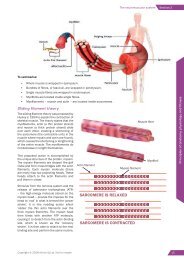
The slid<strong>in</strong>g filament theory and muscle contractions<br />
Perimysium<br />
surround<strong>in</strong>g<br />
the fascicle<br />
Figure 2.2 The structure of a muscle<br />
Muscular contraction beg<strong>in</strong>s with the two contractile prote<strong>in</strong>s (or myofilaments): act<strong>in</strong> and myos<strong>in</strong>. Act<strong>in</strong> (the th<strong>in</strong><br />
filament) is anchored to the ends of the sarcomere and myos<strong>in</strong> (the thick filament) is located <strong>in</strong> the middle of the<br />
sarcomere.<br />
Spirall<strong>in</strong>g from the myos<strong>in</strong> filament is a series of ‘hook-like’ projections referred to as the myos<strong>in</strong> heads. Dur<strong>in</strong>g<br />
muscular contraction, these heads attach themselves to the act<strong>in</strong> and rotate, pull<strong>in</strong>g on the filaments. This causes<br />
the act<strong>in</strong> to be drawn <strong>in</strong>wards, dragg<strong>in</strong>g the ends of the sarcomere together. This shorten<strong>in</strong>g process is referred<br />
to as the slid<strong>in</strong>g filament mechanism. The characteristic contraction of muscles is caused by the simultaneous<br />
shorten<strong>in</strong>g of multiple sarcomeres.<br />
Applied anatomy and physiology for exercise, health and fitness<br />
Figure 2.3 The slid<strong>in</strong>g filament theory<br />
Copyright © 2017 <strong>Active</strong> <strong>IQ</strong> Ltd. Not for resale 19
Section 2: The musculoskeletal system<br />
Unit 1<br />
Common postural distortions<br />
Kyphosis refers to the outward curve of the thoracic sp<strong>in</strong>e.<br />
Lordosis refers to the <strong>in</strong>ward curve of the lumbar sp<strong>in</strong>e.<br />
These terms are often used to describe excessive curvature,<br />
but normal curvature of the thoracic region is kyphotic <strong>in</strong><br />
nature and normal curvature of the lumbar region is lordotic.<br />
For this reason, the follow<strong>in</strong>g terms can be used to dist<strong>in</strong>guish<br />
dysfunction:<br />
POINT OF<br />
INTEREST<br />
ROOT<br />
WORDS<br />
• Hypo = ‘under’ or ‘less’<br />
• Hyper = ‘over’, ‘above’ or<br />
‘too much’<br />
• Hyperkyphosis – an excessive curve of the thoracic sp<strong>in</strong>e.<br />
• Hypokyphosis – a flattened curve of the thoracic sp<strong>in</strong>e.<br />
• Hyperlordosis – an excessive curve of the lumbar sp<strong>in</strong>e, also known as a ‘hollow back’.<br />
• Hypolordosis – a flattened curve of the lumbar sp<strong>in</strong>e.<br />
The term scoliosis is of Greek orig<strong>in</strong> and means ‘crooked’ or ‘bent’. A scoliotic sp<strong>in</strong>e has an excessive lateral<br />
curvature.<br />
Upper crossed syndrome<br />
Upper crossed syndrome refers to a hyperkyphotic sp<strong>in</strong>al position comb<strong>in</strong>ed with protracted shoulders and a<br />
forward-pok<strong>in</strong>g head. The shortened and lengthened areas caused by this syndrome can be divided by a cross<br />
shape (see Figure 2.6), which is where this condition gets its name.<br />
Shortened/dom<strong>in</strong>ant/<br />
overactive muscles<br />
• Scalenes<br />
• Sternocleidomastoid<br />
• Upper trapezius<br />
• Levator scapula<br />
• Pectoralis major<br />
• Anterior deltoids<br />
• Latissimus dorsi<br />
• Teres major<br />
• Subscapularis<br />
• Pectoralis m<strong>in</strong>or<br />
Lower crossed syndrome<br />
Shortened<br />
Lengthened<br />
Lengthened<br />
Shortened<br />
Lengthened/<strong>in</strong>hibited/<br />
underactive muscles<br />
• Deep neck flexors<br />
(longus capitis and colli)<br />
• Serratus anterior<br />
• Lower trapezius<br />
• Middle trapezius<br />
• Teres m<strong>in</strong>or<br />
• Infrasp<strong>in</strong>atus<br />
• Suprasp<strong>in</strong>atus<br />
• Thoracic erector sp<strong>in</strong>ae<br />
Figure 2.6 Upper crossed syndrome<br />
The shortened and lengthened regions caused by lower crossed syndrome can aga<strong>in</strong> be divided by a cross shape.<br />
This condition is characterised by an anterior pelvic tilt and hyperlordosis (see Figure 2.7).<br />
Applied anatomy and physiology for exercise, health and fitness<br />
Shortened/dom<strong>in</strong>ant/<br />
overactive muscles<br />
• Iliopsoas<br />
• Lumbar erector sp<strong>in</strong>ae<br />
• Rectus femoris<br />
• Adductors<br />
• Tensor fasciae latae<br />
• Quadratus lumborum<br />
• Lumbar multifidus<br />
Shortened<br />
Lengthened<br />
Lengthened<br />
Shortened<br />
Lengthened/<strong>in</strong>hibited/<br />
underactive muscles<br />
• Rectus abdom<strong>in</strong>is<br />
• Obliques<br />
• Transversus abdom<strong>in</strong>is<br />
• Gluteus maximus<br />
• Gluteus medius<br />
• Gluteus m<strong>in</strong>imus<br />
Figure 2.7 Lower crossed syndrome<br />
Copyright © 2017 <strong>Active</strong> <strong>IQ</strong> Ltd. Not for resale 33
Section 4: The endocr<strong>in</strong>e system<br />
Unit 1<br />
Section 4: The endocr<strong>in</strong>e<br />
system<br />
The role of the endocr<strong>in</strong>e system<br />
Along with the nervous system, the endocr<strong>in</strong>e system helps to ma<strong>in</strong>ta<strong>in</strong> homeostasis. Instead of us<strong>in</strong>g action<br />
potentials, however, the endocr<strong>in</strong>e system exerts its <strong>in</strong>fluence via hormones (chemical messengers).<br />
Hormones are chemicals released <strong>in</strong>to the bloodstream to help control and manage the <strong>in</strong>ternal environment of the<br />
body. Hormones are derived from am<strong>in</strong>o acids, steroids or occasionally fatty acids and are released from various<br />
glands around the body, known as the endocr<strong>in</strong>e glands. Different hormones have different chemical shapes which<br />
determ<strong>in</strong>e the effects they will have.<br />
The chemical messages from hormones are slower than the electrical messages (action potentials) of the nervous<br />
system. Cell response times to a specific hormone can range from a few seconds to 30 m<strong>in</strong>utes, depend<strong>in</strong>g on<br />
concentration levels. However, although neural stimulus is very rapid, it does not last; endocr<strong>in</strong>e stimulus can last<br />
for hours, days or even longer.<br />
How hormones work<br />
KEY<br />
POINT<br />
Hormones are chemical messengers (the<br />
key) that affect specific target cells (the lock).<br />
To fully understand how the endocr<strong>in</strong>e system works,<br />
the way hormones function around the body must first<br />
be appreciated. The process beg<strong>in</strong>s when an endocr<strong>in</strong>e<br />
gland receives a stimulus that requires a response. The<br />
response is <strong>in</strong>itiated by releas<strong>in</strong>g a hormone <strong>in</strong>to the<br />
surround<strong>in</strong>g bloodstream. Hormones are transported<br />
around the body, seek<strong>in</strong>g out specific target cells. Each<br />
type of hormone is attracted to particular receptors<br />
with<strong>in</strong> the target cells which, <strong>in</strong> turn, will only be<br />
triggered by the ‘right’ hormone (<strong>in</strong> the same way that<br />
a lock can only be opened with the right key). Once the<br />
released hormone reaches a target cell, it docks at the<br />
cell’s receptor site; this <strong>in</strong>itiates the desired response <strong>in</strong><br />
the cell or group of cells.<br />
F<strong>in</strong>ally, when the hormone response has had the desired<br />
effect, there is usually a feedback loop between the<br />
targeted tissue and the <strong>in</strong>itiat<strong>in</strong>g endocr<strong>in</strong>e gland which<br />
reduces or stops the hormone production.<br />
Applied anatomy and physiology for exercise, health and fitness<br />
Figure 4.1 How hormones work<br />
Copyright © 2017 <strong>Active</strong> <strong>IQ</strong> Ltd. Not for resale 45
Unit 1<br />
Section 6: The digestive system<br />
Mouth and<br />
pharynx<br />
• Chew<strong>in</strong>g (mastication) – mechanical breakdown.<br />
• Produces saliva – conta<strong>in</strong>s enzyme salivary amylase – beg<strong>in</strong>s breakdown<br />
of carbohydrate. Also moistens food and protects teeth aga<strong>in</strong>st decay.<br />
• Food is swallowed and passes from the mouth <strong>in</strong>to the pharynx.<br />
• Rhythmical <strong>in</strong>voluntary muscular contractions (peristalsis) push food <strong>in</strong>to<br />
the stomach.<br />
Oesophagus<br />
Stomach<br />
• Produces gastric juices conta<strong>in</strong><strong>in</strong>g hydrochloric acid (kills bacteria) and the<br />
enzyme peps<strong>in</strong> to break down prote<strong>in</strong>.<br />
Pancreas<br />
• Secretes pancreatic juice conta<strong>in</strong><strong>in</strong>g digestive enzymes<br />
that help further the digestion and absorption of nutrients:<br />
• LIPASE – breaks down fat.<br />
• AMYLASE – breaks down carbohydrate <strong>in</strong>to<br />
glucose.<br />
• TRYPSIN – breaks down prote<strong>in</strong> <strong>in</strong>to am<strong>in</strong>o acids.<br />
• Liver produces bile acids which enable fats to mix with<br />
water (emulsification).<br />
• Gallbladder acts as storage for bile which is released <strong>in</strong>to<br />
the small <strong>in</strong>test<strong>in</strong>e.<br />
Liver and<br />
gallbladder<br />
Small <strong>in</strong>test<strong>in</strong>e<br />
• Receives bile juice from gallbladder and pancreatic juice.<br />
• Primary site for digestion and absorption.<br />
• A comb<strong>in</strong>ation of <strong>in</strong>ner surface folds and f<strong>in</strong>ger-like projections (villi and<br />
microvilli) provide large surface area for absorption.<br />
• Digested food is able to pass <strong>in</strong>to the blood vessels <strong>in</strong> the wall of the<br />
<strong>in</strong>test<strong>in</strong>e through the process of diffusion.<br />
Large <strong>in</strong>test<strong>in</strong>e<br />
(colon)<br />
• Absorbs water, vitam<strong>in</strong>s and m<strong>in</strong>erals.<br />
• Conta<strong>in</strong>s bacteria, which produce vitam<strong>in</strong>s<br />
and help prevent <strong>in</strong>fection <strong>in</strong> the <strong>in</strong>test<strong>in</strong>e.<br />
• Stores faeces.<br />
Rectum<br />
• Open<strong>in</strong>g for elim<strong>in</strong>ation of<br />
waste.<br />
Anus<br />
Figure 6.1 The digestive process<br />
60<br />
Copyright © 2017 <strong>Active</strong> <strong>IQ</strong> Ltd. Not for resale
Section 4: Us<strong>in</strong>g nutrients to fuel activity<br />
Unit 2<br />
POINT OF<br />
INTEREST<br />
Aerobic activities <strong>in</strong>clude:<br />
• Jogg<strong>in</strong>g.<br />
• Distance runn<strong>in</strong>g.<br />
• Distance cycl<strong>in</strong>g.<br />
• Distance swimm<strong>in</strong>g.<br />
• Exercise to music.<br />
• Distance row<strong>in</strong>g.<br />
• Brisk walk<strong>in</strong>g.<br />
• Ski<strong>in</strong>g.<br />
Aerobic activities<br />
Dur<strong>in</strong>g aerobic activities (low-<strong>in</strong>tensity, longer-duration) the<br />
demand for energy is slower. ATP is produced us<strong>in</strong>g oxygen,<br />
so this type of exercise can be kept up for longer. Fats can<br />
also be used to produce energy for aerobic activities as<br />
they can only be broken down with oxygen present. The<br />
body has a larger store of fat than carbohydrate.<br />
Most sports and activities <strong>in</strong>volve a mixture of both<br />
anaerobic and aerobic exercise, e.g. football, hockey and<br />
rugby conta<strong>in</strong> short bursts of strenuous activity (spr<strong>in</strong>ts,<br />
kicks, throws) <strong>in</strong>terspersed with longer periods of less<br />
strenuous activity (jogg<strong>in</strong>g, walk<strong>in</strong>g).<br />
Fuel<br />
Creat<strong>in</strong>e<br />
phosphate<br />
Muscle<br />
glycogen<br />
Liver glycogen +<br />
blood glucose<br />
Maximal<br />
short bursts<br />
High<br />
<strong>in</strong>tensity<br />
<strong>in</strong>termittent<br />
Intensity of exercise<br />
High<br />
<strong>in</strong>tensity<br />
150 m<strong>in</strong><br />
Low <strong>in</strong>tensity<br />
Great. Very great. Moderate. Moderate. Moderate. Negligible.<br />
Slight. Moderate. Moderate. Very great. Very great. Negligible.<br />
Negligible. Slight. Slight. Moderategreat.<br />
Great-very<br />
great.<br />
Slight.<br />
Fat Negligible. Slight. Negligible. Negligible. Slight. Slight.<br />
Energy to start exercise<br />
Table 4.2 Depletion of fuel for different types of exercise<br />
At the start of exercise, energy is produced without oxygen. As the heart and lungs work harder, carbohydrates and<br />
fats can be broken down. If the exercise becomes aerobic <strong>in</strong> nature, more oxygen is delivered around the body and<br />
fats start to be broken down <strong>in</strong>to fatty acids, taken to muscle cells and used as energy. For the first 5-15 m<strong>in</strong>utes<br />
(depend<strong>in</strong>g on fitness level) carbohydrate (glycogen) is used for fuel. As time goes on, the use of carbohydrate<br />
lessens and more fat is utilised for energy.<br />
Fatigue<br />
The fitter the <strong>in</strong>dividual is, the longer it takes to fatigue or run out of glycogen. After three or more hours of exercise,<br />
fatigue is caused by the depletion of glycogen <strong>in</strong> the muscles and liver, as well as low blood glucose levels.<br />
The pr<strong>in</strong>ciples of nutrition and their application to exercise and health<br />
No matter what type of exercise is completed, or how fit the<br />
<strong>in</strong>dividual is, the body will always need glycogen. The amount<br />
of glycogen <strong>in</strong> the muscles (and liver) before exercise is<br />
crucial, as the size of a person’s store dictates how long they<br />
will be able to exercise for before fatigu<strong>in</strong>g. The message is<br />
‘always start with a full glycogen store’.<br />
A diet rich <strong>in</strong> carbohydrates ensures high glycogen stores.<br />
The amount of glycogen <strong>in</strong> the muscles dictates how hard<br />
and for how long an athlete can exercise. If the <strong>in</strong>dividual eats<br />
a high-carbohydrate diet prior to tra<strong>in</strong><strong>in</strong>g and competitions,<br />
their stores will be full.<br />
• Low glycogen levels would only allow for low-<strong>in</strong>tensity<br />
exercise and cause early fatigue.<br />
• High glycogen levels mean an athlete can tra<strong>in</strong> harder<br />
and longer.<br />
Copyright © 2017 <strong>Active</strong> <strong>IQ</strong> Ltd. Not for resale 99