

Evidence base and patients' perspective - World Journal of ...
Evidence base and patients' perspective - World Journal of ...
Evidence base and patients' perspective - World Journal of ...

Create successful ePaper yourself
Turn your PDF publications into a flip-book with our unique Google optimized e-Paper software.
Chen JG et al . Colorectal cancer screening<br />
MATERIALS AND METHODS<br />
Study materials<br />
The stool specimen collection, colonoscopy <strong>and</strong> pathologic<br />
examination were performed in the Eighth Hospital<br />
<strong>of</strong> Wuhan City which is a hospital specializing in anorectal<br />
diseases. GFOBT, IFOBT <strong>and</strong> Tf kits were purchased<br />
from Baso Diagnostics Inc <strong>and</strong> WHPM Inc.<br />
Study group<br />
From January 2010 to September 2010, 861 subjects at<br />
highrisk (a personal history <strong>of</strong> curativeintent resection<br />
<strong>of</strong> CRC or intestinal polyps; family history <strong>of</strong> colorectal<br />
cancer; having the following two or more: chronic diarrhea,<br />
chronic constipation, abdominal pain, dark stool,<br />
blood or mucus on stool) were recruited. The inclusion<br />
criteria were as following: age over 14 years, male or female.<br />
Subjects with age < 14 years were excluded. All<br />
participants provided written informed consent <strong>and</strong> were<br />
instructed on diet <strong>and</strong> drug restrictions three days before<br />
<strong>and</strong> during the period <strong>of</strong> stool collection.<br />
Fecal samples collection <strong>and</strong> IFOBT <strong>and</strong> Tf analysis<br />
All fecal samples were collected the day before colonoscopy<br />
<strong>and</strong> processed in accordance with manufacturer’<br />
s instructions. We applied the fecal sample on the strip<br />
<strong>and</strong> the result was read out within 5 min (the result was<br />
invalid after 5 min). A red bar in control area (C) only was<br />
considered as negative. A red bar in both the testing area<br />
(T) <strong>and</strong> the control area (C), was considered as positive.<br />
If there was no red bar in the control area(C), the test was<br />
considered invalid. Tf, IFOBT <strong>and</strong> gFOBT were performed<br />
simultaneously on the same stool. To minimize<br />
falsenegative cases, all subjects with negative samples<br />
were asked to provide an additional stool specimen for<br />
a second test; if the second test still gave negative result,<br />
a third test would be conducted. As long as one <strong>of</strong> the<br />
three tests showed positive results, the subject was considered<br />
to have a positive sample. If the results were all negative<br />
after testing three repeated samples, the subject was<br />
considered a true negative. Approximately 10% <strong>of</strong> the<br />
samples were repeated <strong>and</strong> the concordance was 100%.<br />
Statistical analysis<br />
The positive rate <strong>of</strong> Tf alone, IFOBT alone, Tf combined<br />
with IFOBT (Tf + IFOBT), Tf <strong>and</strong> IFOBT combined<br />
with gFOBT (Tf + IFOBT + gFOBT), as well as<br />
their respective specificity, likelihood ratio, odd ratio <strong>and</strong><br />
95% confidence interval were calculated to compared the<br />
sensitivity <strong>of</strong> Tf, IFOBT, Tf+ IFOBT <strong>and</strong> Tf + IFOBT<br />
+ gFOBT in detecting CRC <strong>and</strong> precancerous lesions.<br />
χ 2 <strong>and</strong> McNemar’ s test were conducted to determine the<br />
significance <strong>of</strong> difference. P < 0.05 in a twotailed test<br />
was considered statistically significant. Analyses were performed<br />
using SPSS version 17.0.<br />
RESULTS<br />
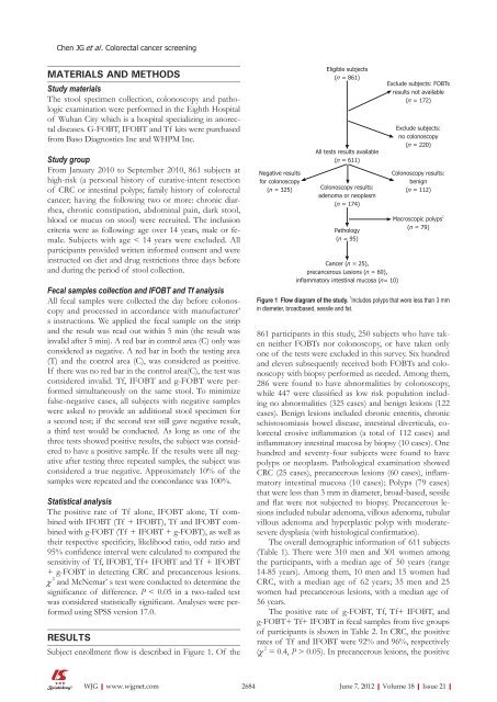
Subject enrollment flow is described in Figure 1. Of the<br />
WJG|www.wjgnet.com<br />
Negative results<br />
for colonoscopy<br />
(n = 325)<br />
Eligible subjects<br />
(n = 861)<br />
All tests results available<br />
(n = 611)<br />
Colonoscopy results:<br />
adenoma or neoplasm<br />
(n = 174)<br />
Pathology<br />
(n = 95)<br />
Cancer (n = 25),<br />
precancerous Lesions (n = 60),<br />
inflammatory intestinal mucosa (n = 10)<br />
Exclude subjects: FOBTs<br />
results not available<br />
(n = 172)<br />
Exclude subjects:<br />
no colonoscopy<br />
(n = 220)<br />
Colonoscopy results:<br />
benign<br />
(n = 112)<br />
Macroscopic polyps 1<br />
(n = 79)<br />
Figure 1 Flow diagram <strong>of</strong> the study. 1 Includes polyps that were less than 3 mm<br />
in diameter, broad<strong>base</strong>d, sessile <strong>and</strong> fat.<br />
861 participants in this study, 250 subjects who have taken<br />
neither FOBTs nor colonoscopy, or have taken only<br />
one <strong>of</strong> the tests were excluded in this survey. Six hundred<br />
<strong>and</strong> eleven subsequently received both FOBTs <strong>and</strong> colonoscopy<br />
with biopsy performed as needed. Among them,<br />
286 were found to have abnormalities by colonoscopy,<br />
while 447 were classified as low risk population including<br />
no abnormalities (325 cases) <strong>and</strong> benign lesions (122<br />
cases). Benign lesions included chronic enteritis, chronic<br />
schistosomiasis bowel disease, intestinal diverticula, colorectal<br />
erosive inflammation (a total <strong>of</strong> 112 cases) <strong>and</strong><br />
inflammatory intestinal mucosa by biopsy (10 cases). One<br />
hundred <strong>and</strong> seventyfour subjects were found to have<br />
polyps or neoplasm. Pathological examination showed<br />
CRC (25 cases), precancerous lesions (60 cases), inflammatory<br />
intestinal mucosa (10 cases); Polyps (79 cases)<br />
that were less than 3 mm in diameter, broad<strong>base</strong>d, sessile<br />
<strong>and</strong> flat were not subjected to biopsy. Precancerous lesions<br />
included tubular adenoma, villous adenoma, tubular<br />
villous adenoma <strong>and</strong> hyperplastic polyp with moderatesevere<br />
dysplasia (with histological confirmation).<br />
The overall demographic information <strong>of</strong> 611 subjects<br />
(Table 1). There were 310 men <strong>and</strong> 301 women among<br />
the participants, with a median age <strong>of</strong> 50 years (range<br />
1485 years). Among them, 10 men <strong>and</strong> 15 women had<br />
CRC, with a median age <strong>of</strong> 62 years; 35 men <strong>and</strong> 25<br />
women had precancerous lesions, with a median age <strong>of</strong><br />
56 years.<br />
The positive rate <strong>of</strong> gFOBT, Tf, Tf+ IFOBT, <strong>and</strong><br />
g-FOBT+ Tf+ IFOBT in fecal samples from five groups<br />
<strong>of</strong> participants is shown in Table 2. In CRC, the positive<br />
rates <strong>of</strong> Tf <strong>and</strong> IFOBT were 92% <strong>and</strong> 96%, respectively<br />
(χ 2 = 0.4, P > 0.05). In precancerous lesions, the positive<br />
2684 June 7, 2012|Volume 18|Issue 21|


