

Evidence base and patients' perspective - World Journal of ...
Evidence base and patients' perspective - World Journal of ...
Evidence base and patients' perspective - World Journal of ...

Create successful ePaper yourself
Turn your PDF publications into a flip-book with our unique Google optimized e-Paper software.
Terzin V et al . Serum IgG4 in autoimmune diseases<br />
Immunological examinations in AIP patients have demonstrated<br />
high incidences <strong>of</strong> hypergammaglobulinemia<br />
(43%), increased serum levels <strong>of</strong> immunoglobulin G (IgG)<br />
(62%-80%) <strong>and</strong> IgG4 (68%-92%), <strong>and</strong> the presence <strong>of</strong><br />
antinuclear antibodies (40%-64%) <strong>and</strong> rheumatoid factor<br />
(25%). Among all the serological diagnostic features, an<br />
elevated serum level <strong>of</strong> IgG4 has the highest individual<br />
diagnostic value; however, it is not disease specific. Furthermore,<br />
an elevated serum IgG4 level correlates with<br />
the activity <strong>of</strong> AIP [5,6] . Kamisawa et al [7] reported an association<br />
between serum IgG4 level <strong>and</strong> extrapancreatic<br />
lesions in patients with AIP. AIP patients with a serum<br />
IgG4 level ≥ 2200 mg/L frequently exhibit extrapancreatic<br />
lesions.<br />
The immunologic <strong>and</strong> histologic features <strong>of</strong> AIP <strong>and</strong><br />
the glucocorticosteroid responsiveness suggest an autoimmune<br />
mechanism for the development <strong>of</strong> the disease [8] .<br />
AIP is accompanied by other autoimmune diseases (sclerosing<br />
cholangitis, sclerosing sialadenitis, retroperitoneal<br />
fibrosis, enlarged celiac <strong>and</strong> hilar lymph nodes, chronic<br />
thyroiditis <strong>and</strong> interstitial nephritis, etc.) in 50%-63% <strong>of</strong><br />
cases, suggesting that AIP may be a systemic disorder [1-4] .<br />
The occurrence <strong>of</strong> autoimmune diseases in association<br />
with AIP is well documented [9,10] , but the incidence <strong>of</strong><br />
such associations has not been reported.<br />
The aim <strong>of</strong> the present study was to assess the presence<br />
<strong>of</strong> AIP in different systemic autoimmune diseases<br />
(SAIDs) through measurement <strong>of</strong> the serum IgG4 level<br />
<strong>and</strong> examination <strong>of</strong> the morphology <strong>of</strong> the pancreas.<br />
MATERIALS AND METHODS<br />
Patients <strong>and</strong> diagnosis <strong>of</strong> diseases<br />
Serum samples were obtained from 61 patients with different<br />
SAIDs who had been admitted to our Department<br />
<strong>of</strong> Rheumatology <strong>and</strong> had not participated in glucocorticosteroid<br />
treatment during the past 2 years. One male<br />
<strong>and</strong> 60 females (mean age 54.5 years, range 29-82 years)<br />
were recruited.<br />
Autoimmune diseases were diagnosed according to<br />
st<strong>and</strong>ard diagnostic criteria [11-14] . The diagnosis <strong>of</strong> AIP<br />
was <strong>base</strong>d on the HISORt criteria [15] . The most frequent<br />
diagnosis was Sjögren’s syndrome (SS), but systemic lupus<br />
erythematosus (SLE), Hashimoto thyroiditis, Raynaud’s<br />
syndrome, polymyositis <strong>and</strong> systemic sclerosis also<br />
occurred (Table 1).<br />
Serum samples were additionally obtained from 7 age-<br />
<strong>and</strong> sex-matched healthy subjects, <strong>and</strong> 6 patients with<br />
AIP. In one AIP patient, the AIP was accompanied by<br />
rheumatoid arthritis <strong>and</strong> ankylosing spondylitis.<br />
All participants provided their written informed consent.<br />
The study protocol was approved by the ethics committee<br />
at the University <strong>of</strong> Szeged <strong>and</strong> was carried out<br />
in full accordance with the most recent revisions <strong>of</strong> the<br />
Helsinki Declaration.<br />
IgG4 assay<br />
After collection, serum samples were stored at -70 ℃ until<br />
analyzed. The IgG4 subclass was determined by the ra-<br />
WJG|www.wjgnet.com<br />
Serum immunoglobulin G4 level (mg/L)<br />
Table 1 Distribution <strong>of</strong> gender <strong>and</strong> age in groups <strong>of</strong> patients<br />
dial immunodiffusion (RID) method (The Binding Site<br />
Limited, Birmingham, United Kingdom). The diameters<br />
<strong>of</strong> precipitation rings were measured after 72 h. The results<br />
were read using the RID reference table. The lowest<br />
detection limit was 22.4 mg/L. The intra- <strong>and</strong> interassay<br />
coefficients <strong>of</strong> variation were 3.26 <strong>and</strong> 0.89 CV%,<br />
respectively, as stated by the manufacturer. A cut<strong>of</strong>f value<br />
<strong>of</strong> 400 mg/L was employed.<br />
Patients with a serum IgG4 level <strong>of</strong> > 400 mg/L were<br />
examined by a gastroenterologist. The clinical <strong>and</strong> laboratory<br />
data were reviewed <strong>and</strong> abdominal ultrasonography<br />
(US) <strong>and</strong> computed tomography (CT) were performed.<br />
Anti-SS-A/SS-B autoantibody determination<br />
The presence <strong>of</strong> anti-SS-A/SS-B autoantibodies was determined<br />
by means <strong>of</strong> commercial enzyme-linked immunosorbent<br />
assays, conducted according to the protocols<br />
provided by the manufacturers.<br />
Experimental data were evaluated statistically with<br />
the independent-samples t test. P-values < 0.05 were accepted<br />
as being statistically significant. Statistical data is<br />
expressed as mean ± SD.<br />
RESULTS<br />
No. <strong>of</strong><br />
patients<br />
Male/<br />
female<br />
Age<br />
mean (range)<br />
Sjögren's syndrome 35 1/34 56.7 (29-82)<br />
Systemic lupus erythematosus 22 0/22 50.2 (31-68)<br />
Systemic sclerosis 4 0/4 59.5 (45-80)<br />
Normal subjects 7 4/3 68 (56-80)<br />
Autoimmune pancreatitis 6 3/3 53.7 (27-75)<br />
4000<br />
3500<br />
3000<br />
2500<br />
2000<br />
1500<br />
1000<br />
500<br />
0<br />
Sjögren's syndrome<br />
Systemic lupus erythematosus<br />
Systemic sclerosis<br />
Autoimmune pancreatitis<br />
Control<br />
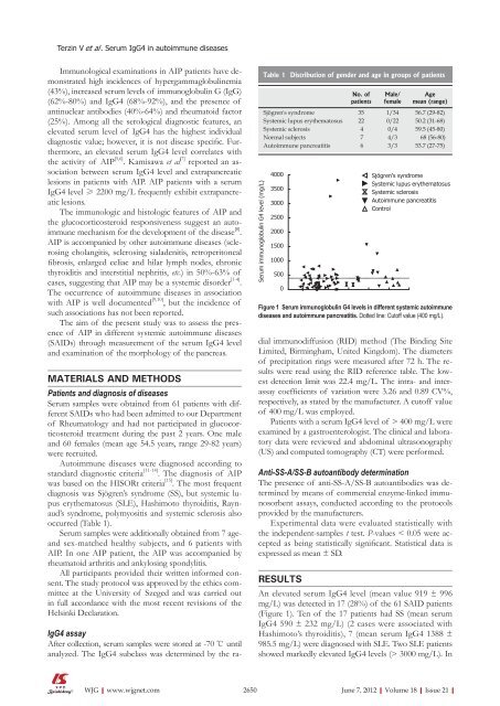
Figure 1 Serum immunoglobulin G4 levels in different systemic autoimmune<br />
diseases <strong>and</strong> autoimmune pancreatitis. Dotted line: Cut<strong>of</strong>f value (400 mg/L).<br />
An elevated serum IgG4 level (mean value 919 ± 996<br />
mg/L) was detected in 17 (28%) <strong>of</strong> the 61 SAID patients<br />
(Figure 1). Ten <strong>of</strong> the 17 patients had SS (mean serum<br />
IgG4 590 ± 232 mg/L) (2 cases were associated with<br />
Hashimoto’s thyroiditis), 7 (mean serum IgG4 1388 ±<br />
985.5 mg/L) were diagnosed with SLE. Two SLE patients<br />
showed markedly elevated IgG4 levels (> 3000 mg/L). In<br />
2650 June 7, 2012|Volume 18|Issue 21|


