Early management of head injury SIGN - Colleges
Early management of head injury SIGN - Colleges
Early management of head injury SIGN - Colleges

Create successful ePaper yourself
Turn your PDF publications into a flip-book with our unique Google optimized e-Paper software.
S I G N<br />
Scottish<br />
Intercollegiate<br />
Guidelines<br />
Network<br />
August 2000<br />
A National Clinical Guideline<br />
46<br />
<strong>SIGN</strong> Publication<br />
Number<br />
<strong>Early</strong> Management <strong>of</strong> Patients<br />
with a Head Injury
KEY TO EVIDENCE STATEMENTS AND GRADES OF RECOMMENDATIONS<br />
The definitions <strong>of</strong> the types <strong>of</strong> evidence and the grading <strong>of</strong> recommendations used in this<br />
guideline originate from the US Agency for Health Care Policy and Research 1 and are set out in<br />
the following tables.<br />
STATEMENTS OF EVIDENCE<br />
Ia Evidence obtained from meta-analysis <strong>of</strong> randomised controlled trials.<br />
Ib Evidence obtained from at least one randomised controlled trial.<br />
IIa Evidence obtained from at least one well-designed controlled study without<br />
randomisation.<br />
IIb Evidence obtained from at least one other type <strong>of</strong> well-designed quasi-experimental<br />
study.<br />
III Evidence obtained from well-designed non-experimental descriptive studies, such<br />
as comparative studies, correlation studies and case studies.<br />
IV Evidence obtained from expert committee reports or opinions and/or clinical<br />
experiences <strong>of</strong> respected authorities.<br />
GRADES OF RECOMMENDATIONS<br />
A Requires at least one randomised controlled trial as part <strong>of</strong> a body <strong>of</strong> literature <strong>of</strong><br />
overall good quality and consistency addressing the specific recommendation.<br />
(Evidence levels Ia, Ib)<br />
B Requires the availability <strong>of</strong> well conducted clinical studies but no randomised<br />
clinical trials on the topic <strong>of</strong> recommendation.<br />
(Evidence levels IIa, IIb, III)<br />
C Requires evidence obtained from expert committee reports or opinions and/or<br />
clinical experiences <strong>of</strong> respected authorities. Indicates an absence <strong>of</strong> directly<br />
applicable clinical studies <strong>of</strong> good quality.<br />
(Evidence level IV)<br />
GOOD PRACTICE POINTS<br />
� Recommended best practice based on the clinical experience <strong>of</strong> the guideline<br />
development group.<br />
Paediatric practice point (see page 2 for further information).
Contents<br />
Guideline development group (i)<br />
Notes for users <strong>of</strong> the guideline (iii)<br />
Abbreviations (iv)<br />
Summary <strong>of</strong> recommendations (v)<br />
Summary <strong>of</strong> paediatric practice points (viii)<br />
1 Introduction<br />
1.1 Background 1<br />
1.2 The need for a guideline 1<br />
1.3 Remit <strong>of</strong> the guideline 1<br />
2 Assessment and classification<br />
2.1 Assessing the patient 3<br />
2.2 The Glasgow Coma Scale and Coma Score 3<br />
3 Indications for referral to hospital 5<br />
4 Principles <strong>of</strong> <strong>management</strong><br />
4.1 Principles <strong>of</strong> advanced trauma life support 7<br />
4.2 Features <strong>of</strong> <strong>head</strong> injured patients attending Scottish hospitals 7<br />
5 Imaging<br />
5.1 Detection <strong>of</strong> traumatic intracranial lesions by imaging 8<br />
5.2 Selection <strong>of</strong> patients for imaging 8<br />
5.3 Indications for skull x-ray 10<br />
5.4 Indications for <strong>head</strong> CT 11<br />
5.5 Interpretation <strong>of</strong> images 13<br />
5.6 Imaging the cervical spine 13<br />
6 Admission or discharge?<br />
6.1 Indications for admission to hospital 16<br />
6.2 Indications for discharge from A&E 17<br />
6.3 Information and instructions on discharge from A&E 18<br />
7 Inpatient observation<br />
7.1 Clinical observation and recording 19<br />
7.2 Frequency <strong>of</strong> observations 20<br />
7.3 Frequency <strong>of</strong> reappraisal 20<br />
7.4 Discharge after observation 21<br />
8 Indications for referral to a neurosurgical unit<br />
8.1 Reasons for consultation 23<br />
8.2 Indications for referral 23<br />
8.3 Transfer between a general hospital and a neurosurgical unit 24<br />
CONTENTS
EARLY MANAGEMENT OF PATIENTS WITH A HEAD INJURY<br />
9 Follow up 25<br />
10 Implications for service delivery 26<br />
11 Recommendations for audit and research<br />
11.1 Audit 27<br />
11.2 Key indicators <strong>of</strong> quality <strong>of</strong> <strong>management</strong> 27<br />
11.3 Recommendations for further research 28<br />
Annexes<br />
1 Details <strong>of</strong> the systematic review undertaken for this guideline 29<br />
2Example <strong>head</strong> <strong>injury</strong> observation chart 30<br />
3 Advice for a person taking a patient home from A&E 32<br />
4 Advice for a person allowed home from A&E 33<br />
5 Observation instructions for parents or carers 34<br />
6 Advice for a person discharged home after hospital admission 35<br />
7 Scottish Trauma Audit Group neurosurgical referral letter 36<br />
References 38<br />
Tables<br />
1 The Glasgow Coma Scale and Score 4<br />
2Level <strong>of</strong> responsiveness in <strong>head</strong> injured patients attending A&E<br />
Departments in Scotland 7<br />
3 Risk <strong>of</strong> an operable intracranial haematoma in <strong>head</strong> injured<br />
patients 9<br />
Figure<br />
1 Use <strong>of</strong> radiographic investigations in patients (>5 years <strong>of</strong> age)<br />
with a <strong>head</strong> <strong>injury</strong> 14
GUIDELINE DEVELOPMENT GROUP<br />
GUIDELINE DEVELOPMENT GROUP<br />
Pr<strong>of</strong>essor Graham Teasdale Pr<strong>of</strong>essor <strong>of</strong> Neurosurgery, University <strong>of</strong> Glasgow<br />
(Chairman) and Consultant Neurosurgeon, Southern General Hospital, Glasgow<br />
Mr Douglas Gentleman Consultant Neurosurgeon, Centre for Brain Injury Rehabilitation,<br />
(Secretary) Royal Victoria Hospital, Dundee<br />
Dr Peter Andrews Consultant Anaesthetist, Western General Hospital, Edinburgh<br />
Mr Chris Blaiklock Clinical Director, Surgical Directorate, Aberdeen Royal Infirmary<br />
Sister Elma Cruickshank Nursing Manager, Aberdeen Royal Infirmary<br />
Mr Tom Donnelly Superintendent Physiotherapist, Southern General Hospital, Glasgow<br />
Mr Patrick Grant Consultant in Accident and Emergency Medicine, Western Infirmary, Glasgow<br />
Dr Peter Hendry Consultant Radiologist, Raigmore Hospital, Inverness<br />
Mr David Johnson Clinical Psychologist, Astley Ainslie Hospital, Edinburgh<br />
Mr John Logie Consultant Surgeon, Raigmore Hospital, Inverness<br />
Dr John Lyon General Practitioner, Taynuilt<br />
Miss Lynn McLeish Manager, Scotcare Brain Injury Rehabilitation Unit, Newmains<br />
Mr Bill Morrison Consultant in Accident and Emergency Medicine, Dundee Royal Infirmary<br />
Dr Leo Murray formerly Consultant in Accident and Emergency Medicine, Ayr Hospital<br />
Mr David Ross Consultant Orthopaedic Surgeon, Stirling Royal Infirmary<br />
Dr David Signorini formerly Senior Statistician, Clinical Neurosciences, University <strong>of</strong> Edinburgh<br />
Mr Ian Swann Consultant in Accident and Emergency Medicine, Glasgow Royal Infirmary<br />
Dr Evelyn Teasdale Consultant Neuroradiologist, Southern General Hospital, Glasgow<br />
Mr Allan Turner Consultant General Surgeon, Queen Margaret Hospital, Dunfermline<br />
Declarations <strong>of</strong> interests were made by all members <strong>of</strong> the guideline development group.<br />
Further details are available on request from the <strong>SIGN</strong> Secretariat.<br />
Additional advice was provided by:<br />
Mr Carl Davis Consultant Paediatric Surgeon, Yorkhill Hospital, Glasgow<br />
Mr David Peck Rehab Clinical Psychologist, Craig Dunain Hospital, Inverness<br />
SPECIALIST REVIEWERS<br />
Dr Tom Beattie Consultant in Paediatric A&E Medicine, Royal Hospital for Sick Children, Edinburgh<br />
Dr Ivan Brenkel Consultant Orthopaedic Surgeon, Victoria Hospital, Kirkcaldy<br />
Dr Clare Campbell General Practitioner, Broxburn, West Lothian<br />
Mr Muftah Eljamel Consultant Neurosurgeon, Ninewells Hospital, Dundee<br />
Mr Paul Fisher Consultant Surgeon, Caithness General Hospital<br />
Dr Peter Freeland Consultant in Accident and Emergency, St Johns Hospital, Livingston<br />
Dr Martin Kirkpatrick Consultant Paediatric Neurologist, Ninewells Hospital, Dundee<br />
Dr Colville Laird General Practitioner, Auchterarder<br />
Dr Andrew Marsden Medical Director, Scottish Ambulance Service<br />
Mr Paul May Consultant Paediatric Neurosurgeon, Alder Hey Hospital, Liverpool<br />
Dr Dermot McKeown Clinical Director <strong>of</strong> Anaesthetics, Royal Infirmary <strong>of</strong> Edinburgh<br />
Pr<strong>of</strong>essor David Mendelow University Department <strong>of</strong> Neurosurgery, Newcastle General Hospital<br />
Dr Allan Merry General Practitioner, Ardrossan<br />
Pr<strong>of</strong>essor Gillian Needham Postgraduate Dean, Aberdeen University Medical School<br />
Dr Peter Raine Consultant Paediatric Surgeon, Royal Hospital for Sick Children, Glasgow<br />
Dr Alasdair Short Consultant Physician in Intensive Care Medicine, Broomfield Hospital, Chelmsford<br />
Mr William Taylor Consultant Neurosurgeon, Southern General Hospital, Glasgow<br />
Dr Joanna Wardlaw Reader, University <strong>of</strong> Edinburgh Department <strong>of</strong> Clinical Neurosciences<br />
Pr<strong>of</strong>essor Jamie Weir Clinical Pr<strong>of</strong>essor <strong>of</strong> Radiology, Grampian University Hospitals<br />
(i)
EARLY MANAGEMENT OF PATIENTS WITH A HEAD INJURY<br />
(ii)<br />
<strong>SIGN</strong> EDITORIAL BOARD<br />
Pr<strong>of</strong>essor James Petrie Chairman <strong>of</strong> <strong>SIGN</strong>, Co-editor<br />
Dr Doreen Campbell CRAG Secretariat, Scottish Executive Department <strong>of</strong> Health<br />
Dr Patricia Donald Royal College <strong>of</strong> General Practitioners<br />
Pr<strong>of</strong>essor Jeremy Grimshaw Health Services Research Unit, University <strong>of</strong> Aberdeen<br />
Mr Douglas Harper Royal College <strong>of</strong> Surgeons <strong>of</strong> Edinburgh<br />
Dr Grahame Howard Royal College <strong>of</strong> Radiologists<br />
Dr Margaret Roberts Royal College <strong>of</strong> Physicians & Surgeons <strong>of</strong> Glasgow<br />
<strong>SIGN</strong> SECRETARIAT<br />
Ms Juliet Miller Director <strong>of</strong> <strong>SIGN</strong>, Co-editor<br />
Ms Anne Borthwick Publications Officer<br />
Ms Francesca Chappell Information Officer<br />
Ms Christine Crack Patient Support Officer<br />
Ms Gail Crosbie Networking Officer<br />
Mrs Lesley Forsyth Conferences Coordinator<br />
Mr Robin Harbour Information Manager<br />
Ms Paula McDonald Senior Guideline Coordinator<br />
Dr Moray Nairn Programme Manager<br />
Mr Richard Nicodème Design and DTP Coordinator<br />
Mrs Judith Proudfoot Adminstrator<br />
Ms Gaynor Rattray Guideline Coordinator<br />
Dr Safia Qureshi Senior Programme Manager
Notes for users <strong>of</strong> the guideline<br />
LOCAL IMPLEMENTATION OF THE GUIDELINE<br />
It is intended that this guideline will be adopted after local discussion involving clinical staff and<br />
<strong>management</strong>. The Area Clinical Effectiveness Committee should be fully involved. Local arrangements<br />
may then be made for the derivation <strong>of</strong> specific local guidelines to implement the national guideline<br />
in individual hospitals, units and practices and for securing compliance with them. This may be done<br />
by a variety <strong>of</strong> means including patient-specific reminders, continuing education and training, and<br />
clinical audit.<br />
<strong>SIGN</strong> consents to the copying <strong>of</strong> this guideline for the purpose <strong>of</strong> implementation in the National Health<br />
Service in Scotland. For details <strong>of</strong> how to order additional copies <strong>of</strong> this or other <strong>SIGN</strong> publications, see<br />
inside back cover.<br />
STATEMENT OF INTENT<br />
This report is not intended to be construed or to serve as a standard <strong>of</strong> medical care. Standards <strong>of</strong><br />
medical care are determined on the basis <strong>of</strong> all clinical data available for an individual case and are<br />
subject to change as scientific knowledge and technology advance and patterns <strong>of</strong> care evolve.<br />
These parameters <strong>of</strong> practice should be considered guidelines only. Adherence to them will not ensure a<br />
successful outcome in every case, nor should they be construed as including all proper methods <strong>of</strong> care<br />
or excluding other acceptable methods <strong>of</strong> care aimed at the same results. The ultimate judgement regarding<br />
a particular clinical procedure or treatment plan must be made by the doctor in light <strong>of</strong> the clinical data<br />
presented by the patient and the diagnostic and treatment options available.<br />
Significant departures from the national guideline as expressed in the local guideline should be fully<br />
documented and the reasons for the differences explained. Significant departures from the local guideline<br />
should be fully documented in the patient’s case notes at the time the relevant decision is taken.<br />
A background paper on the legal implications <strong>of</strong> guidelines is available from the <strong>SIGN</strong> secretariat.<br />
REVIEW OF THE GUIDELINE<br />
This guideline was issued in August 2000 and will be reviewed in 2002 or sooner if new evidence<br />
becomes available. Any updates to the guideline in the interim period will be noted on the <strong>SIGN</strong> web<br />
site. Comments are invited to assist the review process. All correspondence and requests for background<br />
information regarding the guideline should be sent to:<br />
<strong>SIGN</strong> Secretariat<br />
Royal College <strong>of</strong> Physicians<br />
9 Queen Street<br />
Edinburgh EH2 1JQ<br />
Tel: 0131 225 7324<br />
Fax: 0131 225 1769<br />
e-mail: sign@rcpe.ac.uk<br />
www.sign.ac.uk<br />
NOTES FOR USERS OF THE GUIDELINE<br />
(iii)
EARLY MANAGEMENT OF PATIENTS WITH A HEAD INJURY<br />
(iv)<br />
Abbreviations<br />
A&E Accident and Emergency<br />
ATLS Advanced trauma life support<br />
APLS Advanced paediatric life support<br />
CSF Cerebrospinal fluid<br />
CT Computed tomographic<br />
GCS Glasgow Coma Scale and Score<br />
GP General Practitioner<br />
PTA Post-traumatic amnesia<br />
SASM Scottish Audit <strong>of</strong> Surgical Mortality<br />
<strong>SIGN</strong> Scottish Intercollegiate Guidelines Network<br />
STAG Scottish Trauma Audit Group
Summary <strong>of</strong> recommendations<br />
ASSESSMENT AND CLASSIFICATION<br />
B The <strong>management</strong> <strong>of</strong> <strong>head</strong> injured patients should be guided by clinical assessments and protocols<br />
based on the Glasgow Coma Scale and Glasgow Coma Score.<br />
INDICATIONS FOR REFERRAL TO HOSPITAL<br />
B A <strong>head</strong> injured patient should be referred to hospital if any <strong>of</strong> the following is present:<br />
� Impaired consciousness (GCS
EARLY MANAGEMENT OF PATIENTS WITH A HEAD INJURY<br />
(vi)<br />
B CT scanning should be done in a patient who has any <strong>of</strong> the following features:<br />
(1) The patient is eye opening only to pain or does not converse<br />
(CGS 12/15 or less)<br />
(2) A deteriorating level <strong>of</strong> consciousness or progressive focal neurological signs<br />
(3) Confusion or drowsiness (CGS 13 or 14/15) followed by failure to improve within at most<br />
four hours <strong>of</strong> clinical observation<br />
(4) Radiological/clinical evidence <strong>of</strong> a fracture, whatever the level <strong>of</strong> consciousness<br />
(5) New focal neurological signs which are not getting worse<br />
(6) Full consciousness (GCS 15/15) with no fracture but other features, e.g.:<br />
− severe and persistent <strong>head</strong>ache<br />
− nausea and vomiting<br />
– irritability or altered behaviour<br />
– a seizure.<br />
B Skull films should be carried out if any <strong>of</strong> the following apply and if CT is not being performed:<br />
(a) If the patient is alert and orientated and obeying commands (GCS 15/15) but:<br />
− the mechanism <strong>of</strong> <strong>injury</strong> has not been trivial; or<br />
− consciousness has been lost; or<br />
− the patient has loss <strong>of</strong> memory or has vomited; or<br />
− the scalp has a full thickness laceration or a boggy haematoma; or<br />
− the history is inadequate.<br />
or<br />
(b) If the level <strong>of</strong> consciousness is impaired (GCS ≤14/15).<br />
B Imaging <strong>of</strong> the cervical spine, including the cervico-thoracic junction should be carried out:<br />
� in a fully conscious patient (GCS 15/15) if clinical symptoms or signs or the mechanism <strong>of</strong><br />
<strong>injury</strong> indicate the possibility <strong>of</strong> <strong>injury</strong><br />
� in a patient with persisting impaired consciousness (GCS 14/15 or less)<br />
� in an unconscious patient, not localising pain (GCS 6/15 or less) CT scanning <strong>of</strong> the<br />
cervical spine down to C2 should be undertaken routinely, at the time <strong>of</strong> <strong>head</strong> scanning.<br />
ADMISSION OR DISCHARGE?<br />
B A patient should be admitted to hospital if:<br />
� the level <strong>of</strong> consciousness is impaired (GCS
B A patient can be discharged from A&E for observation at home if fully conscious (GCS 15/15)<br />
with none <strong>of</strong> the additional risk factors above or other relevant adverse medical and social<br />
factors.<br />
INPATIENT OBSERVATION<br />
B Any <strong>of</strong> the following examples <strong>of</strong> neurological deterioration should prompt urgent reappraisal by<br />
a doctor:<br />
� the development <strong>of</strong> agitation or abnormal behaviour<br />
� a sustained decrease in conscious level <strong>of</strong> at least one point in the motor or verbal response or<br />
two points in the eye opening response <strong>of</strong> the GCS<br />
� the development <strong>of</strong> severe or increasing <strong>head</strong>ache or persisting vomiting<br />
� new or evolving neurological symptoms or signs, such as pupil inequality or asymmetry <strong>of</strong><br />
limb or facial movement.<br />
INDICATIONS FOR REFERRAL TO A NEUROSURGICAL UNIT<br />
B A <strong>head</strong> injured patient should be discussed with a neurosurgeon:<br />
� when a CT scan in a general hospital shows a recent intracranial lesion<br />
� when a patient fulfils the criteria for CT scanning but this cannot be done within an<br />
appropriate period<br />
� irrespective <strong>of</strong> the result <strong>of</strong> any CT scan, when the patient has clinical features that suggest<br />
that neurosurgical assessment, monitoring, or <strong>management</strong> are appropriate.<br />
B Features suggesting that neurosurgical assessment, monitoring, or <strong>management</strong> are appropriate<br />
include:<br />
(1) persisting coma (GCS score 8/15 or less) after initial resuscitation<br />
(2) confusion which persists for more than four hours<br />
(3) deterioration in level <strong>of</strong> consciousness after admission (a sustained drop <strong>of</strong> one point on the<br />
motor or verbal subscales, or two points on the eye opening subscale <strong>of</strong> the GCS)<br />
(4) progressive focal neurological signs<br />
(5) a seizure without full recovery<br />
(6) compound depressed skull fracture<br />
(7) definite or suspected penetrating <strong>injury</strong><br />
(8) a CSF leak or other sign <strong>of</strong> a basal fracture.<br />
B Transfer <strong>of</strong> the patient should follow the principles set out by the Association <strong>of</strong> Anaesthetists <strong>of</strong><br />
Great Britain and Ireland and the Neuro-anaesthesia Society <strong>of</strong> Great Britain and Ireland.<br />
FOLLOW UP<br />
SUMMARY OF RECOMMENDATIONS<br />
B A discharge letter should be sent to the general practitioner about every patient, whether or<br />
not admitted to hospital.<br />
(vii)
EARLY MANAGEMENT OF PATIENTS WITH A HEAD INJURY<br />
(viii)<br />
SUMMARY OF PAEDIATRIC PRACTICE POINTS<br />
ASSESSMENT AND CLASSIFICATION<br />
The Glasgow Coma Scale is difficult to apply to the young (under 5 years) child. Although<br />
modifications exist, great care needs to be taken with its interpretation and this should be done by<br />
those with experience in the <strong>management</strong> <strong>of</strong> the young child.<br />
IMAGING<br />
Skull fractures in children, though significantly associated with an increased risk <strong>of</strong> intracranial<br />
<strong>injury</strong>, are not as discriminating as in adults. In children with a <strong>head</strong> <strong>injury</strong>, significant intracranial<br />
<strong>injury</strong> occurs more frequently in the absence <strong>of</strong> a skull fracture than is the case in adults. Clinical<br />
features (e.g. tense fontanelle) are an equally important factor in determining the need for a CT<br />
scan to rule out intracranial <strong>injury</strong>.<br />
In the absence <strong>of</strong> clinical signs <strong>of</strong> intracranial <strong>injury</strong>, observation by experienced paediatric medical<br />
and nursing staff in an appropriate unit/ward is an alternative to urgent CT scan.<br />
ADMISSION OR DISCHARGE?<br />
Children should be admitted if any <strong>of</strong> the following risk factors apply:<br />
– history <strong>of</strong> loss <strong>of</strong> consciousness<br />
– neurological abnormality or persisting <strong>head</strong>ache or vomiting<br />
– clinical or radiological evidence <strong>of</strong> skull fracture or penetrating <strong>injury</strong><br />
– difficulty in making a full assessment<br />
– suspicion <strong>of</strong> non-accidental <strong>injury</strong><br />
– other significant medical problem<br />
– not accompanied by responsible adult or social circumstances considered unsatisfactory.<br />
In injured children, especially the very young, the possibility <strong>of</strong> non-accidental <strong>injury</strong> must be<br />
considered when findings are not consistent with the explanation given, if the history changes, or<br />
if the family is known to be on the ‘At Risk’ register. In such a case a medical practitioner with<br />
experience in the care <strong>of</strong> children should be involved and should contact the duty social worker to<br />
allow early investigation. Locally agreed guidelines and protocols should be followed.<br />
Children can be discharged from A&E if none <strong>of</strong> the risk factors noted above apply. Clear written<br />
instruction should be given to and discussed with parents or carers before a child is discharged.<br />
INPATIENT OBSERVATION<br />
Children
1 Introduction<br />
1.1 BACKGROUND<br />
About 100,000 people attend hospital every year in Scotland with a <strong>head</strong> <strong>injury</strong>, and<br />
around 20% are admitted, a rate <strong>of</strong> 330 per 100,000 population. 2 Although case fatality<br />
is low (3.2% <strong>of</strong> admissions, 10 per 100,000 per annum), trauma is the leading cause<br />
<strong>of</strong> death under the age <strong>of</strong> 45 and up to a half are due to a <strong>head</strong> <strong>injury</strong>. Furthermore,<br />
sequelae are common in survivors: a recent study in Scotland estimated an annual<br />
incidence <strong>of</strong> between 100 and 150 per 100,000 adults disabled a year after a <strong>head</strong><br />
<strong>injury</strong>. 3<br />
1.2 THE NEED FOR A GUIDELINE<br />
Outcome after <strong>head</strong> <strong>injury</strong> depends upon the initial severity <strong>of</strong> <strong>injury</strong>, the extent <strong>of</strong><br />
any subsequent complications and how these are managed. Much <strong>of</strong> the early hospital<br />
<strong>management</strong> <strong>of</strong> <strong>head</strong> injuries falls upon Accident and Emergency Departments (A&E),<br />
with primary care and ambulance services involved before hospital. Most <strong>of</strong> the large<br />
number <strong>of</strong> patients who attend hospital after a <strong>head</strong> <strong>injury</strong> do not develop life threatening<br />
or disabling complications in the acute stage. However, in a small but important group<br />
<strong>of</strong> patients, outcome is made worse by a failure to detect promptly or to deal adequately<br />
with complications. 4-9<br />
There is no debate about the efficacy or effectiveness <strong>of</strong> the interventions required to<br />
remove a space-occupying intracranial haematoma or to treat complications such as<br />
the correction <strong>of</strong> hypoxia and hypotension. Instead, much <strong>of</strong> the debate about the<br />
early <strong>management</strong> <strong>of</strong> <strong>head</strong> injuries is focused on the methods used to identify the<br />
patients at risk and to provide appropriate care, in terms <strong>of</strong> investigations utilised,<br />
observations performed and where these should be carried out.<br />
The use <strong>of</strong> guidelines in the early <strong>management</strong> <strong>of</strong> <strong>head</strong> injuries was endorsed by a<br />
Department <strong>of</strong> Health Seminar, 10 based on recommendations from neurosurgeons that<br />
were published subsequently in 1984 11 and incorporated in the Report <strong>of</strong> a Working<br />
Party <strong>of</strong> the Royal College <strong>of</strong> Surgeons <strong>of</strong> England in 1986. 12 Since then, services for the<br />
<strong>management</strong> <strong>of</strong> trauma have changed substantially, with much greater availability <strong>of</strong><br />
computed tomographic (CT) scanning in general hospitals. This has led to proposals<br />
for revisions from sources that include the Society <strong>of</strong> British Neurological Surgeons, 13<br />
the Royal College <strong>of</strong> Radiologists, 14 the Royal College <strong>of</strong> Surgeons <strong>of</strong> England, 15 and the<br />
British Paediatric Association / British Association <strong>of</strong> Paediatric Surgeons. 16 The need to<br />
assimilate evidence obtained from a fresh, systematic review with these earlier<br />
recommendations, into a coherent, consistent approach, provided the remit for the <strong>SIGN</strong><br />
Head Injury Guideline Development Group.<br />
1.3 REMIT OF THE GUIDELINE<br />
The purpose <strong>of</strong> this guideline is to make recommendations which will inform the initial<br />
<strong>management</strong> <strong>of</strong> <strong>head</strong> injuries, focusing on topics <strong>of</strong> importance to the <strong>management</strong> <strong>of</strong><br />
patients throughout the National Health Service in Scotland.<br />
1 INTRODUCTION<br />
1
EARLY MANAGEMENT OF PATIENTS WITH A HEAD INJURY<br />
2<br />
The questions the guideline deals with are:<br />
� How should <strong>head</strong> injured patients be assessed and classified?<br />
� What are the indications for referral to hospital <strong>of</strong> a patient with a recent <strong>head</strong> <strong>injury</strong>?<br />
� What are the principles <strong>of</strong> care during transport and during assessment in A&E?<br />
� What are the relative merits <strong>of</strong> skull radiography (x-ray) and CT scanning in the<br />
recently <strong>head</strong> injured patient?<br />
� Who should undergo radiological investigations, and what technique is appropriate?<br />
� Who should be admitted to hospital for observation?<br />
� Who can be discharged from A&E?<br />
� How should observation be continued in hospital or after discharge?<br />
� Who should be discussed with the regional neurosurgical unit?<br />
The guideline does not discuss the detailed <strong>management</strong> <strong>of</strong> more severe injuries, either<br />
pre- or in-hospital, which are already incorporated in publications from the American<br />
College <strong>of</strong> Surgeons, 17 the American Association <strong>of</strong> Neurosurgeons/Brain Trauma<br />
Foundation, 18 the European Brain Injury Consortium, 19 and the Association <strong>of</strong><br />
Anaesthetists/British Neuroanaesthesia Society. 20<br />
The guideline is based on a thorough review <strong>of</strong> available evidence (see Annex 1). A<br />
particular problem in conducting rigorous prospective studies <strong>of</strong> diagnostic<br />
investigations and triage policies in <strong>head</strong> <strong>injury</strong> is that the absolute risk <strong>of</strong> serious or<br />
catastrophic complications is actually relatively low, so that very large numbers <strong>of</strong><br />
patients are required. Many decisions in <strong>head</strong> <strong>injury</strong> <strong>management</strong> are designed to<br />
minimise risks that are rare. The factors relevant to these risks have been identified<br />
and quantified rigorously, but prospectively collected evidence from randomised<br />
studies <strong>of</strong> the consequences <strong>of</strong> different decisions is lacking. Many recommendations<br />
therefore reflect an appraisal <strong>of</strong> what is rational, authoritatively advocated, and<br />
apparently widely accepted.<br />
The guideline development group considers that its recommendations are appropriate<br />
to most <strong>head</strong> injured patients in Scotland, and as relevant to primary care clinicians as<br />
to the staff <strong>of</strong> acute hospitals, but will require interpretation in the light <strong>of</strong> local facilities<br />
and geography.<br />
A number <strong>of</strong> ‘paediatric practice points’ have been included to highlight specific<br />
aspects <strong>of</strong> <strong>management</strong> which may differ in children (age
2 Assessment and classification<br />
How should <strong>head</strong> injured patients be assessed and classified?<br />
2.1 ASSESSING THE PATIENT<br />
The approach to <strong>management</strong> <strong>of</strong> <strong>head</strong> injuries which depended on taking urgent action<br />
following the detection <strong>of</strong> deterioration has been superseded by one based on utilisation<br />
<strong>of</strong> pre-emptive investigation to detect lesions before they lead to neurological<br />
deterioration. The <strong>management</strong> <strong>of</strong> individual <strong>head</strong> injured patients, and the formulation<br />
and application <strong>of</strong> guidelines depends upon the use <strong>of</strong> a widely accepted and applicable<br />
method <strong>of</strong> assessment and classification <strong>of</strong> the so-called ‘level <strong>of</strong> consciousness’. This<br />
provides the most useful indication <strong>of</strong> the initial severity <strong>of</strong> brain damage and its<br />
subsequent changes over time.<br />
The Glasgow Coma Scale 21 and its derivative, the Glasgow Coma Score, 22 are used<br />
widely for assessing patients both before and after arrival at hospital. 23- 25 Extensive<br />
studies have supported their repeatability, 26- 29 validity, 22, 30-34 and other clinimetric<br />
properties.<br />
B The <strong>management</strong> <strong>of</strong> <strong>head</strong> injured patients should be guided by clinical<br />
assessments and protocols based on the Glasgow Coma Scale and Glasgow<br />
Coma Score.<br />
The Glasgow Coma Scale is difficult to apply to the young (under 5 years) child.<br />
Although modifications exist, 36 great care needs to be taken with its interpretation<br />
and this should be done by those with experience in the <strong>management</strong> <strong>of</strong> the<br />
young child.<br />
The AVPU (Alert, Verbal, Painful, Unresponsive) system can provide a rough guide to<br />
37, 38<br />
whether patients need airway protection, but full assessment will still be required.<br />
2.2 THE GLASGOW COMA SCALE AND COMA SCORE<br />
The Glasgow Coma Scale provides a framework for describing the state <strong>of</strong> a patient in<br />
terms <strong>of</strong> three aspects <strong>of</strong> responsiveness: eye opening, verbal response, and best motor<br />
response, each stratified according to increasing impairment. In the first description <strong>of</strong><br />
the Scale for general use, the motor response had only five options, with no demarcation<br />
between ‘normal’ and ‘abnormal’ flexion. The distinction between these movements<br />
can be difficult to make consistently 26, 27 and is rarely useful in monitoring an individual<br />
patient but is relevant to prognosis and is therefore part <strong>of</strong> an extended six option<br />
30, 32, 39<br />
scale used to classify severity in groups <strong>of</strong> patients.<br />
The Glasgow Coma Score is an artificial index; obtained by adding scores for the three<br />
responses. 22 The notation for the score was derived from the extended scale, incorporating<br />
the distinction between normal and abnormal flexion movements, producing a total<br />
score <strong>of</strong> 15 (see Table 1). This score can provide a useful single figure summary and a<br />
basis for systems <strong>of</strong> classification, but contains less information than a description<br />
separately <strong>of</strong> the three responses.<br />
2 ASSESSMENT AND CLASSIFICATION<br />
Evidence level III<br />
3
EARLY MANAGEMENT OF PATIENTS WITH A HEAD INJURY<br />
4<br />
The three responses <strong>of</strong> the original (1974) scale, not the total score, should therefore be<br />
<strong>of</strong> use in describing, monitoring and exchanging information about individual patients.<br />
The guideline development group recommends that the progress <strong>of</strong> the patient should<br />
be recorded on a chart, incorporating the Glasgow Coma Scale and other features. An<br />
example chart which is widely used is included at Annex 2.<br />
Examination <strong>of</strong> the cranial nerves, in particular pupil reactivity, and neurological<br />
examination <strong>of</strong> the limbs, in particular the pattern and power <strong>of</strong> movement, provide<br />
supplementary information about the site and severity <strong>of</strong> local brain damage.<br />
Information about mechanisms <strong>of</strong> <strong>injury</strong>, other injuries and complications should be<br />
also recorded.<br />
Classification <strong>of</strong> <strong>head</strong> injured patients can be made using information from the<br />
Glasgow Coma Scale or Score. In view <strong>of</strong> the widespread use <strong>of</strong> both systems, the<br />
recommendations in this guideline are framed in both terms where appropriate.<br />
� Monitoring and exchange <strong>of</strong> information about individual patients should be<br />
based on three separate responses <strong>of</strong> the Glasgow Coma Scale.<br />
� If a total score is recorded or communicated, it should be based on a sum <strong>of</strong> 15,<br />
and to avoid confusion this denominator should be specified.<br />
� A standard chart should be used to record and display assessments, including the<br />
Glasgow Coma Scale, pupil size and reaction and movements <strong>of</strong> right and left<br />
limbs.<br />
Table 1<br />
THE GLASGOW COMA SCALE AND SCORE<br />
Feature Scale Score<br />
Responses Notation<br />
Eye opening Spontaneous 4<br />
To speech 3<br />
To pain 2<br />
None 1<br />
Verbal response Orientated 5<br />
Confused conversation 4<br />
Words (inappropriate) 3<br />
Sounds (incomprehensible) 2<br />
None 1<br />
Best motor response Obey commands 6<br />
Localise pain 5<br />
Flexion – Normal 4<br />
– Abnormal 3<br />
Extend 2<br />
None 1<br />
TOTAL COMA ‘SCORE’ 3/15 - 15/15
3 Indications for referral to hospital<br />
3 INDICATIONS FOR REFERRAL TO HOSPITAL<br />
What are the indications for referral to hospital <strong>of</strong> a patient<br />
with a recent <strong>head</strong> <strong>injury</strong>?<br />
An apparently minor blow to the <strong>head</strong> is a common event in every day life and many<br />
patients do not require hospital referral. The principal reasons for hospital referral are<br />
the existence or potential for brain damage or the presence <strong>of</strong> a wound that may<br />
require surgical repair.<br />
Impairment <strong>of</strong> conscious level may indicate brain <strong>injury</strong> and is an indication for referral<br />
to hospital. Impairment may be transient, shown by a loss <strong>of</strong> awareness to onlookers<br />
and/or by a period for which the patient has no recall (amnesia). Although there is<br />
acceptance that the increasing duration <strong>of</strong> such a period relates to increased likelihood<br />
<strong>of</strong> intracranial <strong>injury</strong> there is insufficient evidence to establish a minimum duration,<br />
below which hospital referral is unnecessary. Persisting impaired consciousness (GCS<br />
total score less than 15/15) has been correlated with an increased risk <strong>of</strong> brain <strong>injury</strong><br />
40, 41<br />
and so is always an indication for hospital referral.<br />
Patients with a recent <strong>head</strong> <strong>injury</strong> who have not had impaired consciousness or have<br />
recovered and are alert, with eyes spontaneously open, orientated and obeying<br />
commands (GCS 15/15) are not a homogeneous group. In addition to a loss <strong>of</strong><br />
consciousness or amnesia, other features that may indicate a risk <strong>of</strong> intracranial damage<br />
include nausea and vomiting, <strong>head</strong>ache, irritability or altered behaviour, a seizure,<br />
pupil changes, focal neurological deficits, a suspected penetrating scalp wound,<br />
intoxication 42 or clinical evidence <strong>of</strong> a skull fracture and in particular a basal skull<br />
fracture. 41-48<br />
A lack <strong>of</strong> an adequate history, uncertainty about diagnosis, or co-morbidity (medical or<br />
social e.g. warfarin therapy, alcohol abuse, extracranial injuries, lack <strong>of</strong> supervision or<br />
49- 52<br />
non-accidental <strong>injury</strong>) also provide reasons for referral to hospital.<br />
B A <strong>head</strong> injured patient should be referred to hospital if any <strong>of</strong> the following is<br />
present:<br />
� Impaired consciousness (GCS
EARLY MANAGEMENT OF PATIENTS WITH A HEAD INJURY<br />
6<br />
For many <strong>of</strong> the 1,000 GPs in Scotland who work in rural or remote settings, arranging<br />
the transfer <strong>of</strong> a <strong>head</strong> injured patient to an acute hospital is a major undertaking because<br />
<strong>of</strong> the distance involved. If reference to the guideline suggests that transfer is necessary<br />
then there should be no hesitation in doing so, but some GPs have the option <strong>of</strong><br />
admitting the patient to a community hospital when the risk <strong>of</strong> intracranial<br />
complications is low. This allows a patient who is not causing clinical concern but<br />
who cannot for practical reasons be supervised at home to be observed locally. Such<br />
hospitals rarely have imaging facilities, so care must be taken when deciding whether<br />
to refer or to keep the patient in a community hospital.<br />
A decision to refer might be determined by:<br />
� GCS
4 Principles <strong>of</strong> <strong>management</strong><br />
What general principles apply in the <strong>management</strong> <strong>of</strong> the<br />
recently <strong>head</strong> injured patient during transport and in A&E?<br />
A detailed review <strong>of</strong> all aspects <strong>of</strong> care <strong>of</strong> <strong>head</strong> injured patients before arrival and in<br />
the A&E Department is not within the scope <strong>of</strong> this guideline. A systematic approach<br />
to the assessment and treatment <strong>of</strong> the injured patient, to identify and to treat first the<br />
greatest threat to life, to do no further harm and to arrange safe transfer to the most<br />
appropriate definitive care facility are widely accepted principles. These have their<br />
clearest application initially but continue to be relevant at all subsequent stages.<br />
4.1 PRINCIPLES OF ADVANCED TRAUMA LIFE SUPPORT<br />
The guideline development group endorses the principles <strong>of</strong> Advanced Trauma Life<br />
Support, the systematic, internationally accepted approach for assessment and<br />
resuscitation developed by the American College <strong>of</strong> Surgeons Committee on Trauma. 17<br />
For children, the Advanced Paediatric Life Support system is recommended. 53<br />
C A <strong>head</strong> injured patient should initially be assessed and managed according to<br />
clear principles and standard practice as embodied in the Advanced Trauma<br />
Life Support (ATLS) system and for children the Advanced Paediatric Life<br />
Support (APLS) system.<br />
4.2 FEATURES OF HEAD INJURED PATIENTS ATTENDING SCOTTISH<br />
HOSPITALS<br />
About half <strong>of</strong> patients attending Accident and Emergency Departments in Scotland with<br />
a <strong>head</strong> <strong>injury</strong> are children under the age <strong>of</strong> 14 years. The very great majority are fully<br />
conscious (see Table 2), without even a history <strong>of</strong> loss <strong>of</strong> consciousness or amnesia or<br />
other signs <strong>of</strong> brain damage. 54-56<br />
Table 2<br />
LEVEL OF RESPONSIVENESS IN 7,656 HEAD INJURED PATIENTS ATTENDING<br />
41, 54-56<br />
A&E DEPARTMENTS IN SCOTLAND<br />
GCS (/15) Adults Children<br />
15 93% 96%<br />
9-14 6% 3.5%<br />
≤ 8 1% 0.5%<br />
4 PRINCIPLES OF MANAGEMENT<br />
Evidence level IV<br />
Evidence level III<br />
7
EARLY MANAGEMENT OF PATIENTS WITH A HEAD INJURY<br />
8<br />
5Imaging<br />
What are the relative merits <strong>of</strong> skull radiography and CT<br />
scanning in the recently <strong>head</strong> injured patient?<br />
5.1 DETECTION OF TRAUMATIC INTRACRANIAL LESIONS BY IMAGING<br />
Intracranial lesions can be detected radiologically before they produce clinical<br />
changes. 57, 58 <strong>Early</strong> imaging, rather than awaiting neurological deterioration, reduces<br />
the delay in the detection and treatment <strong>of</strong> acute traumatic intracranial <strong>injury</strong>. This is<br />
reflected in better outcomes. 59-62 Exclusion or demonstration <strong>of</strong> intracranial <strong>injury</strong> can<br />
also guide decisions about the intensity and duration <strong>of</strong> observation in apparently less<br />
severe injuries. 46<br />
CT scans can directly answer the key question: is there structural intracranial damage?<br />
There is a progressive shift away from skull radiography as a source <strong>of</strong> circumstantial<br />
evidence <strong>of</strong> intracranial damage, towards CT scanning to provide a definitive<br />
answer. 63-67 Disadvantages <strong>of</strong> performing early CT include the possible hazards and<br />
inconvenience <strong>of</strong> transfer to a scanner, 68, 69 the occasional need for general anaesthesia to<br />
obtain clinically useful images, and the development later <strong>of</strong> a new significant lesion<br />
but this is rare after a ‘negative’ early scan. 70- 72 There is also a need to avoid over-use <strong>of</strong><br />
this investigation and there is a general duty to reduce radiation dosage when possible.<br />
The estimated dose for brain CT (2.0 mSv) is equivalent to one year’s background radiation<br />
but is considerably more than the dose for three skull films (0.14 mSv). 73<br />
64, 65, 74<br />
CT scanning is increasingly readily available to patients in General Hospitals.<br />
Authoritative sources recommend 24 hour access in all Accident and Emergency<br />
Departments. 14, 15, 75 Nevertheless, the criteria for the use <strong>of</strong> skull x-ray and CT scan in<br />
patients with less severe injuries provoked most controversy in the development <strong>of</strong><br />
this guideline. The <strong>SIGN</strong> guideline development group’s conclusions about the<br />
indications for the selective use <strong>of</strong> skull x-ray and CT scan accord with the<br />
recommendations <strong>of</strong> the Royal College <strong>of</strong> Radiologists, 14 the Society <strong>of</strong> British<br />
Neurological Surgeons, 13 and the Royal College <strong>of</strong> Surgeons <strong>of</strong> England. 15<br />
C CT scanning should be readily available, on a 24 hour basis, to A&E Departments<br />
responsible for assessing <strong>head</strong> injured patients.<br />
5.2 SELECTION OF PATIENTS FOR IMAGING<br />
In the absence <strong>of</strong> evidence from randomised comparisons <strong>of</strong> the results <strong>of</strong> different<br />
imaging modalities, indications depend upon an assessment <strong>of</strong> the likely return in<br />
different categories <strong>of</strong> patient. The return can be considered either in terms <strong>of</strong> the<br />
radiological lesions demonstrated or the yield <strong>of</strong> clinically significant abnormalities,<br />
48, 76- 81<br />
which are typically 10-20% <strong>of</strong> the former.<br />
Evidence level III<br />
Evidence levels<br />
III and IV<br />
Evidence level IV
5.2.1 RISK FACTORS FOR AN INTRACRANIAL LESION<br />
The most firmly established factors that correlate with the occurrence <strong>of</strong> a surgically<br />
significant intracranial haematoma are the level <strong>of</strong> consciousness and the presence or<br />
absence <strong>of</strong> a skull fracture. 40-43, 47, 48, 70, 78, 79, 81-92 The risks <strong>of</strong> an intracranial haematoma<br />
requiring operative evacuation in <strong>head</strong> injured patients based on findings on arrival at<br />
A&E in one large study are summarised in Table 3. 41<br />
Table 3<br />
RISK OF AN OPERABLE INTRACRANIAL HAEMATOMA* IN HEAD INJURED<br />
PATIENTS (adapted from Teasdale et al, 1990 41 )<br />
GCS (/15) Risk Other features Risk<br />
15 1 in 3,615 None 1 in 31,300<br />
Post-traumatic amnesia (PTA) 1 in 6,700<br />
Skull fracture 1 in 81<br />
Skull fracture and PTA 1 in 29<br />
9-14 1 in 51 No fracture 1 in 180<br />
Skull fracture 1 in 5<br />
3-8 1 in 7 No fracture 1 in 27<br />
Skull fracture 1 in 4<br />
*Data from haematomas that were surgically evacuated<br />
Additional factors modify these risks, but to an extent that has not been quantified.<br />
Features include, age over 60, 50, 79, 80, 93 injured in an assault or being struck by a<br />
vehicle, 50, 80 <strong>head</strong>ache and vomiting, 93 47, 78<br />
the presence <strong>of</strong> focal neurological signs,<br />
and a history <strong>of</strong> intake <strong>of</strong> alcohol, 42, 51, 94 or anticoagulant treatment. 95, 96 The Royal<br />
College <strong>of</strong> Radiologists has used these features to categorise <strong>head</strong> injured patients as<br />
at low, medium, high or very high risk <strong>of</strong> intracranial <strong>injury</strong>. 14 These correspond<br />
broadly to combinations <strong>of</strong> clinical and skull fracture findings:<br />
− low: GCS 15/15, no fracture<br />
− medium: GCS 15/15, no fracture but history <strong>of</strong> loss <strong>of</strong> consciousness<br />
− high: GCS 13 or 14/15, or GCS 15/15 with skull fracture<br />
− very high: GCS 12/15 or less, or clinical deterioration.<br />
Patients whose expectation <strong>of</strong> having radiography is not fulfilled are reported to be less<br />
satisfied about their <strong>management</strong> in A&E and to have longer lasting symptoms.<br />
Nevertheless, this should not interfere with a selective approach.<br />
B Selection for imaging should be based on known ‘risk’ factors for the presence<br />
<strong>of</strong> a skull fracture or an intracranial lesion.<br />
5 IMAGING<br />
Evidence level III<br />
Evidence levels<br />
III and IV<br />
Evidence level III<br />
9
EARLY MANAGEMENT OF PATIENTS WITH A HEAD INJURY<br />
10<br />
5.3 INDICATIONS FOR SKULL X-RAY<br />
The indications for skull films need to be considered in the context <strong>of</strong> the criteria for<br />
CT scanning. If an emergency CT scan is planned, there is no reason to carry out skull<br />
radiography. If CT scanning is not planned, the need for skull radiographs must be<br />
considered.<br />
In the large majority <strong>of</strong> patients who are conscious (spontaneous eye opening, talking<br />
and fully orientated, GCS 15/15) CT scanning is not recommended as a routine unless<br />
there is evidence <strong>of</strong> a skull fracture (see below). In these patients, therefore, it is<br />
necessary to decide between a non-selective policy for skull x-ray or an attempt to<br />
focus investigation on subgroups likely to have a ‘high yield’ <strong>of</strong> fractures. If the patient<br />
has persisting impaired consciousness and CT is not planned, skull radiographs are<br />
considered to be appropriate in all cases. 13-15<br />
Features in the history that increase the prospect <strong>of</strong> x-ray showing a fracture include:<br />
� persisting impairment <strong>of</strong> consciousness or neurological signs<br />
� in those who have recovered, a history <strong>of</strong> loss <strong>of</strong> consciousness or amnesia, nausea<br />
or vomiting<br />
� inadequate history to establish the conscious state.<br />
Clinical signs that correlate with the likelihood <strong>of</strong> a fracture include:<br />
� a ‘full thickness’ scalp laceration<br />
� boggy haematoma<br />
� leak <strong>of</strong> CSF or blood from the nose or ear<br />
41, 44, 48, 98-103<br />
� periorbital bruising.<br />
Information about the violence <strong>of</strong> impact is also relevant, for example a skull fracture is<br />
more likely if there has been a fall <strong>of</strong> more than 60 cm onto a broad hard surface or<br />
impact against a small object. 99 Unfortunately, in features where ‘grading’ is possible,<br />
e.g. length <strong>of</strong> amnesia or severity <strong>of</strong> impact, there is not sufficient evidence <strong>of</strong> robust,<br />
distinct ‘break points’ to establish precise ‘thresholds’ to define when a skull x-ray is<br />
or is not appropriate.<br />
B Skull films should be carried out if any <strong>of</strong> the following apply and if CT is not<br />
being performed:<br />
(a) If the patient is alert and orientated and obeying commands (GCS 15/15)<br />
but:<br />
− the mechanism <strong>of</strong> <strong>injury</strong> has not been trivial; or<br />
− consciousness has been lost; or<br />
− the patient has loss <strong>of</strong> memory or has vomited; or<br />
− the scalp has a full thickness laceration or a boggy haematoma; or<br />
− the history is inadequate.<br />
or<br />
(b) If the level <strong>of</strong> consciousness is impaired (GCS ≤14/15).<br />
Evidence level IV<br />
Evidence level III
5.4 INDICATIONS FOR HEAD CT<br />
There is a view in some countries that a CT scan should be done in every <strong>head</strong> injured<br />
46, 50, 104, 105<br />
patient in whom there is any concern whatsoever about an intracranial lesion.<br />
However, over-use <strong>of</strong> CT scanning has disadvantages that include the time and effort<br />
expended in providing emergency immediate 24 hour access, and the possible hazards<br />
<strong>of</strong> transfer and the occasional need for general anaesthesia to obtain satisfactory images.<br />
Appropriate use <strong>of</strong> resources is also a consideration and a realistic balance has to be<br />
14, 82, 84<br />
found.<br />
The pragmatic view taken by the guideline development group is that CT scanning is<br />
appropriate, either as a primary investigation or after a skull x-ray, if the likelihood <strong>of</strong><br />
yielding an abnormality is 10% or more. This corresponds to the likelihood <strong>of</strong><br />
identifying a lesion for which surgery is required <strong>of</strong> at least 1-2%.<br />
The data summarised in Table 3 (see page 9) and data obtained from other studies (see<br />
section 5.2) show that this condition is fulfilled either if the patient is conscious but has<br />
a skull fracture or has impaired consciousness (GCS 14/15 or less, e.g. confusion or<br />
failure to obey commands).<br />
5.4.1 FULLY CONSCIOUS PATIENTS<br />
In the large number <strong>of</strong> patients who are fully conscious (GCS 15/15), the risk <strong>of</strong> an<br />
intracranial haematoma is less than 1%. CT scanning is appropriate only if a higher<br />
risk is indicated by the presence <strong>of</strong> other features. The additional feature most clearly<br />
justifying CT scanning is the presence <strong>of</strong> a skull fracture. In the absence <strong>of</strong> evidence <strong>of</strong><br />
a skull fracture, other features that may make CT scanning appropriate include persisting<br />
severe <strong>head</strong>ache, vomiting or developing neurological signs, and treatment with<br />
anticoagulants (see section 5.2). There is some evidence that the features that indicate<br />
that a skull x-ray is appropriate can also be used to justify a CT scan without a skull<br />
x-ray. 90, 92, 108 However, the guideline development group considers that this has not to<br />
date been established as appropriate in Scottish practice.<br />
5.4.2 PATIENTS WITH IMPAIRED CONSCIOUSNESS (GCS 3-14/15)<br />
CT scanning shows an abnormality in at least 20% <strong>of</strong> cases <strong>of</strong> patients with impairment<br />
<strong>of</strong> consciousness. Some recommend performing CT in all these patients 13, 77, 88, 104 (see<br />
section 5.2). However, the ‘positive’ yield is increased by selecting those with more<br />
pr<strong>of</strong>oundly impaired consciousness. The debate is about the speed <strong>of</strong> obtaining a CT<br />
scan in patients with a lesser degree <strong>of</strong> impaired consciousness.<br />
Patients who are confused and/or open their eyes only after a sound stimulus (GCS 13<br />
or 14/15) show an abnormality on CT scanning in about 20-30% <strong>of</strong> cases. If there is a<br />
skull fracture, the yield is even higher. It is therefore widely accepted that if a patient has<br />
any impaired consciousness and clinical or radiological evidence <strong>of</strong> a skull fracture, CT<br />
is indicated as an emergency. 11, 13-15 When the patient does not have a fracture and<br />
especially if there is strong suspicion that consciousness is impaired as a result <strong>of</strong> alcohol<br />
or other drugs, rather than <strong>injury</strong>, the yield is less and scanning may be more difficult.<br />
An option is to decide after assessing the patient’s progress during a short period (no<br />
more than four hours) <strong>of</strong> clinical observation. CT scanning is then carried out urgently if<br />
the patient fails to improve or there is neurological deterioration.<br />
When the patient’s consciousness is markedly depressed (GCS 12/15 or less), there is a<br />
sufficiently high likelihood <strong>of</strong> finding an abnormality that CT scanning is appropriate in<br />
all cases.<br />
5 IMAGING<br />
Evidence levels<br />
III and IV<br />
Evidence level III<br />
Evidence level III<br />
11
EARLY MANAGEMENT OF PATIENTS WITH A HEAD INJURY<br />
12<br />
In considering the rapidity with which CT scanning should be performed, it can be<br />
useful to make a broad distinction between scanning as an emergency and urgent<br />
scanning. By analogy with use <strong>of</strong> these terms in surgery, emergency indicates that<br />
arrangements for scanning should be initiated immediately and performed as soon as<br />
possible after completion <strong>of</strong> appropriate resuscitation/stabilisation – which will always<br />
take priority. Patients who fulfil the criteria for urgent scanning should, if needed, be<br />
investigated ‘out <strong>of</strong> hours’, and at the most within four hours from the first observation.<br />
The investigation should not be delayed until ‘normal working hours’ because this<br />
may detract from the potential benefit resulting from the wider availability <strong>of</strong> CT.<br />
The above criteria for scanning as an emergency correspond closely with those <strong>of</strong> the<br />
Royal College <strong>of</strong> Radiologists; likewise, the criteria for scanning urgently correspond<br />
with the circumstances in which the Royal College <strong>of</strong> Radiologists states that CT<br />
scanning should be carried out within four hours <strong>of</strong> admission. 14<br />
B CT scanning should be done in a patient who has any <strong>of</strong> the following features:<br />
(1) The patient is eye opening only to pain or does not converse<br />
(GCS 12/15 or less)<br />
(2) A deteriorating level <strong>of</strong> consciousness or progressive focal neurological signs<br />
(3) Confusion or drowsiness (GCS 13/15 or 14/15) followed by failure to improve<br />
within at most four hours <strong>of</strong> clinical observation<br />
(4) Radiological/clinical evidence <strong>of</strong> a fracture, whatever the level <strong>of</strong> consciousness<br />
(5) New focal neurological signs which are not getting worse<br />
(6) Full consciousness (GCS 15/15) with no fracture but other features, e.g.<br />
– severe and persistent <strong>head</strong>ache<br />
– nausea and vomiting<br />
– irritability or altered behaviour<br />
– a seizure.<br />
� Patients in categories (1) and (2) should be scanned as an emergency.<br />
� Patients in categories (3) - (6) should be scanned urgently.<br />
� When clinical features point strongly to an intracranial haematoma (e.g. the<br />
emergence <strong>of</strong> focal signs, or a deterioration in consciousness level), there should<br />
be discussion with a neurosurgeon about the benefits <strong>of</strong> transfer promptly to a<br />
location which has both CT scanning facilities and an emergency neurosurgical<br />
service.<br />
Skull fractures in children, though significantly associated with an increased risk<br />
<strong>of</strong> intracranial <strong>injury</strong>, are not as discriminating as in adults. In children with a<br />
<strong>head</strong> <strong>injury</strong>, significant intracranial <strong>injury</strong> occurs more frequently in the absence<br />
<strong>of</strong> a skull fracture than is the case in adults. Clinical features (e.g. tense fontanelle)<br />
are an equally important factor in determining the need for a CT scan to rule out<br />
intracranial <strong>injury</strong>.<br />
In the absence <strong>of</strong> clinical signs <strong>of</strong> intracranial <strong>injury</strong>, observation by experienced<br />
paediatric medical and nursing staff in an appropriate unit/ward is an alternative<br />
to urgent CT scan.<br />
Evidence level IV
5.4.3 REMOTE COMMUNITIES<br />
In remote communities other factors must be taken into account when considering the<br />
usefulness <strong>of</strong> skull films as a triage tool. Where CT is not available locally the finding <strong>of</strong><br />
a skull fracture (carrying increased risk) may influence the decision to evacuate a patient.<br />
However, if a helicopter or plane is needed even to obtain skull films then it is more<br />
appropriate to arrange transfer directly to a centre with a CT scanner and neurosurgical<br />
facilities.<br />
Recommendations for use <strong>of</strong> radiographic investigations in patients (>5 years old) with<br />
a <strong>head</strong> <strong>injury</strong> are illustrated in Figure 1 overleaf.<br />
5.5 INTERPRETATION OF IMAGES<br />
Casualty <strong>of</strong>ficers have been found to miss 10% <strong>of</strong> skull fractures in x-rays reviewed by<br />
radiologists. 56<br />
B Doctors who interpret and make clinical decisions based upon skull films or<br />
scans should be trained to do so. All imaging should be reviewed by an<br />
experienced radiologist as soon as possible.<br />
The growth in CT scanning <strong>of</strong> <strong>head</strong> injured patients at general hospitals has been<br />
accompanied by consultation about patients whose scans are sent to a tertiary centre<br />
for a second opinion. This may be by physical transport <strong>of</strong> the films or transmission by<br />
teleradiology. There is evidence that image transfer influences decision-making and<br />
may reduce unnecessary transfers <strong>of</strong> <strong>head</strong> injured patients and promote more rapid<br />
transfer in appropriate cases. 107-109<br />
B Transport or transmission <strong>of</strong> images should be used to communicate about<br />
patients in whom the appropriate <strong>management</strong> is not otherwise clear.<br />
5.6 IMAGING THE CERVICAL SPINE<br />
A <strong>head</strong> <strong>injury</strong> may be accompanied by a cervical <strong>injury</strong>. Even though this is an infrequent<br />
event, the need to consider the possibility <strong>of</strong> spinal <strong>injury</strong> and to take measures to ‘clear<br />
the cervical spine’ are well-established components <strong>of</strong> assessment <strong>of</strong> a <strong>head</strong> injured<br />
patient. The approach depends upon whether or not the patient is conscious and talking<br />
and hence able to report any symptoms and co-operate in clinical examination.<br />
Cervical spine films are not considered necessary in conscious patients who are not<br />
complaining <strong>of</strong> pain in the cervical spine, have no neck tenderness, have no clinical<br />
evidence <strong>of</strong> cervical <strong>injury</strong> or neurological deficit, have not had a ‘distracting <strong>injury</strong>’,<br />
(an <strong>injury</strong> to another part <strong>of</strong> the body which draws attention away from the neck <strong>injury</strong>)<br />
and who have a full range <strong>of</strong> pain-free neck movement. 110-113<br />
Immobilisation and imaging <strong>of</strong> the cervical spine is recommended: 17<br />
� if the patient is conscious, but describes the relevant symptoms<br />
� if the mechanism <strong>of</strong> <strong>injury</strong> suggests a high risk <strong>of</strong> cervical <strong>injury</strong><br />
� if the patient has neurological signs suggesting spinal <strong>injury</strong><br />
� if the patient has impaired consciousness.<br />
5 IMAGING<br />
Evidence level III<br />
Evidence level III<br />
Evidence level III<br />
Evidence level III<br />
Evidence level IV<br />
13
EARLY MANAGEMENT OF PATIENTS WITH A HEAD INJURY<br />
Figure 1<br />
USE OF RADIOGRAPHIC INVESTIGATIONS IN PATIENTS<br />
(>5 YEARS OF AGE) WITH A HEAD INJURY
The risk <strong>of</strong> spinal <strong>injury</strong> is reported to be increased in patients injured as a result <strong>of</strong> a<br />
fall from a height, in a road traffic accident or in other circumstances associated with<br />
high velocity, severe violence or multiple injuries. 110 In patients injured in this way,<br />
the frequency <strong>of</strong> spinal <strong>injury</strong> in those with a <strong>head</strong> <strong>injury</strong> (3.5%) may not be greater<br />
than in those without a <strong>head</strong> <strong>injury</strong> and may not be influenced by severity <strong>of</strong> the <strong>head</strong><br />
<strong>injury</strong>. 114<br />
Plain radiographs <strong>of</strong> the cervical spine can detect most, but not all cervical spinal<br />
injuries and exclusion <strong>of</strong> <strong>injury</strong> can be complex, sometimes requiring additional CT<br />
<strong>of</strong> the spine. Detection <strong>of</strong> unstable ligamentous injuries may depend upon additional<br />
flexion and extension radiographs and/or magnetic resonance imaging. In current<br />
practice, a balance is usually made between the perceived likelihood <strong>of</strong> a spinal<br />
<strong>injury</strong> and the extent <strong>of</strong> investigation employed.<br />
Plain cervical spinal radiographs should be taken (lateral, anteroposterial and transoral)<br />
as a single lateral cervical spine radiograph is not sufficient to exclude spinal <strong>injury</strong>. 110,<br />
113, 115 It is important to visualise the C7-T1 region and if this is not demonstrated on<br />
the plain radiographs the need for computed tomography should be considered. 110,<br />
113, 116 This may be conveniently performed at the same time as CT <strong>head</strong> scanning. In<br />
patients in deep coma, it is reported that fractures <strong>of</strong> the upper cervical spine are<br />
commonly detected by CT scanning more <strong>of</strong>ten than expected from the plain films<br />
and therefore the occipito cervical region should be imaged along with the <strong>head</strong> in<br />
patients with GCS
EARLY MANAGEMENT OF PATIENTS WITH A HEAD INJURY<br />
16<br />
6 Admission or discharge?<br />
Who should be admitted to hospital for observation<br />
and who can be discharged home from A&E?<br />
6.1 INDICATIONS FOR ADMISSION TO HOSPITAL<br />
Surveys show that only some 20% <strong>of</strong> the patients who attend hospital with a <strong>head</strong> <strong>injury</strong><br />
are admitted. 54, 119 The reasons for admission include evidence that the patient has not<br />
recovered from the effects <strong>of</strong> the <strong>injury</strong> and/or any brain damage already sustained or<br />
that there are features that indicate the risk that further complications are likely. Some<br />
patients have a <strong>head</strong> <strong>injury</strong> that in itself would not require admission but this is necessary<br />
because they have serious injuries elsewhere, medical problems, or social factors that<br />
indicate that discharge is inappropriate. 120<br />
If a patient has persisting impaired consciousness, there is a clear need for continuing<br />
observation and care. Debate, about where and how care should be provided, can<br />
arise if it is suspected that the patient’s condition is not due to a <strong>head</strong> <strong>injury</strong> but to<br />
another factor such as the effects <strong>of</strong> the intake <strong>of</strong> alcohol or drugs. 121 If there is doubt,<br />
the appropriate course usually is to regard the patient’s condition as due to a <strong>head</strong><br />
121, 122<br />
<strong>injury</strong>.<br />
If a patient has apparently recovered from the effects <strong>of</strong> a <strong>head</strong> <strong>injury</strong>, so that concern<br />
is only about the possibility <strong>of</strong> a delayed complication, the benefits <strong>of</strong> admission to<br />
hospital are less clear. 93, 101, 123 The potential advantage lies in the possibility <strong>of</strong> carrying<br />
out repeated observation by trained staff, so that neurological deterioration due to<br />
delayed complication could be detected and appropriate action taken promptly. Against<br />
this has to be set the reality that this event is rare (frequency <strong>of</strong> development <strong>of</strong> an<br />
intracranial haematoma in a patient with a Glasgow Coma Score <strong>of</strong> 15 has been<br />
estimated as 1 in 3,615 41 (see section 5.2.1). In addition to the cost, in terms <strong>of</strong> resources,<br />
being disproportionately high, 123 it has been argued that observation in hospital is<br />
more likely to be effective if it is focused on patients selected to be at higher risk,<br />
whereas well conducted home observation can be appropriate in low risk<br />
cases. 124-126<br />
In a patient who is fully conscious after an <strong>injury</strong>, the guideline development group<br />
consider that clear indications for admission include persistence <strong>of</strong> symptoms or signs<br />
that develop as a consequence <strong>of</strong> the <strong>injury</strong> or the finding <strong>of</strong> abnormalities on a skull<br />
x-ray or CT scan.<br />
The necessity to admit a patient who has recovered, but who has a clear history <strong>of</strong><br />
having lost consciousness or who has amnesia for the circumstances <strong>of</strong> the <strong>injury</strong>,<br />
remains controversial. There is evidence that such patients are at very low risk <strong>of</strong> an<br />
intracranial complication but also a view that extended periods <strong>of</strong> unconsciousness<br />
or amnesia in themselves may merit admission. The precise duration <strong>of</strong> amnesia or<br />
unconsciousness that enables the distinction to be made has not been established; the<br />
guideline development group’s view was that a period <strong>of</strong> more than five minutes should<br />
be considered an indication for admission. The concept that admission for 24 hours <strong>of</strong><br />
those with a brief period <strong>of</strong> amnesia might reduce the occurrence <strong>of</strong> post-concussional<br />
syndrome has not been confirmed. 127<br />
Evidence level III<br />
Evidence levels<br />
III and IV
B A patient should be admitted to hospital if:<br />
� the level <strong>of</strong> consciousness is impaired (GCS
EARLY MANAGEMENT OF PATIENTS WITH A HEAD INJURY<br />
18<br />
B A patient can be discharged from A&E for observation at home if fully conscious<br />
(GCS 15/15) with none <strong>of</strong> the additional risk factors noted in section 6.1 above or<br />
other relevant adverse medical and social factors.<br />
� The following social criteria must be met prior to discharge:<br />
� a responsible adult is available and willing to observe the patient for at least 24<br />
hours<br />
� verbal and written instructions about observations to be made and action to be<br />
taken are given to and discussed with that adult<br />
� there is easy access to a telephone<br />
� the home is within reasonable distance <strong>of</strong> medical advice<br />
� transport home is available.<br />
Children can be discharged from A&E if none <strong>of</strong> the risk factors noted in section<br />
6.1 apply.<br />
6.3 INFORMATION AND INSTRUCTION ON DISCHARGE FROM A&E<br />
If a patient is to be observed at home, compliance with observations and awakening<br />
advice is best if the verbal and written instructions are given directly to a responsible<br />
adult who can understand them. 125, 128 Several different versions <strong>of</strong> instruction sheets<br />
have been described and many hospitals have locally devised versions. 128 The<br />
development <strong>of</strong> a standard uniform approach has been advocated. 126 Example advice<br />
sheets for the person taking responsibility for the patient and for the patient are<br />
provided as Annexes 3 and 4.<br />
� Patients and carers should be given verbal and written advice and encouraged to<br />
seek prompt advice from their general practitioner or hospital emergency department<br />
by phone about any worrying symptoms or other concerns.<br />
Clear written <strong>head</strong> <strong>injury</strong> observation instructions should be given to and discussed<br />
with parents or carers before a child is discharged (see Annex 5).
7 Inpatient observation<br />
What practices are appropriate during inpatient observation<br />
and at subsequent discharge <strong>of</strong> <strong>head</strong> injured patients not<br />
admitted to specialist neurosurgical or intensive care?<br />
7.1 CLINICAL OBSERVATION AND RECORDING<br />
Careful, repeated observation forms a major part <strong>of</strong> the care <strong>of</strong> patients admitted to a<br />
general (i.e. non neurosurgical) ward according to the criteria described in section<br />
6.1. The aim is to detect promptly patients who deteriorate neurologically who may<br />
need referral to a neurosurgical unit, and to confirm satisfactory recovery and to enable<br />
discharge in the majority <strong>of</strong> patients. The process <strong>of</strong> admission to a hospital ward<br />
requires good verbal and written communication and record-keeping.<br />
� Accident and Emergency medical and nursing staff should communicate details<br />
<strong>of</strong> the mechanism and type <strong>of</strong> <strong>injury</strong> and maintain a chart <strong>of</strong> the neurological<br />
progress since arrival in A&E.<br />
� Nursing staff should carry out a neurological assessment on arrival in the ward<br />
and compare it with that obtained in A&E. Any discrepancy between these<br />
assessments, suggesting deterioration, or other concerns about the patient’s<br />
condition should be discussed immediately with the relevant medical staff.<br />
Children
EARLY MANAGEMENT OF PATIENTS WITH A HEAD INJURY<br />
20<br />
� Other features monitored should be pupil size and reactivity, limb movements,<br />
respiratory rate, heart rate, blood pressure and temperature.<br />
� Observations should be recorded on a chart <strong>of</strong> a design common to Scottish<br />
hospitals, a copy <strong>of</strong> which must go with the patient throughout the different<br />
departments during the patient’s hospital stay.<br />
An example observation recording chart is included as Annex 2.<br />
7.2 FREQUENCY OF OBSERVATIONS<br />
How <strong>of</strong>ten observations should be made has not been rigorously studied, but<br />
should relate to the estimated risk <strong>of</strong> clinically influential findings. The risk <strong>of</strong> rapid<br />
deterioration is higher during the first six hours and diminishes as the time since <strong>injury</strong><br />
57, 96, 95, 133<br />
increases.<br />
The guideline development group recommends that the factors to be considered include:<br />
� the history <strong>of</strong> post traumatic amnesia<br />
� the time lapse since <strong>injury</strong><br />
� the pattern <strong>of</strong> previous findings in GCS<br />
� the findings on skull x-ray or CT scan<br />
� other risk factors, e.g. warfarin therapy.<br />
� Head injured patients, who warrant admission, should have neurological<br />
observations carried out at least in the following frequency starting after initial<br />
assessment in A&E:<br />
− ½ hourly for 2 hours<br />
− 1 hourly for 4 hours<br />
− 2 hourly for 6 hours<br />
− 4 hourly thereafter until fit for discharge.<br />
� It is necessary for medical staff to know the patient’s condition on admission and<br />
to review progress. Medical staff should assess the patient on admission to the<br />
ward and should re-assess the patient at least once within the next 24 hours.<br />
Assessment should include examination for the GCS, neck movement, limb power,<br />
pupil reactions, other cranial nerves and signs <strong>of</strong> basal skull fracture.<br />
7.3 FREQUENCY OF REAPPRAISAL<br />
Head injured patients can develop a wide range <strong>of</strong> secondary complications, both<br />
intracranial and extracranial. 134 The occurrence <strong>of</strong> such complications may be indicated<br />
clinically either if a patient fails to improve at the expected rate or if there is evidence <strong>of</strong><br />
clinical worsening. In either circumstance the patient should be reappraised by a member<br />
<strong>of</strong> the medical staff in order to confirm the clinical features, to consider how they may<br />
be explained and to arrange for appropriate investigations and intervention. 101<br />
Evidence level III<br />
Evidence levels<br />
III and IV
Although neurological changes direct attention to the possibility <strong>of</strong> intracranial<br />
complication, more <strong>of</strong>ten the cause is an extracranial complication and the priority is to<br />
ensure that the airway is clear, oxygenation adequate, etc. 17 The effects <strong>of</strong> alcohol or<br />
other drugs may be a factor in persisting impairment <strong>of</strong> consciousness 119, 122 but these<br />
effects are usually short-lasting (less than four hours) and the role <strong>of</strong> estimation <strong>of</strong><br />
alcohol level is controversial. 94 Sequelae <strong>of</strong> alcohol withdrawal can also contribute to<br />
neurological impairment.<br />
B Any <strong>of</strong> the following examples <strong>of</strong> neurological deterioration should prompt urgent<br />
reappraisal by a doctor:<br />
� the development <strong>of</strong> agitation or abnormal behaviour<br />
� a sustained decrease in conscious level <strong>of</strong> at least one point in the motor or<br />
verbal response or two points in the eye opening response <strong>of</strong> the GCS<br />
� the development <strong>of</strong> severe or increasing <strong>head</strong>ache or persisting vomiting<br />
� new or evolving neurological symptoms or signs, such as pupil inequality or<br />
asymmetry <strong>of</strong> limb or facial movement.<br />
If reappraisal confirms a neurological deterioration, many factors need to be evaluated<br />
but the first step is to ensure the airway is clear, and that oxygenation and circulation<br />
are adequate.<br />
� Clinical signs <strong>of</strong> shock in a <strong>head</strong> injured patient should be assumed, until proven<br />
otherwise, to be due to hypovolaemia caused by associated injuries.<br />
� Whilst an intoxicating agent may confuse the clinical picture, the assumption<br />
that deterioration or failure to improve is due to drugs or alcohol must be resisted.<br />
� If systemic causes <strong>of</strong> deterioration such as hypoxia, fluid and electrolyte<br />
imbalance, or hypoglycaemia can be excluded, then resuscitation should continue<br />
according to ATLS principles while anaesthetic help and neurosurgical advice are<br />
sought (see section 8).<br />
7.4 DISCHARGE AFTER OBSERVATION<br />
Every patient needs a discharge plan. After inpatient observation, the need for home<br />
observation is less, and asking the family to wake the patient at intervals is usually not<br />
125, 135<br />
appropriate.<br />
Whenever possible, relatives should be involved in the patient’s ongoing care and<br />
written advice should be given, modified from that given when a patient is discharged<br />
from A&E without admission (see section 6.2.1 and Annex 6). A careful assessment<br />
should be made <strong>of</strong> previous health and home circumstances, particularly in the elderly,<br />
who may have an associated illness or be taking medication which may have<br />
contributed to a fall, and a referral to the care for the elderly service may reduce the<br />
future risk <strong>of</strong> <strong>injury</strong>. 120<br />
� Written discharge information should be given to the patient or a relative prior<br />
to leaving the ward and they should demonstrate an understanding <strong>of</strong> the<br />
information given.<br />
7 INPATIENT OBSERVATION<br />
Evidence levels<br />
III and IV<br />
Evidence level IV<br />
Evidence level III<br />
21
EARLY MANAGEMENT OF PATIENTS WITH A HEAD INJURY<br />
22<br />
� Before discharge from the ward a patient with a <strong>head</strong> <strong>injury</strong> must be assessed by<br />
an experienced doctor, who must establish that all the following criteria have<br />
been met:<br />
(a) Consciousness has recovered fully and is sustained at the pre-<strong>injury</strong> state.<br />
(b) The patient is eating normally and not vomiting.<br />
(c) Neurological symptoms/signs have either resolved, or are minor and<br />
resolving or are amenable to simple advice/treatment, (e.g. <strong>head</strong>ache relieved<br />
by simple analgesia, or momentary positional vertigo due to vestibular<br />
disturbance).<br />
(d) The patient is either mobile and self caring or returning to a safe environment<br />
with suitable social support.<br />
(e) The results <strong>of</strong> imaging and other investigations have been reviewed and no<br />
further investigation is required.<br />
(f) Extracranial <strong>injury</strong> has been excluded or treated.<br />
� An immediate discharge document should be sent to the patient’s general<br />
practitioner, in advance <strong>of</strong> the more detailed discharge letter (see section 9).<br />
(See the <strong>SIGN</strong> report on the immediate discharge document, 136 which is under review in<br />
2000.)
8 Indications for referral to<br />
a neurosurgical unit<br />
Only a minority <strong>of</strong> <strong>head</strong> injured patients need assessment and treatment at a neurosurgical<br />
unit. The proportion transferred varies in different regions. These patients have the most<br />
serious injuries and highest risk <strong>of</strong> complications and <strong>of</strong> death. The speed with which<br />
patients who need neurosurgical care are identified, referred and transferred may critically<br />
influence their outcome. 9, 45, 46, 48-51, 59, 82, 87, 137, 138 A survey after the introduction in 1978 <strong>of</strong><br />
guidelines for transfer to neurosurgery showed few deaths in general hospitals occurred<br />
in patients who would clearly have benefited from neurosurgical care. 139 However, there<br />
is evidence that delays and other errors in the early <strong>management</strong> still occur. 140<br />
In the past, the main reason for transfer to a neurosurgical unit was deteriorating<br />
consciousness. If this happens, this is an important reason for transfer. However, the<br />
main aim is to take pre-emptive action.<br />
8.1 REASONS FOR CONSULTATION<br />
The circumstances when consultation about referral is appropriate include when a CT<br />
scan has been done and shows an intracranial lesion potentially appropriate for<br />
neurosurgical <strong>management</strong>, or when a CT scan has not been done but there are<br />
features indicating a high likelihood <strong>of</strong> an intracranial lesion requiring urgent attention.<br />
Occasionally, consultation may be needed if the patient’s condition is causing clinical<br />
concern and this has not been resolved by the findings <strong>of</strong> a CT scan. 13 The benefits <strong>of</strong><br />
neurosurgical care, in addition to the skills and facilities for intracranial surgery, include<br />
expertise and facilities for patient assessment and investigation, as well as the<br />
sophisticated monitoring and <strong>management</strong> <strong>of</strong> intracranial conditions that constitute<br />
specialised neuro-intensive care. There are also benefits in the access to enhanced<br />
knowledge and experience resulting from the concentration <strong>of</strong> experience. 137<br />
The potential disadvantages <strong>of</strong> secondary transfer include the possible exposure to<br />
secondary insults or added delay in action. These factors are <strong>of</strong> most concern to patients<br />
with serious multiple injuries 141 whose continuing care requires ready access to a range<br />
<strong>of</strong> expertise.<br />
In Scotland, each neurosurgical centre is in the campus <strong>of</strong> a general hospital. In only one<br />
is cardiac surgery co-located with neurosurgery, but patients who require both forms <strong>of</strong><br />
surgical intervention are extremely rare in Scottish practice.<br />
8.2 INDICATIONS FOR REFERRAL<br />
8 INDICATIONS FOR REFERRAL TO A NEUROSURGICAL UNIT<br />
B A <strong>head</strong> injured patient should be discussed with a neurosurgeon:<br />
� when a CT scan in a general hospital shows a recent intracranial lesion<br />
� when a patient fulfils the criteria for CT scanning but this cannot be done<br />
within an appropriate period<br />
� irrespective <strong>of</strong> the result <strong>of</strong> any CT scan, when the patient has clinical features<br />
that suggest that neurosurgical assessment, monitoring, or <strong>management</strong> are<br />
appropriate.<br />
Evidence level III<br />
Evidence level IV<br />
Evidence level III<br />
23
EARLY MANAGEMENT OF PATIENTS WITH A HEAD INJURY<br />
24<br />
B Features suggesting that neurosurgical assessment, monitoring, or <strong>management</strong><br />
are appropriate include:<br />
(1) persisting coma (GCS score 8/15 or less) after initial resuscitation<br />
(2) confusion which persists for more than 4 hours<br />
(3) deterioration in level <strong>of</strong> consciousness after admission (a sustained drop <strong>of</strong><br />
one point on the motor or verbal subscales, or two points on the eye opening<br />
subscale <strong>of</strong> the GCS)<br />
(4) progressive focal neurological signs<br />
(5) a seizure without full recovery<br />
(6) depressed skull fracture<br />
(7) definite or suspected penetrating <strong>injury</strong><br />
(8) a CSF leak or other sign <strong>of</strong> a basal fracture.<br />
8.3 TRANSFER BETWEEN A GENERAL HOSPITAL AND A NEUROSURGICAL<br />
UNIT<br />
Patients with impaired consciousness are at risk <strong>of</strong> physiological instability that can<br />
result in secondary insults during transport and a worse outcome. 68, 142 These adverse<br />
events can be minimised by resuscitation before transport and high level monitoring and<br />
20, 69<br />
care in transport.<br />
B Transfer <strong>of</strong> the patient should follow the principles set out by the Association <strong>of</strong><br />
Anaesthetists <strong>of</strong> Great Britain and Ireland and the Neuro-anaesthesia Society <strong>of</strong><br />
Great Britain and Ireland.<br />
Transfer <strong>of</strong> a child to a specialist neurosurgical unit should be undertaken by<br />
staff experienced in the transfer <strong>of</strong> critically ill children − i.e. a (Regional)<br />
Paediatric Transfer Team.<br />
A standard method <strong>of</strong> verbal or written communication between referring doctors and<br />
neurosurgeons facilitates patient care. Good communication between nursing teams<br />
is also important. The guideline development group endorse the pr<strong>of</strong>orma developed<br />
by the Scottish Trauma Audit Group (see Annex 7).<br />
� To facilitate communication between general hospitals and neurosurgical unit<br />
staff, a pr<strong>of</strong>orma containing the Glasgow Coma Scale and other relevant features<br />
should be used.<br />
The details <strong>of</strong> neurosurgical care are beyond the scope <strong>of</strong> this guideline, but require<br />
an integrated approach which includes operative neurosurgery, neuro-intensive care<br />
(including care <strong>of</strong> potential organ donors), and neuro-rehabilitation. The care <strong>of</strong> severely<br />
<strong>head</strong> injured patients should follow the guidelines described by a group supported by<br />
the Brain Trauma Foundation and recommended by the American Association <strong>of</strong><br />
Neurosurgeons, 18 and the guidelines <strong>of</strong> the European Brain Injury Consortium. 19<br />
Evidence level III
9 Follow up<br />
A <strong>head</strong> <strong>injury</strong> can be followed by a wide variety <strong>of</strong> problems, and advice on the detailed<br />
<strong>management</strong> <strong>of</strong> these is beyond the scope <strong>of</strong> this guideline. The value <strong>of</strong> specialist<br />
multidisciplinary follow up and rehabilitation services for <strong>head</strong> injured patients may be<br />
a subject for future guidelines; at present, the availability <strong>of</strong> these services is fragmentary<br />
and varies widely in different localities.<br />
Information on the common physical and neuro-psychological sequelae <strong>of</strong> <strong>head</strong> <strong>injury</strong><br />
(e.g. memory problems, dizziness, anosmia, epilepsy), and advice on return to driving,<br />
work, or sport should be available and <strong>of</strong>fered at the time <strong>of</strong> discharge. It may be<br />
helpful (either then or later) to make a referral to a multidisciplinary health/social<br />
services team or an appropriate community support agency. Advice on alcohol and<br />
drug use and contact numbers for victim support groups should be <strong>of</strong>fered where<br />
appropriate. It may be appropriate to provide this information both to the family and<br />
to the patient.<br />
Follow up arrangements may be co-ordinated by the primary care team or the hospital.<br />
Patients who have post traumatic amnesia <strong>of</strong> over an hour, a fractured skull, or<br />
neuropsychological or neurological symptoms at discharge from hospital are more<br />
likely to have residual problems. 143 Simple structured advice can help them and their<br />
families to cope with these sequelae.<br />
Head injured patients may benefit from advice and treatment given by a variety <strong>of</strong><br />
experts, <strong>of</strong>ten working as a team: neuro-rehabilitation physician, clinical neuropsychologist,<br />
rehabilitation nurse, physiotherapist, occupational therapist, speech and<br />
language therapist, and medical social worker/care manager. 143-146 Continuity <strong>of</strong> care and<br />
information about the ability <strong>of</strong> the patient and family to cope in the community can be<br />
obtained by home visits from liaison social workers, occupational therapists, or other<br />
<strong>head</strong> <strong>injury</strong> workers. The more severe the <strong>head</strong> <strong>injury</strong> the more useful an interdisciplinary<br />
and goal-orientated approach to the patient’s problems is likely to be, but even moderately<br />
and mildly <strong>head</strong> injured patients may benefit. Further evaluation <strong>of</strong> this approach is<br />
needed.<br />
B A discharge letter should be sent to the general practitioner about every patient,<br />
whether or not admitted to hospital. At this time an <strong>of</strong>fer should be made to<br />
review the patient in an appropriate clinic if progress is not as expected or if<br />
unforeseen problems develop.<br />
Children suffering significant <strong>head</strong> <strong>injury</strong> should be followed-up by a specialist<br />
multidisciplinary team to assess the need for rehabilitation and should include<br />
notification to the school medical service and the primary health care team.<br />
9 FOLLOW UP<br />
Evidence level Ib<br />
Evidence levels Ib,<br />
III and IV<br />
25
EARLY MANAGEMENT OF PATIENTS WITH A HEAD INJURY<br />
26<br />
10 Implications for service delivery<br />
The following good practice and paediatric practice points relating to service delivery,<br />
training, and organisation <strong>of</strong> care from the earlier sections <strong>of</strong> the guideline are grouped<br />
together here to provide a basis for local discussion <strong>of</strong> implementation <strong>of</strong> the guideline.<br />
� Each neurosurgical department should take a lead in establishing local<br />
implementation <strong>of</strong> the guidelines, ensuring that they are widely known and<br />
regularly reviewed with staff in referring hospitals.<br />
� Staff caring for <strong>head</strong> injuries should be given training to ensure ability to carry out<br />
reliable assessments, and their pr<strong>of</strong>iciency documented.<br />
The increased use <strong>of</strong> investigations may reduce hospital admissions, but there is no<br />
evidence yet <strong>of</strong> a substantial change <strong>of</strong> practice. This area is an important target for<br />
research and audit (see section 11).<br />
� All A&E Departments which receive <strong>head</strong> injured patients should have rapid<br />
access, round the clock, to facilities for skull films and CT scanning. Staff trained<br />
to interpret the results <strong>of</strong> these investigations should be available.<br />
� Patients admitted to a general hospital for observation should be cared for in<br />
wards where observation and care <strong>of</strong> <strong>head</strong> <strong>injury</strong> patients is a regular part <strong>of</strong> the<br />
workload, and where staff have sufficient training and time to provide appropriate<br />
care during admission and to arrange for appropriate aftercare following discharge.<br />
Children requiring admission should be admitted under the care <strong>of</strong> a paediatric<br />
surgeon or paediatrician with experience in the care <strong>of</strong> children with a <strong>head</strong> <strong>injury</strong><br />
and those children should be observed in a children’s ward.<br />
Decision-making about patients who need transfer to a second hospital with a neurosurgical<br />
unit can be difficult, particularly in the context <strong>of</strong> multiple injuries or where prognosis<br />
appears poor.<br />
� General hospitals in Scotland should have facilities for electronic transmission <strong>of</strong><br />
images and consultation with the regional neurosurgical unit.<br />
Children
11 Recommendations for audit and research<br />
11.1 AUDIT<br />
The <strong>SIGN</strong> guideline development group’s deliberations have been given focus by<br />
information provided by the Scottish Trauma Audit Group (STAG) on all patients with<br />
<strong>head</strong> <strong>injury</strong> admitted to participating hospitals who either die or remain in hospital for<br />
three or more days. Analysis <strong>of</strong> the information on these patients was underway at the<br />
time <strong>of</strong> development <strong>of</strong> the guideline. Preliminary information confirms that problems<br />
in assessment, diagnosis, triage and transfer continue to be important potential adverse<br />
factors. Audit <strong>of</strong> these guidelines and their impact on clinical and radiological practice<br />
should continue.<br />
11.2 KEY INDICATORS OF QUALITY OF MANAGEMENT<br />
11 RECOMMENDATIONS FOR AUDIT AND RESEARCH<br />
1.Has a comprehensive assessment been documented?<br />
(a) Have guidelines been followed in decision-making about investigation?<br />
(b) Have guidelines been followed in decisions about admission or discharge for<br />
home observation?<br />
(c) Have guidelines for neurosurgical consultation been appropriately used?<br />
(d) Have decisions about neurosurgical consultation or transfer been carried out<br />
appropriately?<br />
2.Have clear and appropriate instructions been given about <strong>management</strong>?<br />
3.Have appropriate instructions been given to patients discharged?<br />
4.Has progress in hospital been appropriately documented?<br />
5.Is there evidence <strong>of</strong> untoward incidents in patient <strong>management</strong>?<br />
The above indicators concentrate on process rather than outcome because death after a<br />
<strong>head</strong> <strong>injury</strong> is a relatively rare occurrence and information about disability, although<br />
common in survivors, is not routinely collected. Nevertheless, patients who die after<br />
a <strong>head</strong> <strong>injury</strong> are an important and readily identifiable group, and form an appropriate<br />
target for a Scotland-wide audit in every instance. STAG is currently developing a<br />
robust peer review process for such patients, and this work should be actively<br />
encouraged.<br />
Deaths may also be included in the Scottish Audit <strong>of</strong> Surgical Mortality (SASM), but<br />
the focus <strong>of</strong> the latter has, so far, been on the role <strong>of</strong> operative and perioperative<br />
factors and the value <strong>of</strong> the data from this source in auditing other aspects <strong>of</strong><br />
<strong>management</strong> should be expanded.<br />
27
EARLY MANAGEMENT OF PATIENTS WITH A HEAD INJURY<br />
28<br />
11.3 RECOMMENDATIONS FOR FURTHER RESEARCH<br />
Rigorous, prospective studies are needed to increase the quality <strong>of</strong> evidence upon<br />
which to base future revisions <strong>of</strong> these guidelines.<br />
Important topics include:<br />
� the merits <strong>of</strong> the use <strong>of</strong> skull x-ray or CT scanning in the triage <strong>of</strong> less severe injuries<br />
� the ‘trade-<strong>of</strong>f’ between CT scanning in the A&E Department with discharge after<br />
normal findings, against routine admission for observation in patients with apparently<br />
minor <strong>head</strong> <strong>injury</strong><br />
� the ‘trade-<strong>of</strong>f’ between early CT scanning in patients admitted to hospital against<br />
scanning selected patients after a period <strong>of</strong> observation<br />
� triage <strong>of</strong> <strong>head</strong> injured patients by paramedical crews to select patients for transport<br />
to different levels <strong>of</strong> hospital<br />
� the extent to which recommendations based on data from adults can apply safely to<br />
children, and how this is influenced by the age <strong>of</strong> the child<br />
� the efficacy and effectiveness <strong>of</strong> different arrangements for follow up.
Annex 1<br />
DETAILS OF THE SYSTEMATIC REVIEW UNDERTAKEN FOR THIS GUIDELINE<br />
The evidence base for this guideline was synthesised in accordance with <strong>SIGN</strong> methodology. An initial<br />
systematic review <strong>of</strong> the literature was carried out using an explicit search strategy devised by the <strong>SIGN</strong><br />
Information Officer in collaboration with members <strong>of</strong> the guideline development group.<br />
Searches were mostly restricted to meta analyses, systematic reviews, and randomised controlled trials,<br />
but were extended to cover observational studies in areas where other types <strong>of</strong> study were weak or nonexistent.<br />
Inclusion criteria were the <strong>management</strong>, diagnosis and treatment <strong>of</strong> injuries to the <strong>head</strong> or neck<br />
from accident site through to discharge or transfer from the Accident and Emergency Department. Intensive<br />
care <strong>of</strong> <strong>head</strong> injured patients, or maxil<strong>of</strong>acial injuries, were specifically excluded.<br />
Initial searches were carried out on the Cochrane Library, Embase, Healthstar, and Medline from 1985<br />
through to September 1997. The main searches were supplemented by material identified by individual<br />
members <strong>of</strong> the development group and the evidence base for the guideline was updated during the<br />
course <strong>of</strong> development.<br />
All selected papers were evaluated using standard methodological checklists before conclusions were<br />
considered as evidence.<br />
ANNEXES<br />
29
EARLY MANAGEMENT OF PATIENTS WITH A HEAD INJURY<br />
30<br />
Annex 2<br />
EXAMPLE HEAD INJURY OBSERVATION CHART
NAME<br />
RECORD No.<br />
COMA SCALE<br />
Eyes<br />
open<br />
Best<br />
verbal<br />
response<br />
Best<br />
motor<br />
response<br />
Pupil scale<br />
(mm)<br />
LIMB MOVEMENT<br />
PUPILS<br />
Arms<br />
Legs<br />
1<br />
2<br />
3<br />
4<br />
5<br />
6<br />
7<br />
8<br />
Spontaneously<br />
To speech<br />
To pain<br />
None<br />
Orientated<br />
Confused<br />
Inapproriate words<br />
Incomprehensible<br />
sounds<br />
None<br />
Obey commands<br />
Localise pain<br />
Flexion to pain<br />
Extension to pain<br />
Blood pressure and Pulse rate<br />
Respiration<br />
Right<br />
Left<br />
None<br />
240<br />
230<br />
220<br />
210<br />
200<br />
190<br />
180<br />
170<br />
160<br />
150<br />
140<br />
130<br />
120<br />
110<br />
100<br />
90<br />
80<br />
70<br />
60<br />
50<br />
40<br />
30<br />
20<br />
10<br />
Size<br />
Reaction<br />
Size<br />
Reaction<br />
Normal power<br />
Mild weakness<br />
Severe weakness<br />
Spastic flexion<br />
Extension<br />
No response<br />
Normal power<br />
Mild weakness<br />
Severe weakness<br />
Extension<br />
No response<br />
SOUTHERN GENERAL HOSPITAL NHS TRUST<br />
INSTITUTE OF NEUROLOGICAL SCIENCES, GLASGOW<br />
OBSERVATION CHART<br />
DATE<br />
TIME<br />
ANNEXES<br />
Eyes closed by<br />
swelling<br />
= C<br />
Endotracheal<br />
tube or<br />
tracheostomy<br />
= T<br />
Usually record<br />
the best arrival<br />
response<br />
40<br />
39<br />
38<br />
37<br />
36<br />
35<br />
34<br />
33<br />
32<br />
31<br />
30<br />
Temperature °C<br />
+ reacts<br />
-- no reaction<br />
c eye closed<br />
Record right (R)<br />
and left (L)<br />
separately if<br />
there is a<br />
difference<br />
between the two<br />
sides
EARLY MANAGEMENT OF PATIENTS WITH A HEAD INJURY<br />
32<br />
Annex 3<br />
EXAMPLE ADVICE LEAFLET FOR PERSON TAKING A PATIENT HOME FROM A&E<br />
IMPORTANT THINGS TO LOOK FOR AFTER A HEAD INJURY<br />
Advice for the person taking a patient home from the A&E<br />
Department<br />
[Name] has suffered a <strong>head</strong> <strong>injury</strong>, but does not need to be admitted to<br />
a hospital ward. We have examined the patient, and believe that the <strong>injury</strong> is not serious.<br />
Very rarely complications can develop as a result <strong>of</strong> the <strong>injury</strong>, so please watch the patient<br />
closely over the next day or so and rouse gently every couple <strong>of</strong> hours, and follow this<br />
advice:<br />
1. Do not leave the patient alone in the home.<br />
2. Make sure that there is a nearby telephone, and that the patient stays within easy reach <strong>of</strong><br />
medical help.<br />
3. Symptoms to look out for:<br />
� Is it difficult to wake the patient up?<br />
� Is the patient very confused?<br />
� Does the patient complain <strong>of</strong> a very severe <strong>head</strong>ache?<br />
� Has the patient:<br />
− vomited?<br />
− had any fits?<br />
− lost consciousness?<br />
− complained <strong>of</strong> weakness or numbness in an arm or a leg?<br />
− complained about not seeing normally?<br />
− had any watery fluid coming out <strong>of</strong> their ear or nose?<br />
If the answer to any <strong>of</strong> these questions is ‘Yes’ or you are worried about anything else, you<br />
should telephone the Accident and Emergency Department on:<br />
TEL. NO.: ....................................................<br />
Or if you are very worried take the patient straight back to the Accident and Emergency<br />
Department.
Annex 4<br />
EXAMPLE ADVICE LEAFLET FOR PATIENT ALLOWED HOME FROM A&E<br />
ADVICE FOR A PATIENT ALLOWED HOME FROM A&E FOLLOWING HEAD INJURY<br />
Do you still feel unwell?<br />
Often people can feel unwell after a <strong>head</strong> <strong>injury</strong> even when they are back home.<br />
Common symptoms are:<br />
� mild <strong>head</strong>ache<br />
� dizziness<br />
� memory problems<br />
� poor concentration<br />
� irritability or being easily annoyed<br />
� tiredness<br />
� poor sleep.<br />
If you have any <strong>of</strong> these symptoms, do not worry because they should clear up in time<br />
without any treatment.<br />
But if you still have any <strong>of</strong> the symptoms after two weeks you should see your own<br />
doctor.<br />
Some extra advice to help you get well:<br />
Following this advice will help you to recover from your <strong>head</strong> <strong>injury</strong> more quickly, and it may<br />
stop some <strong>of</strong> the symptoms from happening.<br />
� DO have plenty <strong>of</strong> rest and avoid stressful and noisy situations.<br />
� DO NOT take any alcohol.<br />
� DO NOT take sleeping pills, sedatives or tranquillisers unless they are given by a doctor.<br />
� DO NOT play any contact sport (e.g. football or squash) for at least three<br />
weeks without talking to your doctor first.<br />
ANNEXES<br />
33
EARLY MANAGEMENT OF PATIENTS WITH A HEAD INJURY<br />
34<br />
Annex 5<br />
EXAMPLE OBSERVATION INSTRUCTIONS FOR PARENTS AND CARERS<br />
HEAD INJURY OBSERVATION INSTRUCTIONS FOR PARENTS AND CARERS<br />
Following a <strong>head</strong> <strong>injury</strong>, your child should be kept under observation for the next 24 hours.<br />
If any concern arises that he/she is developing a problem, please seek advice from your GP<br />
or this Accident and Emergency Department or, if necessary, make arrangements to bring<br />
him/her back to hospital by ambulance, taxi or car.<br />
The signs that you should look out for are:<br />
� increasing drowsiness or confusion<br />
� persisting <strong>head</strong>ache<br />
� vomiting<br />
� weakness <strong>of</strong> one or more limbs<br />
� difficulty in seeing or breathing<br />
� continuous discharge or bleeding from ear, nose or mouth<br />
� fits<br />
� any other abnormal behaviour.<br />
When your child is sleeping, you should arrange to observe him/her at two-hour<br />
intervals to establish:<br />
� Does he/she appear to be breathing normally?<br />
� Is he/she sleeping in a normal posture?<br />
� Does he/she make the expected response when you rouse him/her gently?<br />
(e.g. pulling up sheets, cuddling teddy-bear)<br />
� If you cannot satisfy yourself that your child is sleeping normally, he/she<br />
should be wakened fully to be checked.
Annex 6<br />
EXAMPLE ADVICE LEAFLET FOR PATIENT DISCHARGED HOME AFTER ADMISSION<br />
Whenever possible, this leaflet should be given to a responsible adult taking the patient home, as well<br />
as to the patient.<br />
IMPORTANT ADVICE FOR A PATIENT WITH HEAD INJURY WHEN YOU GET HOME<br />
Ward ___________________ Ward telephone number: __________________<br />
We think that it is OK for you to leave hospital now. We have kept a close eye on you since<br />
your <strong>head</strong> <strong>injury</strong>, and you seem to be well on the road to recovery. So when you get home you<br />
will probably not have any serious symptoms. But if any <strong>of</strong> the following symptoms do happen,<br />
you should return to the hospital or telephone the ward for advice.<br />
Important symptoms to look out for:<br />
� severe <strong>head</strong>ache which is not helped by pain-killers such as paracetamol<br />
� vomiting (being sick)<br />
� confusion (not knowing where you are, getting things muddled up)<br />
� drowsiness (feeling very sleepy all the time)<br />
� fits (collapsing and feeling a bit out <strong>of</strong> touch afterwards)<br />
� passing out suddenly<br />
� fluid coming out <strong>of</strong> your ear or nose<br />
� not seeing as well as usual.<br />
Mild Symptoms<br />
People who have had a <strong>head</strong> <strong>injury</strong> sometimes have mild symptoms. You may feel a slight<br />
<strong>head</strong>ache, dizziness, memory problems, poor concentration, irritability (being easily annoyed),<br />
tiredness, or poor sleep. These mild symptoms usually clear up after two weeks or so without<br />
any treatment, so do not worry about them.<br />
But if these symptoms do not clear up after two weeks, you must go and see your own<br />
doctor.<br />
Some extra advice to help you get well:<br />
Following this advice will help you to recover from your <strong>head</strong> <strong>injury</strong> more quickly, and it may<br />
stop any symptoms from happening.<br />
� DO have plenty <strong>of</strong> rest and avoid stressful and noisy situations.<br />
� DO NOT take any alcohol.<br />
� DO NOT take sleeping pills, sedatives or tranquillisers unless they are given by a doctor.<br />
� DO NOT play any contact sport (e.g. football or squash) for at least three<br />
weeks without talking to your doctor first.<br />
ANNEXES<br />
35
EARLY MANAGEMENT OF PATIENTS WITH A HEAD INJURY<br />
36<br />
Annex 7<br />
SCOTTISH TRAUMA AUDIT GROUP (STAG) NEUROSURGICAL REFERRAL LETTER 147
ANNEXES<br />
NEUROSURGICAL REFERRAL LETTER Scottish Trauma Audit Group<br />
Referring Hospital: Consultant:<br />
Name:<br />
Address:<br />
History:<br />
Physiological Observations<br />
on arrival<br />
on transfer<br />
Cranial Injuries:<br />
Interventions<br />
Receiving<br />
Neurosurgeon:<br />
Time HR BP RR O 2 sat<br />
GCS<br />
eye motorverbal<br />
A&E Number:<br />
Sex: Male Female<br />
Date <strong>of</strong> Incident: Time <strong>of</strong> Incident: : Time <strong>of</strong> Admission: :<br />
NB: Have you excluded all possible sites <strong>of</strong> blood loss?<br />
Age:<br />
RIGHT Pupil LEFT Pupil<br />
reacts size reacts size<br />
Extra-Cranial Injuries: (proven or suspected) CT Scan at referring hospital: Yes No<br />
C-spine:<br />
Chest:<br />
Pelvis:<br />
Thoracolumbar:<br />
Limbs:<br />
Other: (specify)<br />
Past Medical History:<br />
Current Medication:<br />
Abdomen:<br />
Face/Neck:<br />
Airway: Guedel ETT Other None<br />
Ventilation: Spontaneous IPPV<br />
Nasogastric tube: Yes No<br />
Urinalysis:<br />
Urinary catheter: Yes No<br />
Drugs given<br />
Tetanus Toxoid<br />
Time<br />
pO2 pCO2 H +<br />
HCO3 Dose Time IV Fluids<br />
crystalloid<br />
colloid<br />
blood<br />
Time<br />
Hb<br />
WCC<br />
Platelets<br />
Glucose<br />
X-match<br />
Next <strong>of</strong> kin: Tel. No:<br />
Notified: yes no<br />
Valuables: patient relatives police none<br />
Clothing: patient relatives police none<br />
Transfer with the patient: Observation charts Medical notes X-rays<br />
Signed: Print: Grade:<br />
Volume<br />
Time<br />
Na +<br />
DOB:<br />
Grade: Transfer Time: :<br />
K +<br />
C-- HCO3 Urea<br />
Creat.
EARLY MANAGEMENT OF PATIENTS WITH A HEAD INJURY<br />
38<br />
References<br />
1 US Department <strong>of</strong> Health and Human Services. Agency for Health Care Policy and Research. Acute pain <strong>management</strong>:<br />
operative or medical procedures and trauma. Rockville (MD): The Agency; 1993. Clinical Practice Guideline No.1.<br />
AHCPR Publication No. 92-0023. p.107.<br />
2 Jennett B, MacMillan R. Epidemiology <strong>of</strong> <strong>head</strong> <strong>injury</strong>. BMJ 1981; 282: 101-7.<br />
3 Thornhill S, Teasdale GM, Murray GD, McEwen J, Roy CW, Penny KI. Disability in young people and adults one year<br />
after <strong>head</strong> <strong>injury</strong>: prospective cohort study. BMJ 2000; 320: 1631-5.<br />
4 Rose J, Valtonen S, Jennett B. Avoidable factors contributing to death after <strong>head</strong> <strong>injury</strong>. BMJ 1977; 2: 615-8.<br />
5 Jennett B, Carlin J. Preventable mortality and morbidity after <strong>head</strong> <strong>injury</strong>. Injury 1978; 10: 31-9.<br />
6 Rockswold GL, Leonard PR, Nagib MG. Analysis <strong>of</strong> <strong>management</strong> in thirty-three closed <strong>head</strong> <strong>injury</strong> patients who<br />
‘talked and deteriorated’. Neurosurgery 1987; 21: 51-5.<br />
7 Mendelow AD, Karma MZ, Paul KS, Fuller GA, Gillingham FJ. Extradural haematoma: effect <strong>of</strong> delayed treatment.<br />
BMJ 1979; I: 1240-2.<br />
8 Klauber MR, Marshall LF, Luerssen TG, Frankowski R, Tabaddor K, Eisenberg HM. Determinants <strong>of</strong> <strong>head</strong> <strong>injury</strong><br />
mortality: importance <strong>of</strong> the low risk patient. Neurosurgery 1989; 24: 31-6.<br />
9 O’Sullivan MG, Gray WP, Buckley TF. Extradural haematoma in the Irish Republic: an analysis <strong>of</strong> 82 cases with<br />
emphasis on delay. Br J Surg 1990; 77: 1391-4.<br />
10 The <strong>management</strong> <strong>of</strong> acute <strong>head</strong> <strong>injury</strong>: seminar papers. London: DHSS 1983.<br />
11 Guidelines for initial <strong>management</strong> after <strong>head</strong> <strong>injury</strong> in adults. Suggestions from a group <strong>of</strong> neurosurgeons. BMJ 1984;<br />
288: 983-5.<br />
12 Report <strong>of</strong> the Working Party on Head Injuries. London: Royal College <strong>of</strong> Surgeons <strong>of</strong> England Commission on the<br />
Provision <strong>of</strong> Surgical Services; 1986.<br />
13 Bartlett J, Kett-White R, Mendelow AD, Miller JD, Pickard J, Teasdale G. Recommendations from the Society <strong>of</strong><br />
British Neurological Surgeons. Br J Neurosurg 1998; 12(4): 349-52.<br />
14 The Royal College <strong>of</strong> Radiologists. Making the best use <strong>of</strong> a department <strong>of</strong> clinical radiology: guidelines for Doctors.<br />
4th Ed. London; The College; 1998.<br />
15 Royal College <strong>of</strong> Surgeons <strong>of</strong> England. Report <strong>of</strong> the Working Party on the Management <strong>of</strong> Patients with Head Injuries.<br />
London: The College; 1999.<br />
16 Working Party <strong>of</strong> the British Paediatric Association and British Association <strong>of</strong> Paediatric Surgeons, Joint Standing<br />
Committee in Childhood Accidents. Guidelines on the Management <strong>of</strong> Head Injuries in Childhood. London: British<br />
Paediatric Association; 1991.<br />
17 American College <strong>of</strong> Surgeons, Committee on Trauma. Advanced Trauma Life Support Manual. 6th Ed. Chicago:<br />
American College <strong>of</strong> Surgeons; 1997.<br />
18 Brain Trauma Foundation, American Association <strong>of</strong> Neurological Surgeons, Joint Sectin on Neurotrauma and Critical<br />
Care. Guidelines for the <strong>management</strong> <strong>of</strong> severe <strong>head</strong> <strong>injury</strong>. J. Neurotrauma 1996; 13: 641-734.<br />
19 Maas AI, Dearden M, Teasdale GM, Braakman R, Cohadon F, Iannotti F, et al. EBIC - guidelines for <strong>management</strong> <strong>of</strong><br />
severe <strong>head</strong> <strong>injury</strong> in adults. European Brain Injury Consortium. Acta Neurochir (Wein) 1997; 139: 286-94.<br />
20 Working Party <strong>of</strong> the Neuroanaesthesia Society and Association <strong>of</strong> Anaesthetists. Recommendations for the transfer <strong>of</strong><br />
patients with acute <strong>head</strong> injuries to Neurosurgical Units. London: Neuroanaesthesia Society <strong>of</strong> Great Britain and<br />
Ireland and the Association <strong>of</strong> Anaesthetists <strong>of</strong> Great Britain and Ireland; 1996.<br />
21 Teasdale G, Jennett B. Assessment <strong>of</strong> coma and impaired consciousness. A practical scale. Lancet 1974; 2: 81-4.<br />
22 Teasdale G, Murray G, Parker L, Jennett B. Adding up the Glasgow Coma Score. Acta Neurochir (Wien) 1979;<br />
28: 13-6.<br />
23 Gentleman D, Teasdale G. Adoption <strong>of</strong> Glasgow Coma Scale in the British Isles. BMJ 1981; 283: 408.<br />
24 Ingersoll GL, Leyden DB. The Glasgow Coma Scale for patients with <strong>head</strong> injuries. Crit Care Nurse 1987; 7: 26-32.<br />
25 Winkler JV, Rosen P, Alfry EJ. Prehospital use <strong>of</strong> the Glasgow Coma Scale in severe <strong>head</strong> <strong>injury</strong>. J Emerg Med 1984;<br />
2: 1-6.<br />
26 Teasdale G, Knill-Jones R, van der Sande J. Observer variability in assessing impaired consciousness and coma.<br />
J Neurol Neurosurg Psychiatry 1978; 41: 603-10.
27 Braakman R, Avezaat CJ, Maas AI, Roel M, Schouten HJ. Inter observer agreement in the assessment <strong>of</strong> the motor<br />
response <strong>of</strong> the Glasgow ‘coma’ scale. Clin Neurol Neurosurg 1977; 80: 100-6.<br />
28 Rowley G, Fielding K. Reliability and accuracy <strong>of</strong> the Glasgow Coma Scale with experienced and inexperienced users.<br />
Lancet 1991; 337: 535-8.<br />
29 Fielding K, Rowley G. Reliability <strong>of</strong> assessments by skilled observers using the Glasgow Coma Scale. Aust J Adv Nurs<br />
1990; 7: 13-7.<br />
30 Braakman R, Gelpke GJ, Habbema JD, Maas AI, Minderhoud JM. Systematic selection <strong>of</strong> prognostic features in<br />
patients with severe <strong>head</strong> <strong>injury</strong>. Neurosurgery 1980; 6: 362-70.<br />
31 Pal J, Brown R, Fleiszer D. The value <strong>of</strong> the Glasgow Coma Scale and Injury Severity Score: predicting outcome in<br />
multiple trauma patients with <strong>head</strong> <strong>injury</strong>. J Trauma 1989; 29: 746-8.<br />
32 Marshall LF, Gautille T, Klauber MR, Eisenberg HM, Jane JA, Luerssen TG, et al. The outcome <strong>of</strong> severe closed <strong>head</strong><br />
<strong>injury</strong>. J Neurosurg 1991; 75: S28-36.<br />
33 Gennarelli TA, Champion HR, Copes WS, Sacco WJ. Comparison <strong>of</strong> mortality, morbidity and severity <strong>of</strong> 59,713 <strong>head</strong><br />
injured patients with 114,447 patients with extracranial injuries. J Trauma 1994; 37: 962-8.<br />
34 Signorini DF, Andrews PJ, Jones PA, Wardlaw JM, Miller JD. Predicting survival using simple clinical variables: a case<br />
study in traumatic brain <strong>injury</strong>. J Neurol Neurosurg Psychiatry 1999; 66: 20-5.<br />
35 Prasad K. The Glasgow Outcome Scale: a critical appraisal <strong>of</strong> its clinimetric properties. J Clin Epidemiol 1996; 49:<br />
755-63.<br />
36 Reilly PL, Simpson DA, Sprod R, Thomas L. Assessing the conscious level in infants and young children: a paediatric<br />
version <strong>of</strong> the Glasgow Coma Scale. Childs Nerv Syst 1988; 4: 30-3.<br />
37 Mackway-Jones K, Maurice S. The Unconscious patient. In: Greaves I, Porter KM (editors) Pre-hospital medicine – the<br />
principles and practice <strong>of</strong> immediate care. London: Arnold, 1999 p.146.<br />
38 Gentleman D. Head and neck injuries. In: Greaves I, Porter KM (editors) Pre-hospital medicine – the principles and<br />
practice <strong>of</strong> immediate care. London: Arnold; 1999. pp. 291-303.<br />
39 Jennett B, Teasdale G, Braakman R, Minderhoud J, Heiden J, Kurze T. Prognosis <strong>of</strong> patients with severe <strong>head</strong> <strong>injury</strong>.<br />
Neurosurgery 1979; 4: 283-9.<br />
40 Mendelow AD, Teasdale G, Jennett B, Bryden J, Hessett C, Murray G. Risks <strong>of</strong> intracranial haematoma in <strong>head</strong><br />
injured adults. BMJ 1983; 287: 1173-6.<br />
41 Teasdale GM, Murray G, Anderson E, Mendelow AD, MacMillan R, Jennett B, et al. Risks <strong>of</strong> acute traumatic<br />
intracranial haematoma in children and adults: implications for managing <strong>head</strong> injuries. BMJ 1990; 300: 363-7.<br />
42 Schynoll W, Overton D, Krome R, Wesolowski D, Wang AM, Wilson A, et al. A prospective study to identify<br />
high-yield criteria associated with acute intracranial computed tomography findings in <strong>head</strong>-injured patients.<br />
Am J Emerg Med 1993; 11: 321-6.<br />
43 Goh KY, Ahuja A, Walkden SB, Poon WS. Is routine computed tomographic (CT) scanning necessary in suspected<br />
basal skull fractures? Injury 1997; 28: 353-7.<br />
44 Masters SJ, McClean PM, Arcarese JS, Brown RF, Campbell JA, Freed HA, et al. Skull x-ray examinations after <strong>head</strong><br />
trauma. Recommendations by a multidisciplinary panel and validation study. N Engl J Med 1987; 316: 84-91.<br />
45 Richless LK, English K, Heller MB, Rachlin J, McClean P, Auble TE. A prospective evaluation <strong>of</strong> radiologic criteria for<br />
<strong>head</strong> <strong>injury</strong> patients in a community emergency department. Am J Emerg Med 1993; 11: 327-30.<br />
46 Stein SC, Ross SE. Mild <strong>head</strong> <strong>injury</strong>: a plea for routine early CT scanning. J Trauma 1992; 33: 11-3.<br />
47 Miller JD, Murray LS, Teasdale GM. Development <strong>of</strong> a traumatic intracranial haematoma after a ‘minor’ <strong>head</strong> <strong>injury</strong>.<br />
Neurosurgery 1990; 27: 669-73.<br />
48 Hsiang JN, Yeung T, Yu AL, Poon WS. High-risk mild <strong>head</strong> <strong>injury</strong>. J Neurosurg 1997; 87: 234-8.<br />
49 Duus BR, Lind B, Christensen H, Nielsen OA. The role <strong>of</strong> neuroimaging in the initial <strong>management</strong> <strong>of</strong> patients with<br />
minor <strong>head</strong> <strong>injury</strong>. Ann Emerg Med 1994; 23: 1279-83.<br />
50 Jeret JS, Mandell M, Anziska B, Lipitz M, Vilceus AP, Ware JA, Zesiewicz TA. Clinical predictors <strong>of</strong> abnormality<br />
disclosed by computed tomography after mild <strong>head</strong> trauma. Neurosurgery 1993; 32: 9-15.<br />
51 Reinus WR, Erickson KK, Wippold FJ. Unenhanced emergency cranial CT: optimising patient selection with univariate<br />
and multivariate analyses. Radiology 1993; 186: 763-8.<br />
52 Arienta C, Caroli M, Balbi S. Management <strong>of</strong> <strong>head</strong>-injured patients in the emergency department: a practical protocol.<br />
Surg. Neurol 1997; 48: 213-9.<br />
53 Advanced Life Support Group. Advanced paediatric life support – a practical approach. 2nd ed. London: BMJ<br />
publishers; 1997.<br />
REFERENCES<br />
39
EARLY MANAGEMENT OF PATIENTS WITH A HEAD INJURY<br />
40<br />
54 Strang I, MacMillan R, Jennett B. Head injuries in accident and emergency departments at Scottish hospitals. Injury<br />
1978; 10: 154-9.<br />
55 Swann IJ, MacMillan R, Strong I. Head injuries at an inner city accident and emergency department. Injury 1981; 12:<br />
274-8.<br />
56 Thillainayagam K, MacMillan R, Mendelow AD, Brookes MT, Mowat W, Jennett B. How accurately are fractures <strong>of</strong><br />
the skull diagnosed in an accident and emergency department. Injury 1987; 18: 319-21.<br />
57 Servadei F, Faccani G, Roccella P, Seracchioli A, Godano R, Ghadirpour R, et al. Asymptomatic extradural haematomas.<br />
Results <strong>of</strong> a multicenter study <strong>of</strong> 158 cases in minor <strong>head</strong> <strong>injury</strong>. Acta Neurochir 1989; 96: 39-45.<br />
58 Servadei F, Vergoni G, Nasi MT, Staffa G, Donati R, Arista A. Management <strong>of</strong> low-risk <strong>head</strong> injuries in an entire area:<br />
results <strong>of</strong> an 18 month survey. Surgi Neurol 1993; 39: 269-75.<br />
59 Teasdale G, Galbraith S, Murray L, Ward P, Gentleman D, McKean M. Management <strong>of</strong> traumatic intracranial<br />
haematoma. BMJ 1982; 285: 1695-7.<br />
60 Miller JD, Tocher JL, Jones PA. Extradural haematoma - earlier detection, better results. Brain Inj 1988; 2: 83-6.<br />
61 Cordobes F, Lobato RD, Rivas JJ, Munoz MJ, Chillon D, Portillo JM, et al. Observations on 82 patients with<br />
extradural hematoma. Comparison <strong>of</strong> results before and after the advent <strong>of</strong> computed tomography. J Neurosurg 1981;<br />
54: 179-86.<br />
62 Bricolo AP, Pasut LM. Extradural hematoma: toward zero mortality. A prospective study. Neurosurgery 1984;<br />
14: 8-12.<br />
63 Jennett B, Macpherson P. Implications <strong>of</strong> scanning recently <strong>head</strong> injured patients in general hospitals. Clin Radiol<br />
1990; 42: 88-90.<br />
64 Cranshaw J, Hughes G, Clancy M. Computerised tomography and acute traumatic <strong>head</strong> <strong>injury</strong>: time for change?<br />
J Accid Emerg Med 1996; 13: 80-5.<br />
65 MacNamara AF, Evans PA. The use <strong>of</strong> CT scanning by accident and emergency departments in the UK: past, present<br />
and future. Injury 1995; 26: 667-9.<br />
66 Carey MJ, van der Post J. Computed tomography scanning and minor <strong>head</strong> injuries. NZ Med J 1994; 107: 500-1.<br />
67 Murshid WR. Role <strong>of</strong> skull radiography in the initial evaluation <strong>of</strong> minor <strong>head</strong> <strong>injury</strong>: a retrospective study. Acta<br />
Neurochir 1994; 129: 11-4.<br />
68 Gentleman D, Jennett B. Hazards <strong>of</strong> inter-hospital transfer <strong>of</strong> comatose <strong>head</strong>-injured patients. Lancet 1981; 2: 853-4.<br />
69 Gentleman D, Jennett B. Audit <strong>of</strong> transfer <strong>of</strong> unconscious <strong>head</strong> injured patients to a neurosurgical unit. Lancet 1990;<br />
335: 330-4.<br />
70 Poon WS, Rehman SU, Poon CY, Li AK. Traumatic extradural hematoma <strong>of</strong> delayed onset is not a rarity. Neurosurgery<br />
1992; 30: 681-6.<br />
71 Smith HK, Miller JD. The danger <strong>of</strong> an ultra-early computed tomographic scan in a patient with an evolving acute<br />
epidural hematoma. Neurosurgery 1991; 29: 258-60.<br />
72 Zahari M, Mohd Ali AK, Chandrasekharan S. Delayed intracranial haemorrhage in <strong>head</strong> <strong>injury</strong>. Singapore Med J<br />
1996; 37: 285-7.<br />
73 Wall BF, Hart D. Revised radiation doses for typical x-ray examinations. Report on a recent review <strong>of</strong> doses to patients<br />
from medical x-ray examinations in the UK by NRPB. National Radiological Protection Board. Br J Radiol 1997; 70:<br />
437-9.<br />
74 Dorman P, Sandercock P. Access to computed tomography in British accident and emergency departments. BMJ<br />
1997; 314: 440-1.<br />
75 Audit Commission for Local Authorities in England and Wales. By accident or design: improving A&E Services in<br />
England and Wales. HMSO; 1996.<br />
76 Culotta VP, Sementilli ME, Gerold K, Watt CC. Clinicopathological heterogeneity in the classification <strong>of</strong> mild <strong>head</strong><br />
<strong>injury</strong>. Neurosurgery 1996; 38: 245-50.<br />
77 Stein SC, Ross SE. The value <strong>of</strong> computed tomographic scans in patients with low-risk <strong>head</strong> injuries. Neurosurgery<br />
1990; 26: 638-40.<br />
78 Shackford SR, Wald SL, Ross SE, Cogbill TH, Hoyt DB, Morris JA, et al. The clinical utility <strong>of</strong> computed tomographic<br />
scanning and neurologic examination in the <strong>management</strong> <strong>of</strong> patients with minor <strong>head</strong> injuries. J Trauma 1992; 33:<br />
385-94.<br />
79 Gomez PA, Lobato RD, Ortega JM, De La Cruz J. Mild <strong>head</strong> <strong>injury</strong>: differences in prognosis among patients with a<br />
Glasgow Coma Scale score <strong>of</strong> 13 to 15 and analysis <strong>of</strong> factors associated with abnormal CT findings. Br J Neurosurg<br />
1996; 10: 453-60.
80 Borczuk P. Predictors <strong>of</strong> intracranial <strong>injury</strong> in patients with mild <strong>head</strong> trauma. Ann Emerg Med 1995; 25: 731-6.<br />
81 Feuerman T, Wackym PA, Gade GF, Becker DP. Value <strong>of</strong> skull radiography, <strong>head</strong> computed tomographic scanning,<br />
and admission for observation in cases <strong>of</strong> minor <strong>head</strong> <strong>injury</strong>. Neurosurgery 1988; 22: 449-53.<br />
82 Miller EC, Derlet RW, Kinser D. Minor <strong>head</strong> trauma: Is computed tomography always necessary? Ann Emerg Med<br />
1996; 27: 290-4.<br />
83 Voss M, Knottenbelt JD, Peden MM. Patients who reattend after <strong>head</strong> <strong>injury</strong>: a high risk group. BMJ 1995; 311:<br />
1395-8.<br />
84 Dunham CM, Coates S, Cooper C. Compelling evidence for discretionary brain computed tomographic imaging in<br />
those patients with mild cognitive impairment after blunt trauma. J Trauma 1996; 41: 679-86.<br />
85 Chan KH, Mann KS, Yue CP, Fan YW, Cheung M. The significance <strong>of</strong> skull fracture in acute traumatic intracranial<br />
hematomas in adolescents: a prospective study. J Neurosurg 1990; 72: 189-94.<br />
86 Servadei F, Ciucci G, Morichetti A, Pagano F, Burzi M, Staffa G, et al. Skull fracture as a factor <strong>of</strong> increased risk in<br />
minor <strong>head</strong> injuries. Indication for a broader use <strong>of</strong> cerebral computed tomography scanning. Surg Neurol 1988; 30:<br />
364-9.<br />
87 Chen SY, Chiu, Hung CC, Lee LS. Relationship between level <strong>of</strong> consciousness, skull fracture and intracranial<br />
haematomata: analysis <strong>of</strong> 16464 cases. In: 11th International Congress <strong>of</strong> Neurological Surgery. Monditore Editore;<br />
1997. 1329-33.<br />
88 Moran SG, McCarthy MC, Uddin DE, Poelstra RJ. Predictors <strong>of</strong> positive CT scans in the trauma patient with minor<br />
<strong>head</strong> <strong>injury</strong>. Am Surg 1994;60: 533-5.<br />
89 Dacey RG, Alves WM, Rimel RW, Winn HR Jane JA. Neurosurgical complications after apparently minor <strong>head</strong><br />
<strong>injury</strong>. Assessment <strong>of</strong> risk in a series <strong>of</strong> 610 patients. J Neurosurg 1986; 65: 203-10.<br />
90 Livingston DH, Loder PA, Koziol J, Hunt CD. The use <strong>of</strong> CT scanning to triage patients requiring admission following<br />
minimal <strong>head</strong> <strong>injury</strong>. J Trauma 1991; 31: 483-7.<br />
91 Reinus WR, Wippold FJ, Erickson KK. Practical selection criteria for noncontrast cranial computed tomography in<br />
patients with <strong>head</strong> trauma. Ann Emerg Med 1993; 22: 1148-55.<br />
92 Harad FT, Kerstein MD. Inadequacy <strong>of</strong> bedside clinical indicators in identifying significant intracranial <strong>injury</strong> in<br />
trauma patients. J Trauma 1992; 32: 359-61.<br />
93 Lee ST, Liu TN, Wong CW, Yeh YS, Tzaan WC. Relative risk <strong>of</strong> deterioration after mild closed <strong>head</strong> <strong>injury</strong>. Acta<br />
Neurochir (Wien) 1995; 135: 136-40.<br />
94 Cook LS, Levitt MA, Simon B, Williams VL. Identification <strong>of</strong> ethanol-intoxicated patients with minor <strong>head</strong> trauma<br />
requiring computed tomography scans. Acad Emerg Med 1994; 1: 227-34.<br />
95 Saab M, Gray A, Hodgkinson D, Irfan M. Warfarin and the apparent minor <strong>head</strong> <strong>injury</strong>. J Accid Emerg Med 1996; 13:<br />
208-9.<br />
96 Hylek EM, Singer DE. Risk factors for intracranial hemorrhage in outpatients taking warfarin. Ann Intern Med 1994;<br />
120: 897-902.<br />
97 McNally E, de Lacey G, Lovell P, Welch T. The effect on patients <strong>of</strong> non-referral for skull radiography following mild<br />
<strong>head</strong> <strong>injury</strong>. Injury 1996; 27: 429-31.<br />
98 Ballantyne E, Mendelow AD, MacMillan R, Brookes M. Relationships between scalp wounds and skull fractures.<br />
IRCS Med Sci 1986; 14: 63-4.<br />
99 Nee PA, Hadfield JM, Yates DW. Biomechanical factors in patient selection for radiography after <strong>head</strong> <strong>injury</strong>. Injury<br />
1993; 24: 471-5.<br />
100 Nee PA, Hadfield JM, Yates DW, Faragher EB. Significance <strong>of</strong> vomiting after <strong>head</strong> <strong>injury</strong>. J Neurol Neurosurg<br />
Psychiatry 1999; 66: 470-3.<br />
101 Swann IJ, Teasdale GM. Current concepts in the <strong>management</strong> <strong>of</strong> patients with so-called ‘minor’ or ‘mild’ <strong>head</strong> <strong>injury</strong>.<br />
Trauma 1999; 1: 143-55.<br />
102 Brookes M, Mendelow AD, MacMillan R. Which <strong>head</strong> injured patients have a skull fracture? First International<br />
conference on Emergency Medicine; 1986 April 14-18; London, England. Arch Emerg Med 1986; 3: 41-2.<br />
103 Bell RS, Loop JW. The utility and futility <strong>of</strong> radiographic skull examination for trauma. N Engl J Med 1971; 284:<br />
236-9.<br />
104 Ingebrigtsen T, Romner B. Routine early CT-scan is cost saving after minor <strong>head</strong> <strong>injury</strong>. Acta Neurol Scand 1996;<br />
93: 207-10.<br />
105 Mohanty SK, Thompson W, Rakower S. Are CT scans for <strong>head</strong> <strong>injury</strong> patients always necessary? J Trauma 1991; 31:<br />
801-4.<br />
REFERENCES<br />
41
EARLY MANAGEMENT OF PATIENTS WITH A HEAD INJURY<br />
42<br />
106 Holmes JF, Baier ME, Derlet RW. Failure <strong>of</strong> the Miller criteria to predict significant intracranial <strong>injury</strong> in patients with<br />
a Glasgow Coma Score <strong>of</strong> 14 after a minor <strong>head</strong> trauma. Acad Emerg Med 1997; 4: 788-92.<br />
107 Spencer JA, Dobson D, Hoare M, Molyneux AJ, Anslow PL. The use <strong>of</strong> a computerised image transfer system linking<br />
a regional neuroradiology centre to its district hospitals. Clin Radiol 1991; 44: 342-4.<br />
108 Lewis M, Moir AT. Medical telematics and telemedicine: an agenda for research evaluation in Scotland. Health<br />
Bulletin 1995; 53: 129-137.<br />
109 Eljamal MS, Nixon T. The use <strong>of</strong> a computer-based image link system to assist inter-hospital referrals. Br J Neurosurg<br />
1992; 6: 559-62.<br />
110 Bachulis BL, Long WB, Hynes GD, Johnson MC. Clinical indications for cervical spine radiographs in the traumatized<br />
patient. Am J Surgery 1987; 153: 473-8.<br />
111 Velmahos GC, Theodorou D, Tatevossian R, Belzberg H, Cornwell EE, Berne TV, et al. Radiographic cervical spine<br />
evaluation in the alert asymptomatic blunt trauma victim: much ado about nothing. J Trauma 1996; 40(5): 768-74.<br />
112 Fischer RP, Carlson J, Perry JF. Post-concussive hospital observation <strong>of</strong> alert patients in a primary trauma centre. J<br />
Trauma 1981; 21: 920-4.<br />
113 Harris MB, Waguespack AM, Kronlage S. ‘Clearing’ cervical spine injuries in polytrauma patients: is it really safe to<br />
remove the collar? Orthopedics 1997; 20: 903-7.<br />
114 Soicher E, Demetriades D. Cervical spine injuries in patients with <strong>head</strong> injuries. B J Surg 1991; 78:<br />
1013-4.<br />
115 Cohn SM, Lyle WG, Linden CH, Lancey RA. Exclusion <strong>of</strong> cervical spine <strong>injury</strong>: a prospective study. J Trauma 1991;<br />
31: 570-4.<br />
116 Schleehauf K, Ross SE, Civil ID, Schwab CW. Computed tomography in the initial evaluation <strong>of</strong> the cervical spine. Ann<br />
Emerg Med 1989; 18: 815-7.<br />
117 Link TM, Schuierer G, Hufendiek A, Horch C, Peters PE. Substantial <strong>head</strong> trauma: value <strong>of</strong> routine CT examination <strong>of</strong><br />
the cervicocranium. Radiology 1995; 196: 741-5.<br />
118 Davis JW, Parks SN, Detlefs CL, Williams GG, Williams JL, Smith RW. Clearing the cervical spine in obtunded<br />
patients: the use <strong>of</strong> dynamic fluoroscopy. J Trauma 1995; 39: 435-8.<br />
119 Brown SR, Raine C, Robertson CE, Swann IJ. Management <strong>of</strong> minor <strong>head</strong> injuries in the accident and emergency<br />
department: the effect <strong>of</strong> an observation ward. J Accid Emerg Med 1994; 11: 144-8.<br />
120 Roy CW, Pentland B, Miller JD. The causes and consequences <strong>of</strong> minor <strong>head</strong> <strong>injury</strong> in the elderly. Injury 1986; 17:<br />
220-3.<br />
121 Gentleman D. In: Shinner D (editor). Cambridge textbook <strong>of</strong> accident and emergency medicine. Cambridge: Cambridge<br />
University Press; 1997. p. 432.<br />
122 Galbraith S, Murray WR, Patel AR, Knill-Jones R. The relationship between alcohol and <strong>head</strong> <strong>injury</strong> and its effect on<br />
the conscious level. Br J Surg 1976; 63: 128-30.<br />
123 Mendelow AD, Campbell DA, Jeffrey RR, Miller JD, Hessett C, Bryden J, et al. Admission after mild <strong>head</strong> <strong>injury</strong>:<br />
benefits and costs. BMJ 1982; 285: 1530-2.<br />
124 Taheri PA, Karamanoukian H, Gibbons K, Waldman N, Doerr RJ, Hoover EL. Can patients with minor <strong>head</strong> injuries<br />
be safely discharged home? Arch Surg 1993; 128: 289-92.<br />
125 Saunders CE, Cota R, Barton CA. Reliability <strong>of</strong> home observation for victims <strong>of</strong> mild closed-<strong>head</strong> <strong>injury</strong>. Ann Emerg<br />
Med 1986; 15: 160-3.<br />
126 Parsley J, Fletcher L, Mabrook AF. Head <strong>injury</strong> instructions: a time to unify. J Accid Emerg Med 1997; 14: 238-9.<br />
127 Lowdon IM, Briggs M, Cockin J. Post-concussional symptoms following minor <strong>head</strong> <strong>injury</strong>. Injury 1989; 20: 193-4.<br />
128 Ward AB, Boughey AM, Aung TS, Barrett K. Use <strong>of</strong> <strong>head</strong> <strong>injury</strong> instruction cards in accident centres. Arch Emerg Med<br />
1992; 9: 314-6.<br />
129 Morris K. Assessment and communication <strong>of</strong> conscious level: an audit <strong>of</strong> neurosurgical referrals. Injury 1993; 6:<br />
369-72.<br />
130 Bassi S, Buxton N, Punt JR, O’Reilly G. Glasgow Coma Scale: A help or hindrance. Proceedings <strong>of</strong> Society <strong>of</strong> British<br />
Neurological Surgeons. Brighton. J Neurology, Neurosurgery and Psychiatry (in press) 2000.<br />
131 MacLaren RE, Ghoorahoo HI, Kirby NG. Use <strong>of</strong> an accident and emergency department observation ward in the<br />
<strong>management</strong> <strong>of</strong> <strong>head</strong> <strong>injury</strong>. Br J Surg 1993; 80: 215-7.<br />
132 Hadden DS, Dearden CH, Rocke LG. Short stay observation patients: general wards are inappropriate. J Accid Emerg<br />
Med 1996; 13: 163-5.
133 Knuckey NW, Gelbard S, Epstein MH. The <strong>management</strong> <strong>of</strong> “asymptomatic” epidural hematomas: A prospective<br />
study. J Neurosurg 1989; 70: 392-6.<br />
134 Miller JD, Becker DP. Secondary insults to the injured brain. J R Coll Surg Edinb 1982; 27: 292-8.<br />
135 Kozak GS, Yura H. A comparison <strong>of</strong> teaching methods for ED discharge instruction after <strong>head</strong> <strong>injury</strong>. J Emerg Nurs<br />
1989; 15: 18-22.<br />
136 Scottish Intercollegiate Guidelines Network (<strong>SIGN</strong>). Interface between the hospital and the community: The immediate<br />
discharge document. Edinburgh: <strong>SIGN</strong>; 1996 (<strong>SIGN</strong> publication no.5).<br />
137 Wester K. Decompressive surgery for ‘pure’ epidural hematomas: does neurosurgical expertise improve the outcome?<br />
Neurosurgery 1999; 44: 495-500.<br />
138 Galbraith S. Misdiagnosis and delayed diagnosis in traumatic intracranial haematoma. BMJ 1976; 1: 1438-9.<br />
139 Gentleman D, Jennett B, MacMillan R. Death in hospital after <strong>head</strong> <strong>injury</strong> without transfer to a neurosurgical unit:<br />
who, when, and why? Injury 1992; 23: 471-4.<br />
140 Seex K, McNeil F, Teasdale G. Anatomy <strong>of</strong> delay in patients undergoing craniotomy for acute traumtic intracranial<br />
haematoma (Abstract). J Neurol Neurosurg Psychiat 1996; 61: 547.<br />
141 Gennarelli TA, Champion HR, Sacco WJ, Copes WS, Alves WM. Mortality <strong>of</strong> patients with <strong>head</strong> <strong>injury</strong> and extracranial<br />
<strong>injury</strong> treated in trauma centers. J Trauma 1989; 29: 1193-201.<br />
142 Andrews PJ, Piper IR, Dearden NM, Miller JD. Secondary insults during intrahospital transport <strong>of</strong> <strong>head</strong>-injured<br />
patients. Lancet 1990; 10: 327-30.<br />
143 Middleboe T, Andersen HS, Birket-Smith M, Friis ML. Minor <strong>head</strong> <strong>injury</strong>: impact on general health after 1 year. A<br />
prospective follow-up study. Acta Neurol Scand 1992; 85: 5-9.<br />
144 Wade DT, Crawford S, Wenden FJ, King NS, Moss NE. Does routine follow up after <strong>head</strong> <strong>injury</strong> help? A randomised<br />
controlled trial. J Neurol Neurosurg Psychiatry 1997; 62: 478-84.<br />
145 Hodgkinson D, Berry E, Yates DW. Mild <strong>head</strong> <strong>injury</strong> - a positive approach to <strong>management</strong>. Eur J Emerg Med 1994;<br />
1: 9-12.<br />
146 King, NS, Crawford S, Wenden FJ, Moss NE, Wade DT. Interventions and service need following mild and moderate<br />
<strong>head</strong> <strong>injury</strong>: the Oxford Head Injury Service.Clinical Rehabilitation 1997; 11: 13-27.<br />
147 Keaney J, Fitzpatrick MO, Beard D, Ritchie DA, Dunn LT. A standardised neurosurgical referral letter for the interhospital<br />
transfer <strong>of</strong> <strong>head</strong> injured patients. J Accident Emerg Med (in press) 2000.<br />
REFERENCES<br />
43
S I G N<br />
<strong>Early</strong> <strong>management</strong> <strong>of</strong> <strong>head</strong> <strong>injury</strong><br />
46<br />
<strong>SIGN</strong> Publication<br />
Number<br />
KEY<br />
Quick Reference Guide<br />
ASSESSMENT AND CLASSIFICATION<br />
B The <strong>management</strong> <strong>of</strong> <strong>head</strong><br />
injured patients should be<br />
guided by clinical assessments<br />
and protocols based on the<br />
Glasgow Coma Scale and<br />
Glasgow Coma score�<br />
The Glasgow Coma Scale is<br />
difficult to apply to the<br />
young (under 5 years) child�<br />
Great care needs to be<br />
taken in interpretation and<br />
this should be done by those<br />
with experience in managing<br />
young children�<br />
INDICATIONS FOR REFERRAL TO HOSPITAL<br />
B<br />
FEATURE SCALE RESPONSES SCORE<br />
Eye opening Spontaneous 4<br />
To speech 3<br />
To pain 2<br />
None 1<br />
Verbal Orientated 5<br />
response Confused conversation 4<br />
Words (inappropriate) 3<br />
Sounds (incomprehensible) 2<br />
None 1<br />
Best motor Obey commands 6<br />
response Localise pain 5<br />
Flexion (normal) 4<br />
Flexion (abnormal) 3<br />
Extend 2<br />
None 1<br />
TOTAL COMA ‘SCORE’3/15 - 15/15<br />
A <strong>head</strong> injured patient should be referred to hospital if any <strong>of</strong> the following is present:<br />
§ Impaired consciousness (GCS
INDICATIONS FOR IMAGING<br />
B<br />
CT SCAN<br />
yes<br />
Clinical evidence<br />
<strong>of</strong> fracture or<br />
new focal signs?<br />
GCS £12/15 GCS 13 or 14/15 GCS 15/15<br />
yes<br />
yes<br />
B<br />
DISCHARGE*<br />
* Patients who have had risk factors for intracranial <strong>injury</strong> should not be discharged until they fulfil the criteria for<br />
discharge from a ward� Further observation is needed for some patients even if consciousness has returned to normal�<br />
no<br />
Deteriorating level<br />
<strong>of</strong> consciousness or<br />
progressive focal<br />
neurological signs?<br />
no<br />
SKULL X-RAY<br />
Fracture?<br />
no<br />
B OBSERVE (max 4 hours)<br />
no yes<br />
Improving?<br />
yes<br />
B<br />
Risk factors<br />
for fracture or<br />
intracranial <strong>injury</strong>?<br />
In children, significant intracranial <strong>injury</strong> occurs more frequently in the absence <strong>of</strong> a skull fracture than in adults�<br />
Clinical features (e�g� tense fontanelle) are an equally important factor in determining the need for CT scan�<br />
ADMISSION OR DISCHARGE?<br />
B<br />
þ<br />
A patient should be admitted to hospital if:<br />
§ level <strong>of</strong> consciousness is impaired<br />
(GCS
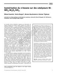
![التجربية الأولي [Read-Only] - KSU](https://img.yumpu.com/15502211/1/190x135/-read-only-ksu.jpg?quality=85)