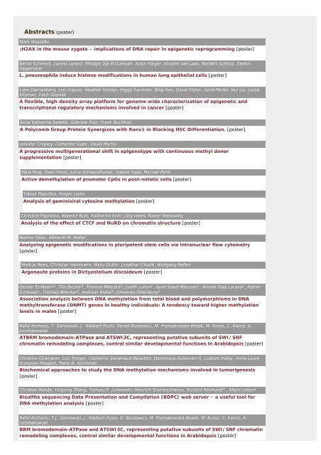
Abstracts (poster) - Wissenschaft Online
Abstracts (poster) - Wissenschaft Online
Abstracts (poster) - Wissenschaft Online

Create successful ePaper yourself
Turn your PDF publications into a flip-book with our unique Google optimized e-Paper software.
<strong>Abstracts</strong> (<strong>poster</strong>)<br />
Mark Wossidlo<br />
γH2AX in the mouse zygote – implications of DNA repair in epigenetic reprogramming [<strong>poster</strong>]<br />
Bernd Schmeck, Janina Lorenz, Philippe Dje N'Guessan, Antje Flieger, Vincent van Laak, Norbert Suttorp, Stefan<br />
Hippenstiel<br />
L. pneumophila induce histone modifications in human lung epithelial cells [<strong>poster</strong>]<br />
Luke Dannenberg, Leo Iniguez, Heather Holster, Peggy Farnham, Bing Ren, David Fisher, Gerd Pfeifer, Hui Liu, Jacob<br />
Kitzman, Fatih Ozsolak<br />
A flexible, high-density array platform for genome-wide characterization of epigenetic and<br />
transcriptional regulatory mechanisms involved in cancer [<strong>poster</strong>]<br />
Anna Katharina Sedello, Gabriele Putz, Frank Buchholz<br />
A Polycomb Group Protein Synergizes with Runx1 in Blocking HSC Differentiation. [<strong>poster</strong>]<br />
Jennifer Cropley, Catherine Suter, David Martin<br />
A progressive multigenerational shift in epigenotype with continuous methyl donor<br />
supplementation [<strong>poster</strong>]<br />
Maja Klug, Sven Heinz, Lucia Schwarzfischer, Sabine Pape, Michael Rehli<br />
Active demethylation of promoter CpGs in post-mitotic cells [<strong>poster</strong>]<br />
Tobias Paprotka, Holger Jeske<br />
Analysis of geminiviral cytosine methylation [<strong>poster</strong>]<br />
Christine Paprotka, Mareike Rust, Katharina Kohl, Jörg Leers, Rainer Renkawitz<br />
Analysis of the effect of CTCF and NuRD on chromatin structure [<strong>poster</strong>]<br />
Nadine Obier, Albrecht M. Müller<br />
Analyzing epigenetic modifications in pluripotent stem cells via intranuclear flow cytometry<br />
[<strong>poster</strong>]<br />
Markus Nees, Christian Hammann, Manu Dubin, Jonathan Chubb, Wolfgang Nellen<br />
Argonaute proteins in Dictyostelium discoideum [<strong>poster</strong>]<br />
Osman El-Maarri1 , Tim Becker2 , Thomas Mikeska3 , Judith Junen1 , Syed Saadi Manzoor1 , Amalia Diaz-Lacava2 , Rainer<br />
Schwaab1 , Thomas Wienker2 , Andreas Waha3 , Johannes Oldenburg1 Association analysis between DNA methylation from total blood and polymorphisms in DNA<br />
methyltransferase (DNMT) genes in healthy individuals: A tendency toward higher methylation<br />
levels in males [<strong>poster</strong>]<br />
Rafal Archacki, T. Sarnowski, J. Halibart-Puzio, Daniel Buszewicz, M. Prymakowska-Bosak, M. Kuras, C. Koncz, A.<br />
Jerzmanowski<br />
ATBRM bromodomain-ATPase and ATSWI3C, representing putative subunits of SWI/SNF<br />
chromatin remodeling complexes, control similar developmental functions in Arabidopsis [<strong>poster</strong>]<br />
Christine Champion, Loïc Ponger, Catherine Senamaud-Beaufort, Dominique Guianvarc'h, Ludovic Halby, Anne-Laure<br />
Guieysse-Peugeot, Paola B. Arimondo<br />
Biochemical approaches to study the DNA methylation mechanisms involved in tumorigenesis<br />
[<strong>poster</strong>]<br />
Christian Rohde, Yingying Zhang, Tomasz P. Jurkowski, Heinrich Stamerjohanns, Richard Reinhardt*, Albert Jeltsch<br />
Bisulfite sequencing Data Presentation and Compilation (BDPC) web server – a useful tool for<br />
DNA methylation analysis [<strong>poster</strong>]<br />
Rafal Archacki, T.J. Sarnowski, J. Halibart-Puzio, D. Buszewicz, M. Prymakowska-Bosak, M. Kuras, C. Koncz, A.<br />
Jerzmanowski<br />
BRM bromodomain-ATPase and ATSWI3C, representing putative subunits of SWI/SNF chromatin<br />
remodeling complexes, control similar developmental functions in Arabidopsis [<strong>poster</strong>]
Katerina Krizova, Miloslava Fojtova, Ann Depicker, Ales Kovarik<br />
Callus-induced epiallelism of an invertedly repeated transgene locus influences its transsilencing<br />
abilities [<strong>poster</strong>]<br />
Britta Wallmen, Simon Wöhrle, Andreas Hecht<br />
Cell specific inducibility of Wnt target genes correlates with epigenetic modifications and<br />
differential promoter occupancy by TCF/LEF proteins [<strong>poster</strong>]<br />
Peter Hemmerich, Stefanie Weidtkamp-Peters, Christian Hoischen, Lars Schmiedeberg, Indri Erliandri, Stephan<br />
Diekmann<br />
CENP-I as a new epigentic mark at centromere chromatin [<strong>poster</strong>]<br />
Claudia Gebhard, Elmar Schilling, Lucia Schwarzfischer-Pfeilschifter, Mathias Ehrich, Michael Rehli<br />
Comparative methylation profiling of tumor samples using methyl-CpG-immuno precipitation<br />
(MCIp) and CpG island microarrays [<strong>poster</strong>]<br />
Ruxandra Farcas, Eberhard Schneider, Ulrich Zechner, Achim Tresch, Hans Zischler, Angelika Daser, Thomas Haaf<br />
Comparison of human and non-human primate methylation status of CpG islands in the promoter<br />
region of CCRK [<strong>poster</strong>]<br />
Martin Herold, Dorte Bohla, Marek Bartkuhn, Imke Panzer, Rainer Renkawitz<br />
CTCF, the highly conserved boundary factor of Drosophila and vertebrates [<strong>poster</strong>]<br />
Jürgen Geisel, Heike Schorr, Gunar H. Heine, Marion Bodis, Ulrich Hübner, Jean-Pierre Knapp, Wolfgang Herrmann<br />
Decreased p66Shc promoter methylation in patients and end-stage renal disease [<strong>poster</strong>]<br />
Martina Dadejova, K. Yoong Lim, Roman Matyasek, Andrew Leitch, Ales Kovarik<br />
Developmental activation of silent rRNA genes is associated with increased transcription activity<br />
of rDNA loci in synthetic hybrids of Nicotiana [<strong>poster</strong>]<br />
OLUSOLA DOKUN, WOLFGANG SCHULZ<br />
DNA hypomethylation of SNCG (synuclein-gamma) in cancer: tumor-specific or cell typespecific?<br />
[<strong>poster</strong>]<br />
Joachim Weitzel<br />
DNA methylation impairs activation of haploid expressed genes in male germ cells. [<strong>poster</strong>]<br />
Christina Klaus, Daniela Kremer, Victoria Kolb-Bachofen<br />
DNA methyltransferases and the influence of cytokines and nitric oxide (NO) on DNA methylation<br />
[<strong>poster</strong>]<br />
Stefanie Stepanow, Kathrin Reichwald, Klaus Huse, Matthias Platzer<br />
Do epigenetic effects at MCHR1 contribute to obesity? [<strong>poster</strong>]<br />
Perrine Gaub, Andrea Tedeschi, Antonio Schmandke, Radhika Puttagunta, Tuan Nguyen, Simone Di Giovanni<br />
Enhancement of neuronal acetylation promotes neurite and axon outgrowth [<strong>poster</strong>]<br />
Jana Krejci, Eva Bartova, Andrea Harnicarova, Roman Hajek, Gabriela Galiova, Stanislav Kozubek<br />
Epigenetic changes in multiple myeloma cells [<strong>poster</strong>]<br />
Georgios J. Vlachojannis, Andreas M. Zeiher, Stefanie Dimmeler<br />
Epigenetic control of the eNOS promoter by DNA methylation in vasculogenic progenitor cell<br />
populations [<strong>poster</strong>]<br />
Maria Elena Torres-Padilla<br />
Epigenetic mechanisms in early mouse development [<strong>poster</strong>]<br />
Michael Michalkiewicz, Teresa Michalkiewicz, Kyle MacGillis<br />
Epigenetic mechanisms in hypertension [<strong>poster</strong>]
Silke Götze, Sonja Sievers, Oliver Müller<br />
Epigenetic regulation in the Wnt signalling pathway [<strong>poster</strong>]<br />
Nadia Sellami, Sabine Adam-Klages, Reiner Siebert, Hans-Jürgen Heidebrecht<br />
Epigenetic Regulation of the Cancer Testis Antigen CT45 [<strong>poster</strong>]<br />
Svend Petersen-Mahrt, Wolf Reik, Siim Pauklin, Heather Coker<br />
Epigenetic Reprogramming of 5-meC via DNA Deamination and DNA Repair [<strong>poster</strong>]<br />
Robert Liefke, Daniela Salat, Jörg Wiedenmann, Franz Oswald, Tilman Borggrefe<br />
ETO, but not AML1/ETO, augments RBP-Jk/Sharp-mediated transcriptional repression of Notch<br />
target genes [<strong>poster</strong>]<br />
Yvonne Möller-Steinbach, Cristina Madeira Alessandre, Vivien Exner, Patti Taranto, Claudia Köhler, Lars Hennig<br />
Function of Polycomb group proteins in the transition to flowering in plants [<strong>poster</strong>]<br />
Cordula Tschuch, Angela Schulz, Armin Pscherer, Meinhard Hahn, Peter Lichter, Daniel Mertens<br />
Functional analysis of candidate genes localized in 13q14.3, a region commonly affected in B-CLL<br />
[<strong>poster</strong>]<br />
Devi Thiagarajan, Sanjeev Khosla<br />
Functional characterisation of mDnmt2 [<strong>poster</strong>]<br />
Soyoung Lim, Johannes Schulte, Hans-Ulrich Schildhaus, Uta Flucke, Phillip Kahl, Roland Schüle, Reinhard Büttner,<br />
Jutta Kirfel<br />
Functional role of Lysine-specific histone methylase-1 in carcinogenesis [<strong>poster</strong>]<br />
Andreas Werner, Mark Carlile<br />
Functional short RNAs from naturally occurring sense/antisense transcripts [<strong>poster</strong>]<br />
Sandra Weiss, Ralf Gilsbach, Frederico Barreto, Achim Lother, Lutz Hein<br />
Heart failure and fibrosis induced by overexpression of methyl-CpG- binding protein 2 (MeCP2) in<br />
transgenic mice [<strong>poster</strong>]<br />
Alexandra Moosmann, Coen Campsteijn, Martina Schmid,, Eric M. Thompson<br />
High diversity of developmental stage-specific histone variants in the larvacean, Oikopleura<br />
dioica [<strong>poster</strong>]<br />
Irene Tiemann-Boege, Christina Curtis, Darryl Shibata, Simon Tavaré<br />
High-throughput analysis of methylation patterns to track cell divisions [<strong>poster</strong>]<br />
Tzvetina Brumbarova, Cecile Doyen, Emilie Bonnefoy, Guillermo Orsi, Pierre Couble, Benjamin Loppin<br />
HIRA functions in Drosophila [<strong>poster</strong>]<br />
Michael Haberland, Rusty Montgomery, Eric N. Olson<br />
Histone deacetylases 1 & 2 control adipogenesis [<strong>poster</strong>]<br />
RAFFAELE TEPERINO, MICHELE LONGO, PAOLA MIRRA, PIETRO FORMISANO, FRANCESCO BEGUINOT, PAOLA UNGARO<br />
HNF4 DIRECTS HISTONE METHYLATION TO SILENCE PED/PEA-15 EXPRESSION IN HUMAN<br />
HEPATOCYTES [<strong>poster</strong>]<br />
Eva Bartova, Abdrea Harnicarova, Jana Krejci, Gabriela Galiova, Stanislav Kozubek<br />
Human embryonic stem cells are characterized by distinct patterns of histone modifications in<br />
comparison with cells of feeder layer [<strong>poster</strong>]<br />
Francesco Nicassio1 , Joseph Vissers4 , Nadia Corrado1 , Liliana Areces1,2 , Steven Bergink3 , Jurgen Marteijn3 , Wim<br />
Vermeulen3 , Maarten van Lohuizen4 , Pier Paolo di Fiore1,2 , Elisabetta Citterio4 Human USP3 is a chromatin modifier required for S-phase progression and genome stability<br />
[<strong>poster</strong>]
Andrea Tedeschi, Tuan Nguyen, Radhika Puttagunta, Perrine Gaub, Simone Di Giovanni<br />
Identification of a novel transcription module for axon outgrowth and regeneration [<strong>poster</strong>]<br />
Japke Polman, E. Ronald de Kloet, Nicole Datson<br />
Identification of binding sites of the Glucocorticoid Receptor in the brain [<strong>poster</strong>]<br />
Philipp Rathert, Arunkumar Dhayalan, Xing Zhang, Renata Jurkowska, Raluca Tamas, Yoichi Shinkai, Xiaodong Cheng,<br />
Albert Jeltsch<br />
Identification of new non-histone targets of the human G9a protein methyltransferase using<br />
peptide arrays [<strong>poster</strong>]<br />
Sylvia Erhardt, Craig M. Betts, Barbara G. Mellone, Gary H. Karpen, Aaron F. Straight<br />
Identification of novel regulators of centromeric chromatin by genome-wide RNAi screening<br />
[<strong>poster</strong>]<br />
Bastian Stielow, Alexandra Sapetschnig, Imme Krüger, Michael Boutros, Guntram Suske<br />
Identification of SUMO-dependent chromatin-associated transcriptional repression components<br />
by a genome-wide RNA interference screen [<strong>poster</strong>]<br />
Harriet Wikman, Michaela Kraemling, Dirk Kemming, Klaus Pantel<br />
Identification of Target Genes in Micrometastatic Lung Cancer by Methylation Arrays [<strong>poster</strong>]<br />
Isabelle GUILLERET, Maria-Chiara OSTERHELD, Richard BRAUNSCHWEIG, Véronique GASTINEAU, Suzanne TAILLENS<br />
Imprinting of tumor-suppressor genes in human placenta. [<strong>poster</strong>]<br />
Alexandre Ceccaldi, Dominique Guianvarc'h, Catherine Senamaud-Beaufort, Renata Jurkowska, Daniel Dauzonne,<br />
Albert Jeltsch, Paola B Arimondo<br />
In quest of DNMT inhibitors [<strong>poster</strong>]<br />
Careen Katryniok, Bernd L. Sorg, Dieter Steinhilber<br />
INDUCTION OF HUMAN 5-LIPOXYGENASE GENE EXPRESSION BY THE HISTONE DEACETYLASE<br />
INHIBITOR TRICHOSTATIN A - INVESTIGATIONS ON THE MECHANISM [<strong>poster</strong>]<br />
Daniela Kremer, Wolfgang Schulz, Victoria Kolb-Bachofen<br />
iNOS-generated NO plasy an critical role in DNA-methylation [<strong>poster</strong>]<br />
Nathalie Jurisch, Bjoern Textor, Peter Angel, Marina Schorpp-Kistner<br />
Involvement of JunB in post-translational HDAC6 regulation and chromatin remodelling [<strong>poster</strong>]<br />
Annette Scharf, Karin Meier, Volker Seitz, Alexander Brehm, Axel Imhof<br />
Kinetics of histone modifications during in vitro chromatin assembly [<strong>poster</strong>]<br />
Fabio Mohn, Michael Weber, Michael Rebhan, Tim Roloff, Jens Richter, Michael Stadler, Miriam Bibel, Dirk Schübeler<br />
Lineage-specific Polycomb targets and de novo DNA methylation define restriction and potential<br />
of neuronal progenitors [<strong>poster</strong>]<br />
Maciej Meglicki, Marta Teperek, Ewa Borsuk<br />
Localization of heterochromatin protein 1α during mouse oogenesis and early embryonic<br />
development [<strong>poster</strong>]<br />
Christian Schmidl, Maja Klug, Tina Böld, Petra Hoffmann, Matthias Edinger, Michael Rehli<br />
Locus-wide detection of cell type specific DNA methylation patterns using comparative methyl-<br />
CpG-Immunoprecipitation (MCIp) [<strong>poster</strong>]<br />
Nicole Happel, Stefan Stoldt, Detlef Doenecke<br />
M-phase specific phosphorylation of histone H1.5 at threonine 10 by GSK3 [<strong>poster</strong>]<br />
Stephanie Jungmichel, Christoph Spycher, Manuel Stucki<br />
Mechanism of MDC1 dimerization [<strong>poster</strong>]
Gunter Reuter, Thomas Rudolph, Sandro Lein, Matthias Walther, Heiko Baisch, Sameer Phalke, Christian Apelt, Sandy<br />
Mietsch<br />
Mechanisms of chromatin differentiation during early embryogenesis of Drosophila [<strong>poster</strong>]<br />
Anette Tippelt<br />
Methyl Primer Express® Software and the Influence of Amplicon Characteristics to the success<br />
rate in DNA Sequencing of bis treated gDNA [<strong>poster</strong>]<br />
Sonja Röhrs, Julia Romani, Wilhelm Dirks, Hans G. Drexler, Hilmar Quentmeier<br />
Methylation profiles of tumour suppressor genes in Hodgkin and non-Hodgkin lymphoma cell<br />
lines [<strong>poster</strong>]<br />
Aditi Kanhere, Vingron Martin, Haas Stefan<br />
Methylation status of promoter depends on its CpG content [<strong>poster</strong>]<br />
Szabolcs Sörös, Wolfgang Fischle<br />
Molecular insights into HP1-chromatin interaction [<strong>poster</strong>]<br />
Renata Jurkowska, Da Jia, Sergey Ragozin, Nils Ansbach, Claus Urbanke, Xing Zhang, Richard Reinhardt, Wolfgang<br />
Nellen, Xiaodong Cheng, Albert Jeltsch<br />
Multimerisation of the Dnmt3L-Dnmt3a complex on DNA and its mechanistic implications [<strong>poster</strong>]<br />
Timo Quante, Lars Tögel, Wolfgang Deppert, Genrich V. Tolstonog<br />
Mutp53 as a modulator of global chromatin organisation [<strong>poster</strong>]<br />
Sascha Tierling, Yingying Zhang, Christian Rohde, Nina Pälmke, Julia Arand, Diana Santacruz, Matthias Platzer, Richard<br />
Reinhardt, Albert Jeltsch, Jörn Walter<br />
NAME21: The National Methylome Project of Human Chromosome 21 [<strong>poster</strong>]<br />
Yingying Zhang, Christian Rohde, Sascha Tierling, Heinrich Stamerjohanns, Matthias Platzer, Richard Reinhardt, Jörn<br />
Walter, Albert Jeltsch<br />
NAME21: The National Methylome Project of Human Chromosome 21 [<strong>poster</strong>]<br />
Franck COURT, Marion BANIOL, Hélène HAGEGE, Julie BORGEL, Jacques PIETTE, Guy CATHALA, Thierry FORNE<br />
NEW INSIGHTS INTO THE IMPRINTED MOUSE Igf2/H19 LOCUS BY 3C-qPCR METHOD [<strong>poster</strong>]<br />
Wibke Peters, Thomas Macherey, Mike Duisken, Sophie Willnow, Bernhard Lüscher, Elmar Weinhold<br />
New S-Adenosyl-L-methionine Analogues to Investigate the Methylome [<strong>poster</strong>]<br />
Andrea Felten, Peter Leister, Karl Heinz Scheidtmann<br />
Novel Coactivators of Androgen Receptor: AATF and ZIP Kinase [<strong>poster</strong>]<br />
Andrea Harni•arová, Eva Bártová, Jana Krej•í, Gabriela Galiová, Stanislav Kozubek<br />
Nuclear location of Oct3/4 and c-myc genes in human embryonic stem cells undergoing<br />
differentiation [<strong>poster</strong>]<br />
Niels Boeckel, Masamichi Koyanagi, Masayoshi Iwasaki, Andreas M. Zeiher, Stefanie Dimmeler<br />
Oct3/4 and Klf4 promoter status in multipotent circulating mesangioblasts [<strong>poster</strong>]<br />
Emilia Jarochowska, Pawe• Krawczyk, Anna •ach, Micha• Krzyszto•<br />
Presentation of Students' Society of Genetics and Epigenetics [<strong>poster</strong>]<br />
Daniel Buszewicz, Marta Teperek, Pawel Krawczyk, Emilia Jarechowska, Michal Krzyszton<br />
Presentation of Students' Society of Genetics and Epigenetics [<strong>poster</strong>]<br />
Arunkumar Dhayalan, Tomasz Jurkowski, Heike Laser, Richard Reinhardt, Da Jia, Xiaodong Cheng, Albert Jeltsch<br />
Protein - protein interaction analysis by Absence of Interference approach [<strong>poster</strong>]<br />
Michael Grzendowski, Markus J. Riemenschneider, Marietta Wolter, Uwe Schlegel, Helmut E. Meyer, Guido<br />
Reifenberger, Kai Stühler<br />
Proteome analysis of human glioma with 1p/19q LOH [<strong>poster</strong>]
Rudolf Engelke, Gerhard Mittler<br />
Proteomic analysis of the nuclear matrix in pre-B cells. [<strong>poster</strong>]<br />
Levin Böhlig, Kurt Engeland<br />
Regulation of an intronic microRNA and its host gene by the tumor suppressor p53 [<strong>poster</strong>]<br />
Huan Shu, Lars Hennig<br />
Restructuring of epigenetic landscapes during plant development [<strong>poster</strong>]<br />
Lin XU, Rozenn MENARD, Alexandre BERR, Denise MEYER, Wen-Hui SHEN<br />
Role of Histone Ubiquitination in Arabidopsis Development [<strong>poster</strong>]<br />
Annelen Schemm, Sabine Neumann, Pamela Strissel, Cord-Michael Becker<br />
Role of NRSF/ hREST4 in Neuronalisation of Tumors [<strong>poster</strong>]<br />
Ernst Aichinger, Aleksandra Erilova, Grigory Makarevich, Claudia Köhler<br />
Role of the Mi-2 homolog PICKLE in repression of Polycomb group target genes in Arabidopsis<br />
[<strong>poster</strong>]<br />
Lorenz Kallenbach, Patrick Heun<br />
Role of the SUMO E3 ligase PIAS in chromosome and nuclear organization in Drosophila<br />
melanogaster [<strong>poster</strong>]<br />
Amos Tanay<br />
Selection and mutation in the evolution of CpG islands [<strong>poster</strong>]<br />
Gabriela Galiová, Eva Bártová, Andrea Harni•arová, Jana Krej•í, Stanislav Kozubek<br />
Single-cell c-myc gene expression in human embryonic stem cells and human teratocarcinoma<br />
NTERA cells [<strong>poster</strong>]<br />
Henriette Franz, Steven A. Jacobs, C. David Allis, Sepideh Khorasanizadeh, Wolfgang Fischle<br />
SPECIFICITY OF THE CDY FAMILY OF CHROMODOMAINS FOR METHYLATED ARKS MOTIFS IN<br />
CHROMATIN [<strong>poster</strong>]<br />
Bastian Stielow, Alexandra Sapetschnig, Christina Wink, Guntram Suske<br />
SUMO-modified transcription factors repress transcription by provoking local heterochromatic<br />
gene silencing [<strong>poster</strong>]<br />
Günter Kahl, Carlos Molina Medina, Björn Rotter, Peter Winter, Hideo Matsumura, Ryohei Terauchi<br />
SuperSAGE: A complete genome-wide quantitative expression profiling platform [<strong>poster</strong>]<br />
Peter R. Lange, Andreas Finke, Claus Wasternack<br />
TFL2 as an epigenetic regulator in Arabidopsis development [<strong>poster</strong>]<br />
Stephan Hupfer, Julia Brill, Cord-Michael Becker, Kristina Becker<br />
The entla mouse - a model for human absence epilepsy [<strong>poster</strong>]<br />
Jennifer Gerke, Özgür Bayram, Gerhard H. Braus<br />
The velvet complex coordinates light, fungal development and secondary metabolism in<br />
Aspergillus nidulans [<strong>poster</strong>]<br />
Filip Senigl, Jiri Plachy, Jiri Hejnar<br />
The CpG island core element protects retroviral vectors from transcriptional silencing [<strong>poster</strong>]<br />
Sarantis Chlamydas, Patrick Heun, Ruggiero Caizzi<br />
The Drosophila melanogaster centromeric region: a chromosomal domain in a dynamic state<br />
[<strong>poster</strong>]<br />
Andrea Just, Falk Butter, Esther Lizano, Michelle Trenkmann, Tony Heitkam, Heike Betat, Mario Mörl
The function of two conserved elements in the bacterial Poly(A)Polymerase and CCA-adding<br />
enzyme [<strong>poster</strong>]<br />
Stefan Ehrentraut, Jan Weber, Ann E. Ehrenhofer-Murray<br />
The HDAC Rpd3 functions in boundary formation by removal of Sir2 substrate [<strong>poster</strong>]<br />
Marcus Buschbeck, Iris Uribesalgo, Luciano Di Croce<br />
The histone variant macroH2A regulates key developmental genes [<strong>poster</strong>]<br />
Andreas May, Daniela Weise, Kurt Reifenberg, Thomas Haaf, Ulrich Zechner<br />
The impact of ovarian stimulation on the cellular epigenome in preimplantation mouse embryos<br />
[<strong>poster</strong>]<br />
Andreas Thomae, Dagmar Pich, Jan Brocher, Christian Berens, Robert Hock, Wolfgang Hammerschmidt, Aloys<br />
Schepers<br />
The interaction between ORC and the high mobility group protein HMGA1a creates site-specific<br />
replication origins [<strong>poster</strong>]<br />
Tomasz Jurkowski, Nils Anspach, Lilia Kulishova, Wolfgang Nellen, Albert Jeltsch<br />
The M.EcoRV DNA methyltransferase uses DNA bending for recognition of an expanded EcoDam<br />
recognition site. [<strong>poster</strong>]<br />
Yamuna Gangadharan, Gary Karpen, Patrick Heun<br />
The role of Drosophila SUMO E3 ligase dPIAS in Chromosome and Nuclear Organization [<strong>poster</strong>]<br />
Tuan Nguyen, Mahmoud Youness, Andrea Tedeschi, Andrew Green, Kirsi Forsberg, Simone Di Giovanni<br />
The role of NFAT in axonal outgrowth and regeneration [<strong>poster</strong>]<br />
Christine Vogler, Tanja Waldmann, Lora Braun, Mirek Dundr, Robert Schneider<br />
The tale of a tail - Histone H2A and its C-terminal tail [<strong>poster</strong>]<br />
Myriam Ekici, Mathias Hohl, Gerald Thiel<br />
Transcription of genes encoding synaptic vesicle proteins in human neural stem cells:chromatin<br />
accessability, histone methylation pattern and essential role of REST [<strong>poster</strong>]<br />
Madeleine Meusburger, Mark Helm, Frank Lyko<br />
tRNA targets methylated by the Dnmt2 methyltransferase [<strong>poster</strong>]<br />
Chandan Goswami, Tim Hucho<br />
TRPV4 Biochemically And Functionally Interacts With The Cytoskeleton [<strong>poster</strong>]<br />
Akuma Divine Saningong, Peter Bayer, Jonathan Wolf Mueller<br />
Unravelling the Function of Human DNA-Binding Protein Par14 in the Cellular Nucleus [<strong>poster</strong>]<br />
Agnieszka Sokol, Aleksandra Kwiatowska, Andrzej Jerzmanowski, Marta Prymakowska-Bosak<br />
Up-regulation of stress-inducible genes in tobacco and Arabidopsis cells in response to abiotic<br />
stresses and ABA treatment correlates with dynamic changes in histone H3 and H4 modifcations<br />
[<strong>poster</strong>]
Mark Wossidlo<br />
γH2AX in the mouse zygote – implications of DNA repair in<br />
epigenetic reprogramming<br />
Wossidlo M, Lepikhov K, Paelmke N, Walter J<br />
University of Saarland, Natural Sciences – Technical Faculty III, FR 8.3, Biological<br />
Sciences, Genetics/Epigenetics, Saarbrücken, Germany<br />
In mammals shortly after the fertilization of the oocyte the paternal genome undergoes<br />
dramatic epigenetic changes. The paternal DNA in mouse zygotes is rapidly<br />
demethylated by an apparently active mechanism, while the maternal DNA stays<br />
methylated. It still remains unknown which enzymes are responsible for the paternal<br />
DNA demethylation, same holds true for the mechanisms behind the process. Therefore<br />
we examined whether this paternal demethylation is mediated by a ubiquitous DNA<br />
repair process. To address this question we used the indirect immunofluorescense<br />
approach to detect the presence of DNA repair associated phosphorylated histone H2AX<br />
(γH2AX) in mouse zygotes at different pronuclear stages.<br />
We found out that the common DNA strand break associated marker γH2AX<br />
preferentially appears in the paternal pronucleus at certain pronuclear stages. In present<br />
work we describe the dynamic changes of γH2AX pattern, which is influenced by DNA<br />
polymerase inhibitor aphidicolin and at certain pronuclear stages independent on<br />
replication. This replication independent preferential localization of γH2AX in the paternal<br />
pronucleus in early zygotic stages indicates that active DNA demethylation in zygotes<br />
might be linked to DNA repair.<br />
contact:<br />
Mark Wossidlo<br />
Universität des Saarlandes<br />
Genetik / Epigenetik<br />
m.wossidlo@mx.uni-saarland.de<br />
Universitäts Campus Geb. A2.4<br />
66123 Saarbrücken (Germany)
Bernd Schmeck, Janina Lorenz, Philippe Dje N'Guessan, Antje Flieger, Vincent van<br />
Laak, Norbert Suttorp, Stefan Hippenstiel<br />
L. pneumophila induce histone modifications in human lung<br />
epithelial cells<br />
Legionella pneumophila causes community- and hospital-acquired pneumonia. Their<br />
effect on histone marks is unknown.<br />
L. pneumophila wildtype strain 130b induced time- and dose-dependently expression of<br />
the important chemoattractant IL-8 and global, genome wide histone modifications in<br />
human lungs epithelial A549 cells. We analyzed the promoter of the important<br />
proinflammatory chemokine IL-8 and found that histone H4 was acetylated and H3 was<br />
phosphorylated at Ser-10 and acetylated at Lys-14, followed by recruitment of<br />
transcription factor NF-κB, and RNA polymerase II as well as gene transcription. L.<br />
pneumophila strain 130b-induced IL-8 expression was decreased by histone<br />
acetyltransferase (HAT) inhibitor anacardic acid and enhanced by the histone<br />
deacetylase (HDAC) inhibitor trichostatin A. Accordingly, after L. pneumophila infection<br />
HATs p300 and CBP were time-dependently recruited to the il8 promoter, whereas<br />
HDAC1 and HDAC5 first vanished and later reappeared at the promoter. Interestingly, L.<br />
pneumophila specifically induced expression of HDAC5 but not of other HDACs in lung<br />
epithelial cells. Furthermore, L. pneumophila-induced cytokine release, promoter specific<br />
histone modifications and Pol-II recruitment were reduced in infection with flagellindeletion<br />
mutants.<br />
In summary, histone acetylation seems to be important for the regulation of<br />
proinflammatory gene expression in L. pneumophila infected lung epithelial cells.<br />
contact:<br />
PD Dr. Bernd Schmeck<br />
Charité - Universitätsmedizin Berlin<br />
Medizinische Klinik m.S. Infektiologie und Pneumologie<br />
Bernd.Schmeck@charite.de<br />
Augustenburger Platz 1<br />
13353 Berlin (Germany)
Luke Dannenberg, Leo Iniguez, Heather Holster, Peggy Farnham, Bing Ren, David<br />
Fisher, Gerd Pfeifer, Hui Liu, Jacob Kitzman, Fatih Ozsolak<br />
A flexible, high-density array platform for genome-wide<br />
characterization of epigenetic and transcriptional regulatory<br />
mechanisms involved in cancer<br />
Epigenetic mechanisms, such as DNA methylation and histone modification, and altered<br />
transcription factor binding play major roles in the development of many human<br />
diseases, most notably cancer. High-density and highly flexible DNA microarrays,<br />
derived from a unique combination of photolithography and a digital micromirror device,<br />
are now allowing researchers the opportunity to examine epigenetic events at an<br />
unprecedented scale and resolution. Phenomenon such as de novo DNA methylation of<br />
tumor suppressor gene promoters silences their expression, hence creating a gateway<br />
for uncontrolled cell division. In addition, the binding of many transcription factors<br />
becomes increased in a cancerous cell to promote the expression of genes involved in<br />
initiating carcinogenesis and metastasis. By looking on a genome-wide scale using highdensity<br />
microarrays, a comprehensive picture of DNA methylation patterns, chromatin<br />
structure, and transcription factor binding can be generated to aid in the characterization<br />
of the differences between normal and cancer cells, different cancer types, the same<br />
cancer from two different individuals, and drug treatment studies. Novel platform<br />
developments, specifically the 2.1 million probe (HD2) long-oligonucleotide array, have<br />
expanded the horizon of genome-wide studies for its application in elucidating epigenetic<br />
and transcriptional regulatory mechanisms involved in cancer. Cancer studies using<br />
NimbleGen 385K microarrays will be presented and discussed as well as data on the HD2<br />
platform.<br />
contact:<br />
Product Manager Luke Dannenberg<br />
Roche NimbleGen<br />
ldannenberg@nimblegen.com<br />
500 S. Rosa Road<br />
53719 Madison, Wisconsin (United States)
Anna Katharina Sedello, Gabriele Putz, Frank Buchholz<br />
A Polycomb Group Protein Synergizes with Runx1 in Blocking<br />
HSC Differentiation.<br />
Our lab previously showed that Runx1 deletion in adult mice leads to a block in<br />
differentiation but not to leukemia development. We used a loss-of-function approach to<br />
identify proteins cooperating with Runx1 in processes determining whether<br />
hematopoietic stem and progenitor cells undergo differentiation or maintain stem-celllike<br />
properties. By transducing lineage negative hematopoietic cells from Runx1Δ/Δ mice<br />
with a pooled shRNA library we obtained cells with a proliferative advantage in a serial<br />
colony-forming-cell assay. Genomic DNA isolated from these immortalized cells revealed<br />
an shRNA targeting a member of the polycomb group protein family, which is involved in<br />
chromatin remodeling. Runx1Δ/Δ cells transduced with shRNA targeting the polycomb<br />
group protein member form distinct colonies for more than double as long as nontransduced<br />
cells in serial colony-forming-cell assays. We hypothesize that block of<br />
differentiation by Runx1 deletion cooperates with a reactivation of self-renewal programs<br />
by knock down of the polycomb group protein to immortalize hematopoietic stem and<br />
progenitor cells, possibly mimicking leukemia.<br />
Literature<br />
Putz G, Rosner A, Nuesslein I, Schmitz N, Buchholz F. AML1 deletion in adult mice<br />
causes splenomegaly and lymphomas. Oncogene (2006) 25, 929-939.<br />
Bernards R, Brummelkamp TR, Beijersbergen RL. shRNA libraries and their use in cancer<br />
genetics. Nature Methods (2006) 3, 701-706.<br />
contact:<br />
Anna Katharina Sedello<br />
Max-Planck Institute of Molecular Cell Biology and Genetics<br />
asedello@mpi-cbg.de<br />
Pfotenhauerstr. 108<br />
01307 Dresden (Germany)<br />
additional information<br />
Gabriele Putz, currently at Osiris Therapeutics, Inc., 7015 Albert Einstein Dr., Colombia, MD 21046,<br />
USA
Jennifer Cropley, Catherine Suter, David Martin<br />
A progressive multigenerational shift in epigenotype with<br />
continuous methyl donor supplementation<br />
The epigenetic state of a locus can be affected by environmental factors such as diet.<br />
The murine A vy (agouti viable yellow) allele is one such locus: dietary supplementation<br />
of pregnant dams with methyl donors changes the epigenetic state of the locus in the<br />
offspring. At A vy , an IAP retrotransposon is inserted upstream of agouti. When<br />
epigenetically active the IAP usurps transcriptional control, driving ectopic expression of<br />
agouti signalling protein to produce the characteristic obese yellow phenotype. The<br />
epigenetic state of the IAP is unstable in the germline, so that isogenic mice show wide<br />
variation in the somatic epigenetic state of the IAP, with resultant broadly variable<br />
penetrance and expressivity. Supplementation of maternal diet with methyl donors<br />
promotes epigenetic silencing of the IAP, shifting the spectrum of offspring phenotypes<br />
away from obese yellow. We have previously shown that methyl donors can affect the<br />
germline epigenetic state of the A vy IAP. Here we show that continual supplementation<br />
of A vy mice over five generations leads to progressive germline stabilisation of the IAP<br />
epigenotype, so that the silent state becomes more strongly heritable and thus<br />
significantly more prevalent in the population. In unsupplemented populations the IAP is<br />
completely silent in 13% of mice. In a supplemented population, successive breeding of<br />
males carrying a silent IAP increases the prevalence of the silent allele almost three-fold<br />
(to 31%) by the fifth generation. These results suggest that long-term exposure to an<br />
environmental stimulus can effect epigenetic changes throughout a population. Such<br />
mechanisms may contribute to adaptive evolution via stable epigenetic silencing in the<br />
germline.<br />
contact:<br />
Dr Jennifer Cropley<br />
Victor Chang Cardiac Research Institute<br />
j.cropley@victorchang.edu.au<br />
384 Victoria st<br />
2010 Darlinghurst (Australia)<br />
additional information<br />
Dr Catherine Suter: Victor Chang Cardiac Research Institute, Darlinghurst, Australia<br />
Prof David Martin: Childrens Hospital Oakland Research Institute, Oakland, CA, USA
Maja Klug, Sven Heinz, Lucia Schwarzfischer, Sabine Pape, Michael Rehli<br />
Active demethylation of promoter CpGs in post-mitotic cells<br />
Within the last decades, it has become increasingly evident that the epigenetic code,<br />
including chromatin structure, DNA methylation, as well as histone modifications, plays<br />
an important role in regulating gene expression. The dynamics of DNA methylation, in<br />
particular the regulated, active removal of methyl-CpG marks, has remained a mystery,<br />
partly due to the lack of appropriate model systems. The differentiation of human blood<br />
monocytes into macrophages or dendritic cells proceeds without proliferation,<br />
representing an excellent model system for analyzing active demethylation processes in<br />
post-mitotic cells. In earlier studies, we observed the strong up-regulation of the CCL13<br />
gene specifically in monocyte-derived dendritic cells. The transcriptional activation<br />
coincides with the demethylation of three defined CpG residues in the CCL13-promoter<br />
during differentiation of monocytes into dendritic cells, whereas the promoter remains<br />
methylated and silent in monocyte-derived macrophages. Here we present a detailed<br />
time-course analysis of the epigenetic/chromatin status of the CCL13 promoter by<br />
quantitative mRNA expression analysis, chromatin immunoprecipitation, methyl-CpG<br />
immunoprecipitation and MNase-hypersensitivity, and restriction enzyme accessibility<br />
assays. We detected a strong correlation between active DNA demethylation,<br />
nucleosome remodeling, Histone H3 lysine 4 methylation and transcription of the CCL13<br />
gene during dendritic cell differentiation. Our data suggest that active demethylation<br />
proceeds in parallel with chromatin remodeling and gene activation.<br />
contact:<br />
Dipl Maja Klug<br />
Uniklinikum Regensburg<br />
Hämatologie/Onkologie<br />
maja.klug@klinik.uni-regensburg.de<br />
Franz-Josef-Strauss Allee 11<br />
93053 Regensurg (Deutschland)
Tobias Paprotka, Holger Jeske<br />
Analysis of geminiviral cytosine methylation<br />
Geminiviruses are important plant pathogens, causing yield losses in crop plants all over<br />
the world. They consist of a single stranded DNA genome which varies in size from 2.5<br />
to 3.0 kb, depending on the virus and is packed into icosahedral twin shaped particles.<br />
Plant defence mechanisms against geminiviruses, including post transcriptional gene<br />
silencing (PTGS) and transcriptional gene silencing (TGS) have been observed and are<br />
competed by the virus with silencing suppressors. The methylation of partial viral DNA<br />
sequences has been shown, but a complete determination of the cytosine methylation<br />
pattern of a viral genome has not yet been accomplished. A recent advance in<br />
geminivirus discovery and diagnostics was the application of rolling circle amplification<br />
(RCA), using the Φ29 polymerase. To analyse the complete methylation pattern of a<br />
geminivirus genome a method based on bisulphite sequencing combined with RCA was<br />
developed. The unmethylated cytosines are converted conventionally, but the<br />
subsequent PCR step is replaced through amplification with RCA. Preferential<br />
amplification of particular sequences is avoided by the use of random hexamer primers.<br />
The obtained DNA is then digested with restriction enzymes that miss Cs or Gs in their<br />
recognition sequence and randomly cloned into pBluescript. After colony PCR or RCA the<br />
cloned fragments can be sequenced directly using universal primers. A diverse pattern<br />
was observed including methylation of CpG, CpNpG and asymmetrical sites, which<br />
indicates the enforcement by the plants TGS machinery. An increase of cytosine<br />
methylation during the course of infection was also detected. This approach therefore<br />
shows the usefulness of RCA in bisulphite sequencing as a beneficial method to observe<br />
methylation patterns of small circular DNA molecules.<br />
contact:<br />
Dipl.-Biol. Tobias Paprotka<br />
University of Stuttgart<br />
Institute of Biology, Department of Molecular Biology and Virology of Plants<br />
tobias.paprotka@bio.uni-stuttgart.de<br />
Pfaffenwaldring 57<br />
70550 Stuttgart (Germany)
Christine Paprotka, Mareike Rust, Katharina Kohl, Jörg Leers, Rainer Renkawitz<br />
Analysis of the effect of CTCF and NuRD on chromatin<br />
structure<br />
CTCF, a ubiquitously expressed transcription factor, is the only protein in vertebrates<br />
known to mediate enhancer blocking. The mechanism of enhancer blocking is still<br />
unclear, but there are several models postulated. All these models share the idea, that<br />
the modification, remodeling and three-dimensional arrangement of chromatin play a<br />
major role.<br />
The NuRD complex is a multi-subunit protein complex with enzymatic activities involving<br />
chromatin remodeling and histone deacetylation. Targeting of NuRD to methylated CpG<br />
sequences leads to gene repression and is mediated through the methyl-binding-domain<br />
proteins MBD2 and MBD3 [1-4].<br />
Here we present a strategy to analyse the influence of CTCF and distinct NuRD<br />
complexes on chromatin structure in vivo at defined genomic regions. For our studies we<br />
utilize a well established system using the ability of the Lac-repressor to bind to the Lacoperator.<br />
LacO repeat clusters stably integrated into the genome have been generated<br />
[5]. Using this system we analyze CTCF and NuRD recruited to the array via LacI-MBD2/-<br />
CTCF. Using immunofluorescence microscopy we can analyze the recruitment and<br />
assembly of NuRD components. In addition histone modification depending on CTCF or<br />
NuRD can be analysed by chromatin immunoprecipitation as well. This allows to address<br />
the question wether CTCF has an influence on chromatin structure and if spreading of<br />
repressed chromatin occures after NuRD binding.<br />
Literature<br />
[1] Brackertz, M., Gong, Z., Leers, J., Renkawitz, R., 2006, Nucleic Acids Res, Vol.34,<br />
397-406<br />
[2] Gong, Z., Brackertz, M., Renkawitz, R. 2006, Mol. Cell. Biol., Vol. 26, 4519-4528<br />
[3] Zhang et al., 2002,Mol. Cell. Biol., Vol 22, 536-546<br />
[4] Guezennec et al., 2006, Mol. Cell. Biol.,Vol 26, 843-851<br />
[5] T. Jegou, K. Rippe, unpublished data<br />
contact:<br />
Christine Paprotka<br />
Justus-Liebig-Universität Giessen<br />
Institut für Genetik<br />
christine.paprotka@gen.bio.uni-giessen.de<br />
Heinrich-Buff-Ring 58 -62<br />
35392 Giessen (Germany)
Nadine Obier, Albrecht M. Müller<br />
Analyzing epigenetic modifications in pluripotent stem cells<br />
via intranuclear flow cytometry<br />
Pluripotency describes the ability of embryonic stem cells (ES cells) to self-renew and to<br />
give rise to all cell types of the developing embryo including the germline. Our goal is to<br />
define pluripotency by identifying the entry point of cells into an irreversible<br />
differentiation state with restricted differentiation potential.<br />
In this regard we developed a FACS-based protocol which can quantitatively display<br />
levels of different histone modifications in diverse cell types, such as in ES cells and in<br />
their differentiated derivatives. Applying this intranuclear flow cytometric method, we<br />
detected differences in global histone H4 acetylation levels between cells that were<br />
either treated or not treated with the HDAC inhibitor TSA. Further, we observed a<br />
distinct reduction of histone H3 lysine 27 tri-methylation (H3K27me3) levels in ES cells<br />
lacking the protein EED, which – as a critical component of the Polycomb-group<br />
repressor complex 2 - is participating in enzymatic methylation of H3K27. Our<br />
preliminary studies on global levels of H3K4me3 and H3K27me3 in combination with ckit-surface<br />
expression revealed that c-kit high immunophenotypic ES cells are also<br />
highly positive for both H3K4m3 and H3K27me3 “chromatin-immunophenotypes”.<br />
Together, we developed a new method for the analysis of global histone modifications by<br />
intra-nuclear flow cytometry. This method represents a promising tool to simultaneously<br />
study cellular properties, such as cell proliferation, apoptosis, surface marker expression<br />
and intranuclear “chromatin-immunophenotype”, on the single cell level of large<br />
quantities of cells.<br />
contact:<br />
M.Sc. Nadine Obier<br />
Universität Würzburg<br />
Institut für medizinische Strahlenkunde und Zellforschung (MSZ)<br />
nadineobier@web.de<br />
Versbacher Str. 5<br />
97078 Würzburg (Germany)
Markus Nees, Christian Hammann, Manu Dubin, Jonathan Chubb, Wolfgang Nellen<br />
Argonaute proteins in Dictyostelium discoideum<br />
M. Nees, C. Hammann, M. Dubin, J. Chubb*, W. Nellen<br />
Department of Genetics, Universität Kassel, D-34132 Kassel, Germany.<br />
* School of Life Sciences, University of Dundee, Dow Street, Dundee DD1 5EH<br />
The genom of Dictyostelium discoideum contains six genes which resemble Argonaute or<br />
Piwi proteins from other organisms. Argonautes are involved in slicing of mRNAs in the<br />
RNAi pathway and in translational inhibition in the miRNA pathway. Piwi proteins are<br />
expressed specifically in the germline where they associate with another class of<br />
approximately 29-nucleotide-long small RNAs, named piRNAs. The function of piRNAs is<br />
not yet fully understood.<br />
To elucidate the function of Argonaute proteins in Dictyostelium discoideum we present<br />
first data on the cellular localization, the expression pattern during the developmemtal<br />
cycle and the phenotype of knock-out mutants.<br />
contact:<br />
Markus Nees<br />
Universität Kassel<br />
Genetik<br />
neemar@web.de<br />
Heinrich-Plett-Straße 40<br />
34132 Kassel (Germany)
Osman El-Maarri 1 , Tim Becker 2 , Thomas Mikeska 3 , Judith Junen 1 , Syed Saadi<br />
Manzoor 1 , Amalia Diaz-Lacava 2 , Rainer Schwaab 1 , Thomas Wienker 2 , Andreas Waha 3 ,<br />
Johannes Oldenburg 1<br />
Association analysis between DNA methylation from total<br />
blood and polymorphisms in DNA methyltransferase (DNMT)<br />
genes in healthy individuals: A tendency toward higher<br />
methylation levels in males<br />
To examine the contribution of polymorphisms in DNMT genes to methylation variations<br />
we performed a search for polymorphisms in coding regions of all known human DNA<br />
methyltransferases genes (DNMT1, 3A, 3B, 3L and 2=TRDMT1) in 96 normal males and<br />
96 normal females. Global methylation was estimated by studying two repetitive DNA<br />
elements, namely Line-1 and Alu repeats, while single loci were investigated at three<br />
differentially methylated regions: PEG3, NESP55 and H19; two additional single loci were<br />
also studied at Xq28 and 19q13.4. All studied CpGs showed a slightly higher methylation<br />
in males (P
Rafal Archacki, T. Sarnowski, J. Halibart-Puzio, Daniel Buszewicz, M. Prymakowska-<br />
Bosak, M. Kuras, C. Koncz, A. Jerzmanowski<br />
ATBRM bromodomain-ATPase and ATSWI3C, representing<br />
putative subunits of SWI/SNF chromatin remodeling<br />
complexes, control similar developmental functions in<br />
Arabidopsis<br />
Among the factors that serve to modify chromatin structure, SWI/SNF chromatin<br />
remodeling complexes define conserved and well-characterized group. However, no<br />
SWI/SNF complex has been purified and characterized in higher plants so far, yet its<br />
existence is highly probable. Four genes encoding homologues of Swi2/Snf2 ATPase<br />
(BRM, SYD, CHR12 and CHR23) and four encoding homologues of Swi3 subunit<br />
(ATSWI3A, ATSWI3B, ATSWI3C and ATSWI3D), as well as a single SNF5 orthologue<br />
(BSH) have been identified in Arabidopsis. This makes a number of possibilities for<br />
assembly of plant SWI/SNF complexes. In the lack of structural and biochemical data,<br />
homology analyses and interpretation of genetic and in vitro interactions are the best<br />
tools for investigating SWI/SNF complex composition and function.<br />
Here we show a comparative analysis of brm and atswi3c null mutants. Both of them<br />
display similar (but not identical) developmental alterations, including semidwarfism,<br />
leaf curling, inhibition of root elongation, homeotic-like changes in flowers, and defects<br />
in pollen development. These observations, together with the results showing that BRM<br />
and SWI3C interact in yeast two-hybrid assay (Farrona et al., 2004), suggest that BRM<br />
and SWI3C proteins exist in the same SWI/SNF chromatin remodeling complex. Our<br />
analyses of brm atswi3c double mutants further support this hypothesis, as the brm<br />
atswi3c plants display brm phenotype. Nonetheless, certain differences between<br />
phenotypic traits of atswi3c and brm mutants, such as complete sterility of brm and the<br />
occurrence of unfused carpels in brm flowers, indicate that the biological functions of<br />
these two SWI/SNF subunits are not completely overlapping.<br />
contact:<br />
M. Sc. Daniel Buszewicz<br />
Warsaw University<br />
Laboratory of Plant Molecular Biology<br />
dbuszewicz@gmail.com<br />
Pawinskiego 5A/F<br />
02-106 Warsaw (Poland)
Christine Champion, Loïc Ponger, Catherine Senamaud-Beaufort, Dominique<br />
Guianvarc'h, Ludovic Halby, Anne-Laure Guieysse-Peugeot, Paola B. Arimondo<br />
Biochemical approaches to study the DNA methylation<br />
mechanisms involved in tumorigenesis<br />
Cancer cells show a highly disturbed epigenetic landscape, with a global<br />
hypomethylation of the genome that induces abnormal expression of genes (such as<br />
oncogenes) and a local hypermethylation of promotors that silences tumor suppressor<br />
genes (TSG). DNA methylation is catalysed by a family of enzymes called DNA<br />
methyltransferases (DNMTs) and only occurs at position 5 of cytosines in CpG<br />
dinucleotides (in Vertebrates) that are not randomly distributed in the genome but<br />
mainly grouped in CpG islands. Yet the mechanism by which specific de novo<br />
methylation is directed to the TSG promotors remains still unknown. Indeed, it would be<br />
of great therapeutic interest to block this specific hypermethylation of TSG promotors in<br />
order to restore in malignant cells their natural ability to block tumorigenesis.<br />
We have chosen to focus on prostate cancer and two TSG for which inactivation is due to<br />
promotor hypermethylation : RASSF1A and RARβ2.<br />
On one hand, we investigate whether short DNA sequences influence the DNA<br />
methylation pattern. By a bioinformatic analysis we have found 10 DNA motifs that are<br />
overrepresented in hypermethylated promotors in prostate cancer versus non<br />
hypermethylated promotors.<br />
On the other hand, we are developing two affinity chromatography approaches to<br />
identify the protein partners of DNMTs involved in DNA methylation.<br />
contact:<br />
PhD Student Christine Champion<br />
Museum National d'Histoire Naturelle<br />
MNHN USM 503, CNRS UMR5153, INSERM U565<br />
christine.champion@mnhn.fr<br />
43, rue Cuvier<br />
75231 Paris cedex 5 (France)<br />
additional information<br />
Dominique Guianvarc'h : CNRS UMR7613-Université Paris VI
Christian Rohde, Yingying Zhang, Tomasz P. Jurkowski, Heinrich Stamerjohanns,<br />
Richard Reinhardt*, Albert Jeltsch<br />
Bisulfite sequencing Data Presentation and Compilation<br />
(BDPC) web server – a useful tool for DNA methylation<br />
analysis<br />
During bisulfite genomic sequencing projects large amount of data is generated. The<br />
BDPC web interface (http://biochem.jacobs-university.de/BDPC/) automatically analyzes<br />
bisulfite datasets prepared using the BiQ Analyzer (Bock et al. 2005, Bioinformatics 21,<br />
4067-8). BDPC provides the following output: 1) MS-Excel compatible files compiling for<br />
each PCR product i) the average methylation level, the number of clones analyzed, and<br />
the percentage of CG sites analyzed (which is an indicator of data quality), ii) the<br />
methylation level observed at each CG site, and iii) the methylation level of each clone.<br />
2) A methylation overview table compiling the methylation of all amplicons in all tissues.<br />
3) Publication grade figures in PNG format showing the methylation pattern for each PCR<br />
product embedded in an HMTL file summarizing the methylation data, the DNA sequence<br />
and some basic statistics. 4) A summary file compiling the methylation pattern of<br />
different tissues, which is linked to the individual HTML result files, and can be directly<br />
used for presentation of the data in the internet. 5) A condensed file, containing all<br />
primary data in simplified format for further downstream data analysis, and 6) a custom<br />
track file for display of the results in the UCSC genome browser.<br />
contact:<br />
Christian Rohde<br />
Jacobs University Bremen<br />
School of Engineering and Science<br />
c.rohde@jacobs-university.de<br />
Campus Ring 1<br />
28759 Bremen (Germany)<br />
additional information<br />
*Max Planck Institute for Molecular Genetics, Ihnestrasse 63-73, D-14195 Berlin-Dahlem, Germany
Rafal Archacki, T.J. Sarnowski, J. Halibart-Puzio, D. Buszewicz, M. Prymakowska-<br />
Bosak, M. Kuras, C. Koncz, A. Jerzmanowski<br />
BRM bromodomain-ATPase and ATSWI3C, representing<br />
putative subunits of SWI/SNF chromatin remodeling<br />
complexes, control similar developmental functions in<br />
Arabidopsis<br />
Among the factors that serve to modify chromatin structure, SWI/SNF chromatin<br />
remodeling complexes define conserved and well-characterized group. However, no<br />
SWI/SNF complex has been purified and characterized in higher plants so far, yet its<br />
existence is highly probable. Four genes encoding homologues of Swi2/Snf2 ATPase<br />
(BRM, SYD, CHR12 and CHR23) and four encoding homologues of Swi3 subunit<br />
(ATSWI3A, ATSWI3B, ATSWI3C and ATSWI3D), as well as a single Snf5 orthologue<br />
(BSH) have been identified in Arabidopsis (1). This makes a number of possibilities for<br />
assembly of plant SWI/SNF complexes. In the lack of structural and biochemical data,<br />
homology analyses and interpretation of genetic and in vitro interactions are the best<br />
tools for investigating SWI/SNF complex composition and function.<br />
Here we show a comparative analysis of brm and atswi3c null mutants. Both of them<br />
display similar (but not identical) developmental alterations, including semidwarfism,<br />
leaf curling, inhibition of root elongation, homeotic-like changes in flowers, and defects<br />
in pollen development. These observations, together with the results showing that BRM<br />
and SWI3C interact in yeast two-hybrid assay (2), suggest that BRM and SWI3C proteins<br />
exist in the same SWI/SNF chromatin remodeling complex. Our analyses of brm atswi3c<br />
double mutants further support this hypothesis, as the brm atswi3c plants display brm<br />
phenotype. Nonetheless, certain differences between phenotypic traits of atswi3c and<br />
brm mutants, such as complete sterility of brm and the occurrence of unfused carpels in<br />
brm flowers, indicate that the biological functions of these two SWI/SNF subunits are not<br />
completely overlapping.<br />
Literature<br />
(1) Jerzmanowski A. SWI/SNF chromatin remodeling and linker histones in plants.<br />
Biochim Biophys Acta. 2007 May-Jun;1769(5-6):330-45<br />
(2) Farrona S, Hurtado L, Bowman JL, Reyes JC. The Arabidopsis thaliana SNF2 homolog<br />
AtBRM controls shoot development and flowering. Development. 2004 Oct;131(20):4965-<br />
75<br />
contact:<br />
M.Sc Rafal Archacki<br />
University of Warsaw<br />
Laboratory of Plant Molecular Biology<br />
rafa@ibb.waw.pl<br />
Pawinskiego 5A<br />
02-106 Warsaw (Poland)<br />
additional information<br />
Affiliation of T.J. Sarnowski, J. Halibart-Puzio, and D. Buszewicz: Polish Academy of Sciences,<br />
Institute of Biochemistry and Biophysics, Pawinskiego 5A, 02-106 Warsaw, Poland<br />
Second affiliation of M. Prymakowska-Bosak and A. Jerzmanowski: Polish Academy of Sciences,<br />
Institute of Biochemistry and Biophysics, Pawinskiego 5A, 02-106 Warsaw, Poland<br />
Affiliation of M. Kuras: University of Warsaw, Department of Ecotoxicology, Miecznikowa 1, 02-096<br />
Warsaw, Poland<br />
Affiliation of C. Koncz: Max-Planck Institut für Züchtungsforschung, Carl-von-Linné-Weg 10, D-<br />
50829 Köln, Germany
Katerina Krizova, Miloslava Fojtova, Ann Depicker, Ales Kovarik<br />
Callus-induced epiallelism of an invertedly repeated transgene<br />
locus influences its trans-silencing abilities<br />
Using a two component transgene system involving two epiallelic variants of an<br />
invertedly repeated silencing locus (1) we have studied stability of trans-silencing<br />
interactions in tobacco cell culture and regenerated plants. In parental hybrids the<br />
posttranscriptionally but not transcriptionally silenced epiallele of locus 1 trans-silenced<br />
and trans-methylated target locus (2). Expression and methylation of both silenced<br />
(Lo1/Lo2) and non-silenced (Lo1E/Lo2) hybrids were stable over several generations in<br />
plants. However, in early Lo1E/Lo2 callus decreased expression of the nptII reporter<br />
gene was observed while the expression in Lo1/Lo2 remained unchanged. Analysis of<br />
small RNA species and coding region methylation suggested that the nptII genes were<br />
silenced by a PTGS mechanism in both cultures. Expression changes were correlated<br />
with changes in locus 1 promoter methylation status: the PTGS variant in Lo1/Lo2 line<br />
acquired methylation while the TGS epiallele in Lo1E/Lo2 line showed reduced<br />
methylation compared to the parental plant. Bisulfite genomic sequencing of locus 1<br />
revealed molecules with no, intermediate and high level of methylation. These data<br />
indicated that a cell culture process brought two epialleles of the silencer locus 1 to the<br />
same epigenetic ground characterized by high epilallelic diversity. In regenerated plants<br />
about 75% of Lo1E/Lo2 individuals returned to the original non-silenced phenotype while<br />
25% of individuals were silenced. From Lo1/Lo2 callus, 25% of regenerated plants<br />
showed increased expression whereas 75% of individuals remained silenced. The results<br />
demonstrated sensitivity of transgenes containing inverted structures towards epigenetic<br />
changes imposed by cell culture.<br />
contact:<br />
Mgr. Katerina Krizova<br />
Academy of Sciences<br />
Institute of Biophysics v.v.i.<br />
krizova@ibp.cz<br />
Kralovopolska 135<br />
CZ-61265 Brno (Czech Republic)
Britta Wallmen, Simon Wöhrle, Andreas Hecht<br />
Cell specific inducibility of Wnt target genes correlates with<br />
epigenetic modifications and differential promoter occupancy<br />
by TCF/LEF proteins<br />
Transcription factors of the T-cell factor (TCF)/lymphoid enhancer factor (LEF) family are<br />
considered to act in conjunction with corepressors and coactivators as bimodal switches<br />
for the activation or repression, respectively, of Wnt/beta-catenin target genes.<br />
Accordingly, TCF/LEF proteins are thought to remain constantly bound to the promoter<br />
regions of their target genes. However, constant promoter occupancy by TCF/LEF factors<br />
does not readily explain how distinct groups of Wnt target genes can be differentially<br />
regulated in a cell-type specific and developmentally controlled manner. We<br />
systematically compared known target genes with respect to Wnt-responsiveness,<br />
promoter occupancy by TCF/LEF proteins and epigenetic features in different cell lines.<br />
In E14 embryonic stem cells, in the neural cell line C17.2 and in C2C12 myogenic cells<br />
we find that Axin2, Cdx1 and T/Brachyury are differentially expressed and regulated.<br />
Activation of these target genes is predominantly mediated by a subset of TCF/LEF<br />
factors. Analysis of DNA methylation patterns and histone modifications at promoter<br />
regions revealed that Wnt-inducibility correlates with DNA hypomethylation and active<br />
histone marks. In contrast, non-responsive promoters showed hypermethylation and<br />
repressive histone marks. Moreover, Wnt-responsiveness correlates with differential<br />
promoter occupancy by TCF/LEF proteins. Notably, in contrast to current models,<br />
TCF/LEF transcription factors are not present at promoter regions of non-responding<br />
genes. We hypothesize that distinct promoter occupancy by TCF/LEF proteins and<br />
epigenetic control mechanisms form a multi-layered control system to achieve<br />
differential regulation of Wnt target gene expression.<br />
contact:<br />
Britta Wallmen<br />
University of Freiburg<br />
Institute of Molecular Medicine and Cell Research<br />
britta.wallmen@mol-med.uni-freiburg.de<br />
Stefan-Meier-Str. 17<br />
79104 Freiburg (Germany)
Peter Hemmerich, Stefanie Weidtkamp-Peters, Christian Hoischen, Lars Schmiedeberg,<br />
Indri Erliandri, Stephan Diekmann<br />
CENP-I as a new epigentic mark at centromere chromatin<br />
Epigenetic marking of a DNA locus may be realized by posttranslational modifications of<br />
nucleosomal histones or by stable binding of a specific protein at that locus. Centromere<br />
identity is believed to be conveyed by CENP-A, a specialized histone H3 analog that<br />
substitutes canonical H3 within centromeric nucleosomes. CENP-A is constitutively<br />
present at centromeres and required for the association of all other kinetochore proteins.<br />
To test whether these epigenetic properties are unique to CENP-A we have assessed the<br />
exchange rates of inner centromere proteins by quantitative microscopy throughout the<br />
cell cycle in living human cells (1). We demonstrate that, in addition to CENP-A, CENP-I<br />
is also a stable centromere component that does at no time exchange with soluble pools<br />
at centromeres. Loading of CENP-I onto centromeric chromatin occurs co-replicationally,<br />
while CENP-A is loaded in early G1. A subfraction of CENP-H (~20%) also stays stably<br />
bound to centromeres throughout the cell cyle. In contrast, CENP-B, CENP-C, and<br />
hMis12 turn over completely at centromeres with residence times ranging between<br />
seconds to hours. Our data reveal a wide range of cell cycle-specific assembly plasticity<br />
of the centromere during the cell-cycle and identify CENP-I as a potentially additional<br />
epigentic marker at centromeres.<br />
Literature<br />
(1) Hemmerich et al., J. Cell Biol. (2008) in press<br />
contact:<br />
PhD Peter Hemmerich<br />
Leibniz Institute for Age Research<br />
Fritz-Lipmann-Institute<br />
phemmer@fli-leibniz.de<br />
Beutenbergstr. 11<br />
07745 Jena (Germany)
Claudia Gebhard, Elmar Schilling, Lucia Schwarzfischer-Pfeilschifter, Mathias Ehrich,<br />
Michael Rehli<br />
Comparative methylation profiling of tumor samples using<br />
methyl-CpG-immuno precipitation (MCIp) and CpG island<br />
microarrays<br />
Department of Hematology, University Hospital Regensburg, 93042 Regensburg,<br />
Germany &<br />
*Sequenom, Inc., San Diego, CA, 92121, USA<br />
Methylation of CpG islands is associated with transcriptional repression and, in cancer,<br />
leads to the abnormal silencing of tumor suppressor genes. We have previously<br />
developed a genome wide methylation profiling assay based on a recombinant, antibodylike<br />
MBD-Fc fusion protein that allows the detection of CpG methylation independent of<br />
chemical DNA modification using bisulfite or methylation-sensitive restriction. Here, we<br />
present an in depth comparison of comparative methylation data obtained with an<br />
optimized MCIp/hybridization procedure and quantitative methylation data obtained by<br />
base-specific cleavage of bisulfite amplification products and Matrix-Assisted Laser<br />
Desorption/Ionization Time-Of-Flight Mass Spectrometry (MALDI-TOF MS). MCIp<br />
methylation profiles of appr. 27 000 CpG islands were obtained from two myeloid<br />
leukemia cell lines using different hybridization conditions and various amounts of<br />
starting material (1-4 µg of genomic DNA). A set of 140 genes (covered by 1300<br />
different amplicons) that were selected based on the array results were analyzed by<br />
MALDI-TOF MS. The comparison of both techniques shows an excellent correlation<br />
between bisulfite and MCIp data sets. Our comprehensive validation study shows that<br />
robust methylation profiles can be obtained with as little as 1 µg of genomic DNA and<br />
demonstrates the high sensitivity and reproducibility of the MCIp approach.<br />
contact:<br />
Elmar Schilling<br />
Uniklinik Regensburg<br />
Hämatologie und Onkologie<br />
elmar.schilling@klinik.uni-regensburg.de<br />
Franz-Josef-Strauss Allee 11<br />
93042 Regensburg (Germany)
Ruxandra Farcas, Eberhard Schneider, Ulrich Zechner, Achim Tresch, Hans Zischler,<br />
Angelika Daser, Thomas Haaf<br />
Comparison of human and non-human primate methylation<br />
status of CpG islands in the promoter region of CCRK<br />
Little is known about how the human brain differs from that of our closest relatives,<br />
although it is known that humans and primates share a high extent of DNA sequence<br />
homology. One explanation are species differences in regulation of gene expression.<br />
Here we focused our attention on differences in promoter DNA methylation in human<br />
and non-human primate brains. For comparative methylation analysis, we performed<br />
bisulphite sequencing of DNA from frontal cortex of 11 humans, one chimpanzee, two<br />
baboons, and one rhesus monkey. Species-specific methylation patterns were found for<br />
the cell-cycle related kinase (CCRK) gene that activates CDK2 and is indispensable for<br />
cell growth. CCRK has an intermediate CpG promoter with tendency to high-CpG<br />
promoter. In the analyzed CpG island, we could distinguish three different regions, two<br />
whose methylation status is conserved and one with differences in the methylation<br />
status between the analyzed species. The first region, an ALU-Sg repeat, was almost<br />
completely methylated in all human and primate samples. The second region, a block of<br />
6 CpGs at the end of the ALU-Sg repeat, was mostly unmethylated in the 11 humans<br />
and rhesus monkey, but highly methylated in chimpanzee and the two baboons. The<br />
third region, corresponding to the end of the CpG island, was completely unmethylated<br />
in all human and primate samples. We conclude that methylation status of the second<br />
region varies between human and rhesus monkey on the one hand and chimpanzee and<br />
baboon on the other hand. In future investigations, we will focus on relating our findings<br />
to gene expression data of CCRK in primate brain.<br />
contact:<br />
Ruxandra Farcas<br />
Johannes Gutenberg University<br />
Institute for Human Genetics<br />
farcas@humgen.klinik.uni-mainz.de<br />
Langenbeckstraße 1<br />
55101 Mainz (Germany)
Martin Herold, Dorte Bohla, Marek Bartkuhn, Imke Panzer, Rainer Renkawitz<br />
CTCF, the highly conserved boundary factor of Drosophila and<br />
vertebrates<br />
Insulator sequences guide the function of distantly located enhancer elements to the<br />
appropriate target genes by blocking inappropriate interactions. In Drosophila dCTCF is<br />
the only insulator binding protein known to be conserved in vertebrates. We found that<br />
the structurally related factors dCTCF and Su(Hw) have distinct binding targets, whereas<br />
the Su(Hw) interacting factor CP190 largely overlaps with dCTCF binding sites. Analysis<br />
of the bithorax complex revealed that six of the borders between the parasegment<br />
specific regulatory domains are bound by dCTCF and CP190 in vivo and some of them<br />
act as insulators (1,2). We have shown that the function of one boundary, Fab-8, is<br />
dependent on binding of dCTCF, as mutations of the dCTCF target sites abolish Fab-8<br />
insulator function (1,3).<br />
Since dCTCF is critical for Fab-8 enhancer blocking in Drosophila S2 cells, we used cells<br />
with integrated reporter constructs. These contain variants of the Fab-8 insulator<br />
(Ciavatta et al., 2007), which allow us to investigate the interaction between dCTCF and<br />
CP190 by RNAi experiments and studies of other dCTCF interaction partners, which we<br />
identified by Flag-tag IPs.<br />
As it is known, that CTCF mediates the interchromosomal colocalization between<br />
Igf2/H19 and Wsb1/Nf1 (Ling et al., 2007) we wanted to investigate the influence of<br />
dCTCF on the 3D conformation of the chromatin. For this purpose we use 3C-assay to<br />
analyse the interaction between dCTCF target sites in the presence and absence of<br />
dCTCF.<br />
Literature<br />
1. Mohan, M., Bartkuhn, M., Herold, M., Philippen, A., Heinl, N., Bardenhagen, I., Leers,<br />
J., White, R. A., Renkawitz-Pohl, R., Saumweber, H., and Renkawitz, R. (2007) Embo J<br />
26(19)<br />
2. Holohan, E. E., Kwong, C., Adryan, B., Bartkuhn, M., Herold, M., Renkawitz, R.,<br />
Russell, S., and White, R. (2007) PloS Genet 3(7)<br />
3. Moon, H. et al, (2005) EMBO Rep 6(2)<br />
contact:<br />
Dipl. Biol. Martin Herold<br />
Justus-Liebig-Universität Giessen<br />
Institut für Genetik<br />
martin.herold@bio.uni-giessen.de<br />
Heinrich-Buff-Ring 58<br />
35392 Giessen (Germany)
Jürgen Geisel, Heike Schorr, Gunar H. Heine, Marion Bodis, Ulrich Hübner, Jean-Pierre<br />
Knapp, Wolfgang Herrmann<br />
Decreased p66Shc promoter methylation in patients and endstage<br />
renal disease<br />
p66Shc is a stress response protein and partially regulated by epigenetic modifications.<br />
Mice lacking p66Shc have reduced atherosclerosis and a prolonged life time. The aim of<br />
the present study is to compare promoter methylation of the p66Shc gene between<br />
healthy controls and patients with end-stage renal disease (ESRD). There are two<br />
reasons for studying patients with ESRD. First, patients with ESRD have a disturbed<br />
homocysteine metabolism and second an increased risk for cardiovascular disease.<br />
In our study we measured fasting levels of homocysteine, S-adenosylmethionine (SAM),<br />
S-adenosylhomocysteine (SAH) and 8-isoprostane in 22 patients and in 26 healthy, age-<br />
and sex-matched controls. The methylation of the p66Shc promoter and Line-1, as<br />
marker of whole genome methylation was quantified in peripheral blood mononuclear<br />
cells.<br />
In comparison to the control group homocysteine, SAM, SAH, 8-isoprostane and whole<br />
genome methylation were significantly elevated in ESRD patients, while the p66Shc<br />
promoter methylation was significantly reduced. A significant correlation was found<br />
between SAH and p66Shc promoter methylation in the patient group. This observation<br />
underlines the role of SAH as a potent inhibitor of methyltransferases. Using backward<br />
regression analysis, we demonstrated that 8-isoprostane has a significant influence on<br />
p66Shc promoter methylation. In the control group and in patients with ESRD increasing<br />
8-isoprostane levels were linked to an elevated promoter methylation.<br />
Under physiological conditions, based on the results of the control group, the p66Shc<br />
expression is more silenced through epigenetic modifications. The atheroclerotic risk is<br />
dramatically increased in ESRD patients; therefore our experimental results of<br />
methylation are in accordance with the clinical situation.<br />
Literature<br />
1)Migliaccio E, Giorgio M, Pelicci PG. Apoptosis and aging: Role of p66Shc redox protein.<br />
Antioxid. Redox Signal. 2006; 8:600-8.<br />
2)Geisel J, Schorr H, Heine GH, Bodis M, Hübner U, Knapp JP, Herrmann W. Decreased<br />
p66Shc promoter methylation in patients and end-stage renal disease. Clin. Chem. Lab.<br />
Med. 2007;45:1764-70<br />
contact:<br />
Prof. Dr. Jürgen Geisel<br />
University of Saarland<br />
Department of Clinical Chemistry<br />
kchjgei@uniklinikum-saarland.de<br />
Kirrbergerstr<br />
66421 Homburg (Germany)
Martina Dadejova, K. Yoong Lim, Roman Matyasek, Andrew Leitch, Ales Kovarik<br />
Developmental activation of silent rRNA genes is associated<br />
with increased transcription activity of rDNA loci in synthetic<br />
hybrids of Nicotiana<br />
The formation of allopolyploid plant is often associated with homogenization and<br />
expression changes of repeated sequences including tandem arrays of units coding for<br />
18-5.8-26S nuclear ribosomal DNA (rDNA). We have studied inheritance and expression<br />
of parental rDNA (nucleolar dominance) in Nicotiana rustica (2n = 4x = 48), which is a<br />
natural 10 000 years-dated allotetraploid between the diploid species N. paniculata (2n<br />
= 2x = 24, maternal P- genome donor) and N. undulata (2n = 2x = 24, paternal U-<br />
genome donor). We also studied synthetic F1 diploid (1n = 2x = 24) and allotetraploid<br />
(2n = 4x = 48) hybrids derived from respective progenitor species. Natural N. rustica<br />
has approx. three times higher number of U-derived genes than expected from gene<br />
additivity due to interlocus homogenization process, in synthetic hybrids structure and<br />
relative amount of P- and U-type units was not altered (Dadejova et al., 2007).<br />
Expression of rDNA (internal transcribed spacers ITS1 and ITS2) loci was examined by<br />
RT-CAPS method. Transcription analysis revealed silencing of P-type units in leaves of<br />
both N. rustica and synthetic hybrids. In roots, calli and floral organs of the synthetic<br />
hybrids the silent P-units were derepressed. Quantitative PCR showed several fold higher<br />
levels of primary ETS-18S-5.8S-26S transcripts in root tips, floral organs and calli than<br />
in leaves.<br />
Several conclusions can be drawn from this study: (i) developmental switches of<br />
nucleolar dominance occur in synthetic Nicotiana hybrids and polyploids with balanced<br />
numbers of parental rRNA genes (ii) uniparental silencing is broken in tissues with<br />
increased transcription of rDNA (iii) no such switches occur in natural N. rustica with<br />
partially homogenized parental units.<br />
Literature<br />
Dadejová M, Lim KY, Sou•ková-Skalická K, Matyášek R, Grandbastien MA, Leitch AR,<br />
Kovarik A. 2007. Transcription activity of rRNA genes correlates with a tendency towards<br />
intergenomic homogenization in Nicotiana allotetraploids. New Phytologist 174: 658-668.<br />
contact:<br />
Mgr. Martina Dadejova<br />
Academy of Sciences<br />
Institute of Biophysics v.v.i.<br />
dadejova@ibp.cz<br />
Kralovopolska 135<br />
CZ-61265 Brno (Czech Republic)
OLUSOLA DOKUN, WOLFGANG SCHULZ<br />
DNA hypomethylation of SNCG (synuclein-gamma) in cancer:<br />
tumor-specific or cell type- specific?<br />
SNCG is one of few single-copy genes reported to be activated by DNA hypomethylation<br />
in human cancers. Accordingly, a microarray comparison of cultured urothelial carcinoma<br />
(UC) and normal cells from the same patient indicated a 5-fold upregulation. Of 13 UC<br />
cell lines, 6 showed overexpression, but 7 very low levels vs. normal cells. Similarly, in<br />
indivdiual carcinoma tissues, both increased and strongly diminished SNCG expressions<br />
were found. Treatment with the DNA methyltransferase inhibitor 5-aza-2-deoxycytidine<br />
induced expression in non-expressing cell lines. Bisulfite sequencing revealed dense<br />
methylation of the SNCG promoter in UC lines without expression, whereas normal<br />
urothelial cells and expressing UC lines lacked methylation. Investigation of a larger<br />
range of cells showed partial methylation of SNCG in fibroblasts, blood leukocytes and<br />
ureteral connective tissue. In fibroblasts low expression was found. The promoter was<br />
unmethylated in normal prostate epithelial cells, but densely methylated in prostate<br />
cancer cell lines. These data demonstrate cell type-specific methylation of the SNCG<br />
promoter, i.e. low methylation in epithelia and partial methylation in connective tissue<br />
and blood. In carcinomas, downregulation of SNCG appears associated with<br />
hypermethylation, whereas upregulation occurs in cells with an already unmethylated<br />
promoter and not by hypomethylation. We propose SNCG as a new example of a gene<br />
subject to tissue-specific methylation.<br />
Literature<br />
Hoffmann MJ, Schulz WA. 2005. Causes and consequences of DNA Hypomethylation in<br />
human cancer. Biochem cell bio. 83:296-321.<br />
•Liu H, Liu W, Wu Y, Zhou Y, Xue R, Luo C, Wang L, Zhao W, Jiang JD, Liu J. 2005. Loss<br />
of epigenetic control of synuclein-gamma gene as a molecular indicator of metastasis in<br />
a wide range of human cancers. Cancer Res 65:7635-7643.<br />
contact:<br />
MR OLUSOLA DOKUN<br />
HEINRICH HEINE UNIVERSITY<br />
DEPT. OF UROLOGY<br />
olusolayakub.dokun@uni-duesseldorf.de<br />
UNIVERSITATSTR. 1<br />
40225 DUESSELDORF (GERMANY)
Joachim Weitzel<br />
DNA methylation impairs activation of haploid expressed<br />
genes in male germ cells.<br />
Spermatogenesis is a complex developmental process starting from a pluripotent<br />
spermatogonial stem cell finally leading to highly specialized spermatozoa. The<br />
developmental program of spermatogenesis is regulated by several testis-specific<br />
transcription factors, e.g. the cAMP response element modulator tau (CREMtau) or the<br />
germ cell nuclear factor (GCNF). CREMtau, a testis-specific transcriptional activator, is<br />
an alternative splice product of the CREM gene belonging to a family of proteins, which<br />
are regulated by cAMP and bind CRE sites. The consensus sequence of the CRE site (5’-<br />
TGACGTCA-3’) contains a central CpG which is a target of DNA methylation. Here we<br />
show that methylation of the consensus CRE site prevents the CREM protein for binding<br />
in electrophoretic mobility shift assays. Furthermore, CREM is unable to activate a<br />
methylated promoter-reporter construct in cell experiments. In silico analysis identified<br />
variants of the CRE site within several haploid expressed gene. One of these modified<br />
CRE sites within the mGPDH gene promoter lost the critical CpG motif; however,<br />
generated an additional binding site for the nuclear receptor GCNF. GCNF is a<br />
transcriptional repressor and belongs to the nuclear receptor superfamily of ligandactivated<br />
transcription factors; however, a ligand for GCNF is unknown. Transcriptional<br />
activity of CREM leads to an activation of a promoter-reporter construct in cell culture<br />
whereas additional activation of GCNF leads to an alleviation of gene transcription. The<br />
activation of CREM and GCNF correlates with modifications of the chromatin structure<br />
(e.g. histone acetylation rates), suggesting a balanced activation of CREM and GCNF for<br />
appropriate gene expression in haploid male germ cells. Since several haploid-expressed<br />
target genes contain CREM and GCNF sites and these sites are sensitive to epigenetic<br />
modifications this regulation schema might be a general regulation mechanism for<br />
several post-meiotically expressed genes implicated in sperm motility and male fertility.<br />
Literature<br />
Rajkovi•,M., Middendorff,R., Wetzel,M.G., Frkovi•,D., Damerow,S., Seitz,H.J. and<br />
Weitzel,J.M. (2004). Germ cell nuclear factor relieves cAMP-response-element modulator<br />
tau-mediated activation of the testis-specific promoter of human mitochondrial glycerol-<br />
3-phosphate dehydrogenase.<br />
Journal of Biological Chemistry 279, 52493-52499.<br />
contact:<br />
Dr Joachim Weitzel<br />
FBN Dummerstorf<br />
Department of Reproductive Biology<br />
weitzel@fbn-dummerstorf.de<br />
Wilhelm-Stahl-Allee 2<br />
18196 Dummerstorf (Germany)
Christina Klaus, Daniela Kremer, Victoria Kolb-Bachofen<br />
DNA methyltransferases and the influence of cytokines and<br />
nitric oxide (NO) on DNA methylation<br />
Chronic inflammation has been recognized to play a role in the regulation of DNA<br />
methylation, but so far no molecular mechanisms are known.<br />
During the inflammation reaction proinflammatory cytokines lead to the expression of<br />
the inducible NO synthase (iNOS) which then generates high output NO synthesis.<br />
In a parallel contribution we describe that NO causes DNA remethylation (see “iNOS –<br />
generated NO plays a critical role in DNA-methylation” by Kremer D., Schulz W. and Kolb-<br />
Bachofen V.) but it is not known in which way NO influences DNA methylation.<br />
We here searched for modulation of Dnmt expression by cytokines and/or by NO.<br />
Expression of Dnmt 1, 3a, 3b and Gadd 45• at the mRNA level as well as the protein<br />
level were maintained by real-time PCR and by Western blotting experiments.<br />
The cell line A549iNOS, stably transfected with the iNOS promoter, was treated with 5aza-2´-deoxycytidine<br />
(Aza) and Trichostatin A (TSA) for three days.<br />
The cells were exposed to the cytokines IL-1•, IFN-• and TNF-• for two days with<br />
continued presence of Aza/TSA.<br />
In some experiments the NOS inhibitor N5-(1-Iminoethyl)-L-ornithine (L-NIO) was also<br />
added and/or the chemical NO donor DetaNO at nontoxic concentrations or alternatively<br />
Deta as control.<br />
In a third experiment the cells were treated with DetaNO or Deta only.<br />
Real-time PCR at 24 hours and 48 hours after treatment with Aza/TSA results in<br />
significant increases of all four mRNA-species. Neither cytokine addition nor NOS<br />
inhibition altered this response, with the exception of Gadd 45• mRNA, which was<br />
significantly reduced by these additions. All four mRNA species were completely<br />
suppressed by the additional presence of the NO donor.<br />
Protein levels of Dnmt 1, Dnmt 3a and Gadd 45• were determined 48 hours after<br />
treatment with the different agents and analyzed in Western blots.<br />
We see no significant alterations in protein levels by the various treatments, except that<br />
the additional presence of the NO donor increases protein levels of Dnmt 1, 3b and Gadd<br />
45•.<br />
In summary, these results demonstrate that indeed Dnmt´s are modulated in their<br />
expression levels by inhibitor treatment and by NO.<br />
Further experiments will have to show, whether an impact or enzyme activity is also<br />
seen.<br />
contact:<br />
Christina Klaus<br />
Heinrich-Heine-Universität Düsseldorf<br />
Arbeitsgruppe Immunbiologie<br />
christina.klaus@uni-duesseldorf.de<br />
Universitätsstrasse 1<br />
40225 Düsseldorf (Germany)
Stefanie Stepanow, Kathrin Reichwald, Klaus Huse, Matthias Platzer<br />
Do epigenetic effects at MCHR1 contribute to obesity?<br />
Obesity is a major cause of morbidity & mortality in western community. It is associated<br />
with an increased risk of type 2 diabetes, heart disease, cancer etc. Melaninconcentrating<br />
hormone receptor 1 (MCHR1) plays a significant role in regulation of<br />
energy balance, food intake and body weight in humans and rodents. Wermter et al.<br />
(2005) detect an association and transmission disequilibrium with obesity for two MCHR1<br />
SNPs (rs133072, rs133073) in obese German children and adolescents. However, this<br />
finding could not be confirmed in four independent study samples from Germany,<br />
France, Denmark & USA. Accordingly, results of two other groups were contradictory.<br />
Gibson et al. (2004) did not find association of these SNPs with obesity in a cohort of<br />
British Caucasians aged 40-65 years. In contrast, Bell et al. (2005) reported a weak<br />
association for rs133072 and obesity in a French Caucasian study group comprising<br />
morbidly obese children and adults. These findings raise the question of age-dependent<br />
mechanisms and/or epigenetic effects.<br />
SNPs rs133072 and rs133073 are in tight linkage and form two haplotypes in which one<br />
allele of either SNP represents a methylation site. We hypothesize that epigenetic factors<br />
contribute to the contradictory results reported for the association of MCHR1 SNPs and<br />
obesity. We have analyzed both SNPs and 16 CpGs in their vicinity with respect to a<br />
haplotype specific DNA methylation in human individuals of different age and will present<br />
initial results.<br />
Literature<br />
Wermter & Reichwald et al. 2005: Mutation analysis of the MCHR1 gene in human<br />
obesity.Eur J Endocrin 152(6): 851-862.<br />
Gibson et al. 2004: Melanin-Concentrating Hormone Receptor Mutations and Human<br />
Obesity: Functional Analysis. Obesity 12: 743-749.<br />
Bell et al. 2005: Association of Melanin-Concentrating Hormone Receptor 1 5'<br />
Polymorphism With Early-Onset Extreme Obesity. Diabetes 54: 3049-3055.<br />
contact:<br />
Stefanie Stepanow<br />
Leibniz institute for age research<br />
Fritz-Lipmann institute<br />
stepanow@fli-leibniz.de<br />
Beutenbergstr. 11<br />
07745 Jena (Germany)
Perrine Gaub, Andrea Tedeschi, Antonio Schmandke, Radhika Puttagunta, Tuan<br />
Nguyen, Simone Di Giovanni<br />
Enhancement of neuronal acetylation promotes neurite and<br />
axon outgrowth<br />
Spontaneous axon regeneration following injury in the central nervous system is<br />
extremely limited due to the presence of an inhibitory environment, mediated by<br />
disrupted myelin and extracellular matrix proteins, and to a deficiency of the intrinsic proaxon<br />
outgrowth gene expression program.<br />
Here, we have employed a novel strategy to switch the intrinsic neuronal genetic<br />
program from a “non permissive” to a “permissive” pattern for neurite/axon outgrowth<br />
by increasing acetylation by facilitating the histone acetyltransferases (HAT) activity. In<br />
fact, HAT regulate the remodelling of chromatin and of the dynamic changes in the<br />
nucleosomal packaging, by transferring acetyl groups to core histone proteins and<br />
transcription factors. These modifications promote chromatin relaxation, accessibility to<br />
chromosomal DNA, and ultimately facilitate transcription.<br />
Previously, increased neuronal acetylation has been shown to protect from apoptosis and<br />
to promote differentiation..<br />
Here, we show that the enhancement of endogenous acetylation in neurons, by using<br />
specific deacetylase inhibitory drugs, leads to increased neurite/axon outgrowth.<br />
Specifically, we demonstrate that increased acetylation enhances axon outgrowth and<br />
reduced growth cone collapse in primary neurons on both permissive (poly-D-lysine) and<br />
non-permissive (myelin, CSPG) substrates. In addition, we show that these effects are<br />
largely dependent upon the HATs CBP/p300 and P/CAF and the acetylation of the<br />
transcription factor p53.<br />
Finally, in vivo experiments in models of axon injury further address the relevance of<br />
acetylation on axon regeneration.<br />
These findings may contribute to developing new strategies for axonal outgrowth and<br />
regeneration after injury.<br />
contact:<br />
MD, PhD Simone Di Giovanni<br />
University of Tuebingen<br />
HIH<br />
simone.digiovanni@medizin.uni-tuebingen.de<br />
otfried mueller strasse 27<br />
72076 Tuebingen (Germany)
Jana Krejci, Eva Bartova, Andrea Harnicarova, Roman Hajek, Gabriela Galiova,<br />
Stanislav Kozubek<br />
Epigenetic changes in multiple myeloma cells<br />
Chromosomal rearrangements can be frequently observed in many cancer cells. Multiple<br />
myeloma represents one such example. Karyotypic abnormalities of MM correlated with<br />
the stage of this disease and drug response. Here, we focused on two MM cell lines;<br />
CD138- ARH-77 and CD138+ MOLP-8 cells which represent interesting models for their<br />
analogy with the bone marrow environment of multiple myeloma patients. CD138- cells<br />
are considered as a physiological component while CD138+ cells are considered as a<br />
malignant cell population. ChIP-PCR analyses of epigenetic profiles such as H3K9<br />
acetylation and H3K9 di-methylation at promoters and coding regions of the c-myc and<br />
CCND1 genes showed specific epigenetic patterns of these histone modifications induced<br />
by cytostatics such as bortezomib, dexamethason, melphalan and after gamma<br />
irradiation. Analyzing H3K9 acetylation at selected promoter regions through the use of<br />
ChIP on chip methodology, we observed interesting differences between the control and<br />
melphalan treated ARH-77 cells. Our preliminary data showed that melphalan causes<br />
changes in H3K9 acetylation at certain region of human chromosomes.<br />
These experiments were supported by grant LC06027 and by other grants<br />
AVOZ50040507 and AVOZ50040702.<br />
contact:<br />
PhD Jana Krejci<br />
Institute of Biophysics AS CR, v.v.i.<br />
krejci@ibp.cz<br />
Kralovopolska 135<br />
61265 Brno (Czech Rebublic)
Georgios J. Vlachojannis, Andreas M. Zeiher, Stefanie Dimmeler<br />
Epigenetic control of the eNOS promoter by DNA methylation<br />
in vasculogenic progenitor cell populations<br />
DNA methylation has been shown to play an essential role in both the transcriptional<br />
regulation and endothelial cell-specific expression of the human endothelial nitric oxide<br />
synthase (eNOS) gene. Further, recent data emphasizes an important role of eNOS in<br />
stem cell biology in particular with regard to mobilization and vasculoprotective<br />
properties. We assessed the hypothesis that stem and vasculogenic progenitor cells will<br />
exhibit different DNA methylation patterns of the eNOS promoter region dependent on<br />
their vascular fate. Genomic DNA from either cultivated cells i.e. endothelial progenitor<br />
cells (EPCs), mesangioblasts, CD34+, HUVECs and microvascular endothelial cells<br />
(MVECs) or sorted stem cell subpopulations (CD34 / KDR) were subjected to sodium<br />
bisulfite treatment. The final PCR products were subcloned and sequenced (5-10 clones).<br />
Whereas the eNOS proximal promoter was either devoid or very lightly methylated in the<br />
human endothelial cell types including HUVECs and MVECs, the promoter was heavily<br />
methylated in the examined progenitor and stem cell types namely CD34+ and<br />
mesangioblasts. Surprisingly, EPCs and CD34+/KDR+ subpopulations exhibit a profound<br />
methylation of the eNOS promoter. Finally, preliminary in vivo data suggest that the<br />
eNOS promoter methylation of in vivo implanted Matrigel-EPCs partly dissolves after 14<br />
days. In conclusion, we have demonstrated that the examined progenitor and stem cells -<br />
although having prospects to a vascular fate - are in contrast to endothelial cell types<br />
heavily methylated in the promoter region of the eNOS gene suggesting epigenetic<br />
silencing at this level of maturation. However, preliminary data show an in vivo effect on<br />
methylation patterns of the eNOS promoter in EPCs.<br />
contact:<br />
Dr. med. Georgios Vlachojannis<br />
J.W.Goethe-University Frankfurt<br />
Molecular Cardiology<br />
georgios.vlachojannis@kgu.de<br />
Theodor-Stern-Kai 7<br />
60590 Frankfurt am Main (Germany)
Maria Elena Torres-Padilla<br />
Epigenetic mechanisms in early mouse development<br />
The early preimplantation mouse embryo is a unique system where it is possible to<br />
explore the foundations of totipotency and differentiation. Following fertilisation, a single<br />
cell, the zygote, will give rise to all tissues of the organism. The first signs of<br />
differentiation in the embryo are evident at the blastocyst stage with the formation of<br />
the trophectoderm, a differentiated tissue that envelopes the inner cell mass. Although<br />
the cells of the preimplantation embryo are totipotent, as the embryo develops some<br />
differences appear to develop between them which are, at least partially, related to the<br />
epigenetic information of each of these cells. We have previously shown that specific<br />
histone marks might predispose cells in the embryo towards the inner cell mass<br />
compartment.We are currently investigating the mechanisms that underlie chromatin<br />
plasticity in the early embryo and the processes that regulate this first cell fate decision.<br />
We are interested in identifying new players involved in the establishment of the<br />
chromatin structure in the preimplantation mouse embryo. We will present our new<br />
results on such players, including chromatin modifiying enzymes and their impact in<br />
embryonic development.<br />
contact:<br />
Dr Maria Elena Torres-Padilla<br />
IGBMC<br />
metp@igbmc.u-strasbg.fr<br />
1 Rue Laurent Fries<br />
67404 Illkirch (France)
Michael Michalkiewicz, Teresa Michalkiewicz, Kyle MacGillis<br />
Epigenetic mechanisms in hypertension<br />
Epigenetic variations may underlie hypertension and in fact, better than variation in DNA<br />
sequence, may explain the late onset and quantitative nature of this disease. Genomewide<br />
DNA methylation patterns in hypertensive Dahl S (SS) and normotensive Brown<br />
Norway (BN) rats were assessed in a high throughput manner by methylated DNA<br />
immunoprecipitation combined with a competitive hybridization on a microarray<br />
(NimbleGene) covering 22K promoters. More than 2K promoters were hypermethylated<br />
in the SS kidney. The heart methylomes were less different. Gene encoding enzymes<br />
and involved in inflammation were the mostly methylated in the hypertensive kidney<br />
whereas the genes involved in signal transmission were the least affected. We then<br />
zoomed in on the known hypertension QTL areas and further narrowed down<br />
hypermethylated targets. We then selected a few candidate genes for a detailed<br />
nucleootide specific quantitative methylation assay using pyrosequencing of bisulfite<br />
converted DNA. Pyrosequencing revealed hypermethylation of the renin promoter (but<br />
not a distal CpG island) in the hypertensive kidney, consistent with the reduced renin<br />
expression in this strain. Thus, an epigenetic mechanism may underlie some forms of<br />
hypertension. However, since the kidney is both the culprit and the victim of<br />
hypertension, the cause-effect relationship between the epigenome and hypertension<br />
has to be established. Support: NIH HL-082798 and AHW MCW.<br />
contact:<br />
Assoc Professor Michael Michalkiewicz<br />
Medical College of Wisconsin<br />
Department of Physiology<br />
mmichalk@mcw.edu<br />
8701 Watertown Plank Road<br />
53226 Milwaukee (USA)
Silke Götze, Sonja Sievers, Oliver Müller<br />
Epigenetic regulation in the Wnt signalling pathway<br />
Epigenetic mechanisms are involved in controlling gene expression. The two main<br />
mechanisms of epigenetic gene regulation are DNA methylation and histone<br />
modification, which affect each other. Histone modifications like acetylation, methylation<br />
and phosphorylation are known to be highly dynamic and are likely to be modulated by<br />
signalling pathways. In contrast very little is known about dynamic changes of DNA<br />
methylation, specifically if they occur and how they might be regulated. Therefore we<br />
want to investigate, which epigenetic changes occur due to Wnt signalling and what<br />
impact these changes have on expression of Wnt target genes.<br />
For this approach we use DMH (differential methylation hybridisation) on a CpG island<br />
microarray to identify changes in the DNA methylation. In a further step we will also<br />
perform a ChIP (chromatin immunoprecipitation) on chip analysis to detect changes in<br />
the histone H3 acetylation.<br />
In a first step we evaluated a proper Wnt model system with a Wnt inactive and a Wnt<br />
active state. We used western blotting and RNA expression analysis to find a suitable<br />
model system. DMH with this model system led to a number of candidate genes, which<br />
showed differential DNA methylation on the microarray. We performed bisulfite<br />
sequencing of some candidate genes but we could not verify the microarray results. The<br />
high number of false positive results was likely caused by incomplete restriction during<br />
DMH. Thus we established internal controls to monitor the restriction during repetition of<br />
the DMH experiment.<br />
contact:<br />
M.Sc. Silke Götze<br />
MPI für molekulare Physiologie<br />
Abteilung Strukturbiologie<br />
Silke.Goetze@mpi-dortmund.mpg.de<br />
Otto-Hahn-Strasse 11<br />
44227 Dortmund (Deutschland)
Nadia Sellami, Sabine Adam-Klages, Reiner Siebert, Hans-Jürgen Heidebrecht<br />
Epigenetic Regulation of the Cancer Testis Antigen CT45<br />
The cancer testis (CT) antigen CT45 is encoded by a family of six highly similar genes<br />
that are clustered in tandem on Xq26.3. (1) The CT45-protein is found in the nucleus of<br />
male germ cells, germ-cell-derived tumors and Hodgkin lymphoma.<br />
The functions of the various CT antigens are highly diverse and little is known about<br />
their regulation. For a set of CT antigens with a CpG-rich promoter, DNA methylation has<br />
been shown to be the primary silencing mechanism. (2) The possibility of epigenetic<br />
regulation of CT45 is indicated by experiments showing that CT45 is inducible in nonexpressing<br />
cells after treatment with the demethylating agent 5-aza-2’-deoxycytidine.<br />
(3)<br />
Our experiments focus on the the methylation profile of the CT45 promoter in CT45expressing<br />
and non-expressing cells to find out whether this CT-X antigen is also<br />
regulated by DNA methylation. Initial experiments show that a small region that contains<br />
3 CpGs in the CT45 transcription start region is differentially methylated. In those, DNA<br />
of CT45-expressing cells from testis or L428 Hodgkin lymphoma cells was about 35 %<br />
methylated whereas DNA of non-expressing cells derived from tonsils, HeLa cancer cells<br />
or cervix was approximately 85 % methylated. After treatment of HeLa cells with 5-aza-<br />
2’-deoxycytidine, around 20 % of the cells expressed CT45. DNA of those cells enriched<br />
by FACS was also less methylated than the CT45-non-expressing cells. We propose that<br />
DNA methylation is a regulatory mechanism for CT45.<br />
Literature<br />
(1) Chen YT et al. Proc Natl Acad Sci U S A 2005;102:7940-5.<br />
(2) De Smet C et al. Mol Cell Biol 1999;19:7327-35.<br />
(3) Heidebrecht HJ et al. Clin Cancer Res 2006;12(16):4804-11.<br />
contact:<br />
Nadia Sellami<br />
Christian-Albrechts-University, Kiel<br />
Institute of Pathology<br />
nsellami@path.uni-kiel.de<br />
Michaelisstr. 11<br />
24105 Kiel (Germany)<br />
additional information<br />
co-author affiliations:<br />
H.J. Heidebrecht: Institute of Pathology<br />
S. Adam-Klages: Institute of Immunology<br />
R. Siebert: Institute of Human Genetics
Svend Petersen-Mahrt, Wolf Reik, Siim Pauklin, Heather Coker<br />
Epigenetic Reprogramming of 5-meC via DNA Deamination and<br />
DNA Repair<br />
Activation-induced cytidine deaminase (AID) coordinates B-cell class switch<br />
recombination and somatic hypermutation, to diversify the expressed immunoglobulin<br />
repertoire of a healthy immune system. Activity of AID at the immunoglobulin locus has<br />
been confirmed by the presence of uracil, the product of AID mediated cytosine<br />
deamination. Other forms of cytosine have not previously been thought to be significant<br />
substrates, however we have demonstrated that in vitro and in E. coli 5’-methyl cytosine<br />
(5’me-C), an important element within epigenetic regulation, is also deaminated by AID.<br />
Such activity results in conversion of 5’me-C to thymine with subsequent repair of the<br />
T:G mismatch not reinstating the methyl cytosine, thus potentially altering the gene’s<br />
epigenetics status. Furthermore, AID is co-expressed with a surrounding cluster of<br />
pluripotency genes during early development, linking expression to function. The<br />
analysis has now been extended to in vivo mouse transgenics. We are able to show that<br />
AID, if targeted to a methylated locus can actively (depending on the catalytic activity of<br />
AID) initiate epigenetic reprogramming. Our current work focuses on the molecular<br />
mechanism on how AID and other DNA deaminases can initiate and process the lesions<br />
at 5’me-C sites in DNA. By comparing and contrasting the different cell milieus for<br />
immunoglobulin diversification and epigenetic reprogramming, as well as identify AID<br />
interacting proteins, our current model states, that AID can alter the balance of the DNA<br />
repair pathways and lead to mutagenic or non-mutagenic repair depending on the status<br />
of the cell.<br />
contact:<br />
Dr Svend Petersen-Mahrt<br />
Cancer Research UK<br />
DNA Editing - Clare Hall Laboratories<br />
skpm@cancer.org.uk<br />
Balnche Lane<br />
EN6 3LD South Mimms (UK)
Robert Liefke, Daniela Salat, Jörg Wiedenmann, Franz Oswald, Tilman Borggrefe<br />
ETO, but not AML1/ETO, augments RBP-Jk/Sharp-mediated<br />
transcriptional repression of Notch target genes<br />
Notch is a transmembrane receptor that determines<br />
cell fates and pattern formation in all animal species.<br />
After ligand binding, proteolytic cleavage steps occur<br />
and the intracellular part of Notch (Notch-IC)<br />
translocates to the nucleus, where it targets the DNA<br />
binding protein RBP-Jκ/CBF1 (reviewed in 1). In the<br />
absence of Notch, RBP-Jκ represses Notch target<br />
genes through recruitment of a corepressor complex.<br />
We and others have identified SHARP, CtIP and CtBP<br />
as a component of this complex (3, 4, 5). Here, we<br />
show that co-repressor ETO, as well as the<br />
leukemogenic fusion protein AML1/ETO, directly<br />
interact with SHARP, that ETO is part of the<br />
endogenous RBP-Jκ-containing co-repressor<br />
complex and that it is found at the Notch target gene<br />
promoters in ChIP experiments. In functional assays<br />
co-repressor ETO, but not leukemogenic fusion<br />
protein AML1/ETO, augments SHARP-mediated<br />
repression. Furthermore, overexpression of<br />
AML1/ETO activates endogenous Notch target genes<br />
whereas knock-down of AML1/ETO leads to<br />
downregulation of Notch target genes. Therefore, we<br />
propose that AML1/ETO can disturb the normal,<br />
repressive function of ETO at Notch target genes.<br />
This activating (derepressing) effect of AML1/ETO<br />
may contribute to its oncogenic potential in myeloid<br />
leukemia.<br />
Literature<br />
1) Bray SJ.. (2006) Nat Rev Mol Cell Biol 7:678-89.<br />
2) Licht JD (2001) Oncogene 20:566079<br />
3) Oswald, F., et al. (2002) Embo J 21:5417-26.<br />
4) Kuroda, K. et al. (2003) Immunity 18:301-12.<br />
5) Oswald, F. et al. (2005) Mol Cell Biol 25:10379-90<br />
contact:<br />
Robert Liefke<br />
Max-Planck Institute of Immunobiology<br />
liefke@immunbio.mpg.de<br />
Stübeweg 51<br />
79108 Freiburg (Germany)
Yvonne Möller-Steinbach, Cristina Madeira Alessandre, Vivien Exner, Patti Taranto,<br />
Claudia Köhler, Lars Hennig<br />
Function of Polycomb group proteins in the transition to<br />
flowering in plants<br />
Polycomb group (PcG) proteins function as silencers of gene transcription by establishing<br />
repressive chromatin environments at specific target loci. In animals, PcG proteins form<br />
protein complexes, such as Polycomb repressive complex PRC1 and PRC2. Plants also<br />
contain PcG proteins, but probably have only PRC2- and no PRC1-homologs. One well<br />
studied function of PcG proteins in plants is to control the decision to flower, which is<br />
based on the integration of several environmental signals. One such key signal is<br />
vernalization, the exposure to prolonged periods of cold, such as typically experienced<br />
during winters. A vernalization requirement enables plants to winter in vegetative phase<br />
before initiating reproduction with approaching spring. The molecular basis of<br />
vernalization involves an epigenetic cellular memory of the duration of cold. Prolonged,<br />
but not short exposures to cold inhibit transcription of FLC, a potent repressor of<br />
flowering. The inactive state of FLC is maintained even if plants are returned to ambient<br />
temperature, and this maintenance requires the VRN2, a homolog of the PRC2 subunit<br />
Su(Z)12. Here, we will present results concerning the composition and function of the<br />
plant PRC2-like VRN2 complex.<br />
contact:<br />
Dipl Biochem Yvonne Möller-Steinbach<br />
ETH Zuerich<br />
Plant Sciences<br />
syvonne@ethz.ch<br />
Universitätstr.2<br />
8092 Zürich (Schweiz)
Cordula Tschuch, Angela Schulz, Armin Pscherer, Meinhard Hahn, Peter Lichter, Daniel<br />
Mertens<br />
Functional analysis of candidate genes localized in 13q14.3, a<br />
region commonly affected in B-CLL<br />
In B-cell Chronic Lymphocytic Leukemia (B-CLL), loss of region 13q14.3 is the most<br />
frequent genomic imbalance. Several characteristics of this region point to an epigenetic<br />
pathomechanism: 1) candidate genes lack point mutations, 2) these genes are<br />
downregulated in tumors, 3) the presence of large non-coding RNA genes is reminiscent<br />
of imprinted regions where only one gene copy is active. Recently, we could show that<br />
the two copies of the critical region are replicating asynchronously suggesting differential<br />
chromatin packaging. Furthermore, a number of genes in the region are monoallelically<br />
expressed in healthy probands. This led us to the functional characterization of genes in<br />
the critical region which will allow identification of their role in the molecular<br />
pathomechanism.<br />
Candidate genes were overexpressed in a hematopoetic cell line and RNAi technology<br />
was used for the knock down of candidate genes. After modulation of expression, RNA<br />
was isolated at different time points to identify effects in downstream target genes via<br />
expression profiling.<br />
After overexpression and knock down of candidate genes, we could identify target genes<br />
involved in pathways for which a tumor suppressor function has been described. Thus,<br />
NOTCH signaling is involved in regulation of cell cycle, apoptosis and survival.<br />
Furthermore, we showed that candidate genes are involved in NF•B signaling. This<br />
pathway has the potential to promote survival of B-cells and plays a major role in B-cell<br />
development.<br />
Our analysis shed light on the function and the pathways involved in the<br />
pathomechanism localized in 13q14.3. The essential role in early B-cell development,<br />
make them highly interesting candidate pathways for elucidation of the pathomechanism<br />
in B-CLL.<br />
contact:<br />
Dr. Cordula Tschuch<br />
German Cancer Research Center<br />
c.tschuch@dkfz.de<br />
Im Neuenheimer Feld 580<br />
69120 Heidelberg (Germany)
Devi Thiagarajan, Sanjeev Khosla<br />
Functional characterisation of mDnmt2<br />
Inspite of having all the signature motifs for a DNA methyl transferase, Dnmt2 seems to<br />
have a weak DNA methyl transferase activity. It has also been shown to have a tRNA<br />
methyltransferase activity whose significance is not clear. The purpose of this study was<br />
to probe the function of the mouse Dnmt2 protein through its interacting partners. We<br />
have identified a few interacting partners of Dnmt2 through Immunoprecipitation and<br />
GST pull down assays. Most of the interacting partners were found to be involved in RNA<br />
metabolism. These interactions have been confirmed through mammalian two hybrid<br />
and reverse Co-Immunoprecipitation experiments. The significance of Dnmt2 interacting<br />
partners, in light of our experiments examining the subcellular localization of Dnmt2 and<br />
its various deletion mutations would be presented in correlation to Dnmt2 function.<br />
contact:<br />
Senior research fellow Devi Thiagarajan<br />
Centre for DNA Fingerprinting and Diagnostics<br />
Centre for DNA Fingerprinting and Diagnostics<br />
devi@cdfd.org.in<br />
ECIL Road, Nacharam<br />
500076 Hyderabad (India)
Soyoung Lim, Johannes Schulte, Hans-Ulrich Schildhaus, Uta Flucke, Phillip Kahl,<br />
Roland Schüle, Reinhard Büttner, Jutta Kirfel<br />
Functional role of Lysine-specific histone methylase-1 in<br />
carcinogenesis<br />
Mutations in the molecular pathways that regulate cell proliferation, differentiation, and<br />
cell death all contribute to cancer formation. Enzymes that covalently modify histones<br />
affect these pathways by controlling the dynamic remodelling of the chromatin structure.<br />
While acetylation of histone is generally related to transcriptional activation, methylation<br />
at the lysine residue either activates or represses gene transcription depending on the<br />
sites of methylation or the number of methyl groups. LSD1 (Lysine-Specific Histone<br />
Demethylase), which is the first discovered lysine-specific histone demethylase, can<br />
function as both a histone diMe H3-K4 and a diMe H3-K9 demethylase. Since the<br />
catalytic domain of LSD1 has sequence homology to monoaminoxidase (MAO) and uses<br />
the same demethylating mechanism, the possible inhibitory effect of MAO inhibitors<br />
(MAOIs) on LSD1 was suggested.<br />
Recently, it has been shown that changes in global levels of individual histone<br />
modification are associated with cancer and that these changes are predictive of clinical<br />
outcome. We showed that LSD1 is highly expressed in different types of cancer,<br />
suggesting the relevance of LSD1 with tumour malignancy in various tumour types. The<br />
potential use of LSD1 as prognostic or even diagnostic marker in different types of<br />
cancer will be evaluated. In addition, the inhibitory effect of clinically used MAOIs will be<br />
tested in vitro and in vivo and discussed.<br />
contact:<br />
Soyoung Lim<br />
Bonn Medical School<br />
Institute of Pathology<br />
limsoyoung@gmx.de<br />
Sigmund-Freud Str. 25<br />
53127 Bonn (Germany)
Andreas Werner, Mark Carlile<br />
Functional short RNAs from naturally occurring<br />
sense/antisense transcripts<br />
Overlapping sense/antisense RNAs transcribed in opposite directions from the same<br />
gene are common in vertebrates but the phenomenon is poorly understood.<br />
We investigated how sense/antisense RNAs of a conserved phosphate transporter gene<br />
(Slc34a) are processed and hypothesize how this may influence gene expression.<br />
Sense/antisense transcripts are transiently co-expressed during zebrafish embryonic<br />
development. We monitored sense/antisense transcript processing using Xenopus<br />
oocytes as an expression system. In the cytoplasm RNAs were stable in whatever<br />
combination expressed. In the nucleus RNA duplexes of >30 bp were degraded into<br />
short RNAs of about 23 bases within 4 hours. The small RNAs triggered degradation of a<br />
reporter RNA. We could also detect small RNAs in zebrafish embryos at stages that show<br />
co-expression of sense/antisense transcripts. Remarkably, both strands of the small RNA<br />
are detectable and the prevalence of sense- or antisense- short RNAs is developmentally<br />
regulated.<br />
Comparable results were obtained with sense/antisense transcripts from the homologous<br />
locus in mouse. Strand selection was non-random with short RNAs complementary to the<br />
antisense transcript detectable in tissues that express the sense encoded Na/phosphate<br />
cotransporter.<br />
We present the first evidence in vertebrates that sense/antisense transcript pairs feed<br />
into an RNA interference related process. The regulated prevalence of sense- or<br />
antisense oriented short RNAs represents a strand specific molecular switch. A model for<br />
the biological role of antisense transcription is presented: We predict that antisense<br />
transcripts provide the tools to perform a dry run of the transcriptome and play an<br />
essential role in genomic quality control of higher eukaryotes.<br />
contact:<br />
Dr Andreas Werner<br />
Newcastle University<br />
Cell and Molecular Biosciences<br />
andreas.werner@ncl.ac.uk<br />
Framlington Place<br />
NE2 4HH Newcastle (United Kingdom)
Sandra Weiss, Ralf Gilsbach, Frederico Barreto, Achim Lother, Lutz Hein<br />
Heart failure and fibrosis induced by overexpression of methyl-<br />
CpG- binding protein 2 (MeCP2) in transgenic mice<br />
Regulation of gene expression by histone acetylation/deacetylation plays a basic role in<br />
the development and progression of cardiac hypertrophy and failure. Genes can be<br />
silenced by histone deacetylation and binding of methyl-CpG-binding proteins to<br />
methylated promoter areas. In the present study, we analyzed expression of MeCP2 in<br />
the heart during the development of heart failure and investigated its cardiac function in<br />
a transgenic mouse model.<br />
Expression of MeCP2 was determined in wild-type (WT) and α 2ABC -adrenoceptordeficient<br />
(α 2ABC KO) mice after induction of cardiac hypertrophy by transverse aortic<br />
constriction (TAC). In WT mice, TAC increased ventricle/body weight ratio by 154±12%<br />
after 8 weeks. In WT TAC mice, cardiac MeCP2 mRNA levels were decreased by 92±5%<br />
as compared to the sham group. Chronic elevation of circulating catecholamines in<br />
α 2ABC KO mice caused a similar decrease in cardiac MeCP2 mRNA levels to 27±15 % of<br />
WT controls. To test if MeCP2 expression affects cardiac myocyte growth, rat neonatal<br />
cardiac myocytes were cotransfected with GFP and MeCP2 or empty vector. Cardiac<br />
myocyte areas increased from 467±26 µm 2 at baseline to 637±44 µm 2 after<br />
phenylephrine treatment and to 755±40 µm 2 after phenylephrine plus MeCP2<br />
transfection. Beyond transgenic mice expressing MeCP2 under control of the α-MHC gene<br />
promoter were generated. Unexpectedly, all MeCP2 transgenic founder mice were lost at<br />
6-8 weeks of age due to a severe cardiomyopathy.<br />
These results reveal that expression of MeCP2 is regulated during heart failure and<br />
cardiac-specific expression in transgenic mice induces severe postnatal cardiomyopathy.<br />
Thus, MeCP2 may play an important role in the control of gene expression during the<br />
development of heart failure.<br />
contact:<br />
Sandra Weiss<br />
Universität Freiburg<br />
Inst. für Experimentelle und Klinische Pharmakologie<br />
sandra.weiss@pharmakol.uni-freiburg.de<br />
Albertstr. 25<br />
79104 Freiburg (Germany)
Alexandra Moosmann, Coen Campsteijn, Martina Schmid,, Eric M. Thompson<br />
High diversity of developmental stage-specific histone<br />
variants in the larvacean, Oikopleura dioica<br />
Histone variants alter the composition of individual nucleosomes and expand the PTM<br />
repertoire. With the exception of histone H4, several different variants of each histone<br />
subtype have been reported for most organisms. Oikopleura dioica is an abundant panglobal<br />
zooplankton with interesting life history features concerning histone gene<br />
organization and regulation, as well as the spatial and temporal use of histone variants.<br />
The life cycle of this urochordate is characterized by a developmental switch between<br />
mitotic and endocycling cells. We are using Oikopleura dioica as a model to study histone<br />
variant expression and their post-translational modifications in mitotic, endocycling and<br />
meiotic nuclei. The Oikopleura histone complement comprises 47 genes encoding 36<br />
different histone isoforms, a number exceeding the library known in mammals and other<br />
organisms. Most mammalian variants are also present in the Oikopleura histone<br />
complement. Performing quantitative RT-PCR we have assessed the expression profile<br />
for all histone genes throughout the Oikopleura development. Histone gene expression in<br />
Oikopleura is coregulated in clusters and several variants are exclusively expressed at<br />
very distinct developmental stages. We also find 14 variants being male specific. Further<br />
we have begun to determine the nuclear localization of the H3 variant family members<br />
with EGFP fusion constructs to investigate their possible functions in Oikopleura.<br />
contact:<br />
Alexandra Moosmann<br />
University of Bergen<br />
Sars International Centre for Marine Molecular Biology<br />
alex.moosmann@gmail.com<br />
Thormøhlensgt. 55<br />
5008 Bergen (Norway)
Irene Tiemann-Boege, Christina Curtis, Darryl Shibata, Simon Tavaré<br />
High-throughput analysis of methylation patterns to track cell<br />
divisions<br />
Errors in epigenetic DNA methylation at non-functional or neutral CpG islands can be<br />
used as a “molecular clock” to track cell replication (1). Methylation errors occur on the<br />
newly replicated DNA strand during the reestablishment of the parental methylation<br />
patterns. Like nucleotide substitutions, methylation of neutral CpG sites is heritable and<br />
cumulative, but occurs at a much higher frequency; thus, the methylation state of 8-12<br />
CpGs in a neutral CpG island can be used as a binary code of information to follow cell<br />
replication ‘a <strong>poster</strong>iori’ in somatic tissues (2-5). A series of studies have already<br />
recorded methylation patterns at 3 neutral CpG islands and modelled the replication<br />
history in different tissues of the human body (2-4). Methylation patterns in these<br />
studies were assessed by sequencing bacterial clones derived from bisulfite treated<br />
genomic DNA. In order to derive a more complete picture of the replication history based<br />
on many more cells and individuals, we developed a high-throughput assay using a new<br />
technology similar to the next generation sequencing platforms. Our assay amplifies<br />
individual molecules on magnetic beads in microscopic aqueous compartments of an oilbuffer<br />
emulsion, resulting in each bead being covered by many identical PCR copies (6,<br />
7). The methylation status of each original molecule can then be assessed by fluorescent<br />
probes under a microscope. With this technology we are capable of interrogating<br />
~400,000 CpG strings in a single experiment. In addition, a tagging strategy allows<br />
analyzing simultaneously many individuals. We have tested the reliability of this<br />
technology on known mixtures of CpG strings. With this technology it should be possible<br />
to capture with great detail the diversity of methylation patterns observed in replicating<br />
tissues.<br />
Literature<br />
1. D. Shibata, S. Tavare, Cell Cycle 5, 610 (Mar, 2006).<br />
2. J. Y. Kim, K. D. Siegmund, S. Tavare, D. Shibata, BMC Med 3, 10 (2005).<br />
3. J. Y. Kim, S. Tavare, D. Shibata, Proc Natl Acad Sci U S A 102, 17739 (Dec 6, 2005).<br />
4. J. Y. Kim, S. Tavare, D. Shibata, BMC Biol 4, 2 (2006).<br />
5. P. Nicolas, K. M. Kim, D. Shibata, S. Tavare, PLoS Comput Biol 3, e28 (Mar 2, 2007).<br />
6. J. Shendure et al., Science 309, 1728 (Sep 9, 2005).<br />
7. M. Li, F. Diehl, D. Dressman, B. Vogelstein, K. W. Kinzler, Nat Methods 3, 95 (Feb,<br />
2006).<br />
contact:<br />
Research Asscociate Irene Tiemann-Boege<br />
University of Cambridge<br />
Cancer Research Cambridge Research Institute<br />
itiemann@gmail.com<br />
Robinson Way<br />
CB2 0RE Cambridge (UK)
Tzvetina Brumbarova, Cecile Doyen, Emilie Bonnefoy, Guillermo Orsi, Pierre Couble,<br />
Benjamin Loppin<br />
HIRA functions in Drosophila<br />
The histone chaperone HIRA is a conserved chromatin assembly factor that is specifically<br />
involved in the assembly of nucleosomes containing the H3 histone variant H3.3. HIRA is<br />
involved in the replication-independent (RI) deposition of core histones, in contrast to<br />
the CAF-1 complex which is responsible for the DNA replication coupled (RC) nucleosome<br />
assembly. Previous work in our group has shown that HIRA has a critical role for the<br />
formation of the male pronucleus during fertilization in Drosophila by allowing the<br />
paternal DNA to recover a nucleosomal chromatin structure and to replicate in<br />
coordination with the maternal DNA [1]. Thus, assembly of paternal chromatin<br />
represents a peculiar case of RI assembly at the scale of a whole nucleus.<br />
Although HIRA is only absolutely required for male pronucleus formation in Drosophila,<br />
analysis of a series of recently generated mutant alleles suggests that this histone<br />
chaperone plays other functions in somatic cells. Data supporting these possible new<br />
roles of HIRA in Drosophila will be presented and discussed.<br />
Literature<br />
[1] Bonnefoy E. et al. (2007) PLoS Genetics 3(10): 1991-2006<br />
contact:<br />
PhD Tzvetina Brumbarova<br />
University of Lyon<br />
tzvetina.brumbarova@cgmc.univ-lyon1.fr<br />
43 Bd du 11 Novembre 1918<br />
69622 Villeurbanne (France)
Michael Haberland, Rusty Montgomery, Eric N. Olson<br />
Histone deacetylases 1 & 2 control adipogenesis<br />
Adipogenesis is a tightly orchestrated process in which mesenchymal precursor cells<br />
differentiate into mature fat cells. This process is under the control of a well established<br />
cascade of transcription factors including the C/EBP family, SREBP and PPARgamma. In<br />
vitro studies have shown that these adipogenic core transcription factors interact with<br />
histone deacetylases, a conserved family of chromatin modifying enzymes that usually<br />
act as transcriptional repressors. Thus, in the classical model of adipocyte differentiation,<br />
HDACs are thought to be inhibitors of the adipogenic program by directly repressing the<br />
transcriptional activity of adipogenic transcription factors.<br />
We used genetic and pharmacological models to test the influence of HDACs on<br />
adipogenesis. Treatment of pre-adipocytes (3T3-L1) and mouse embryonic fibroblasts<br />
(MEFs) with diverse HDAC inhibitors (TSA, SAHA, Scriptaid) lead to a robust block of<br />
adipocyte differentiation in vitro. Time course analyses indicated that distinct phases in<br />
the differentiation program are sensitive to HDAC inhibition. In order to elucidate targets<br />
that are responsible for the observed phenotype, we used MEFs with conditionally<br />
targeted alleles for HDAC1, HDAC2 and HDAC 8. Efficient deletion of the floxed alleles<br />
was obtained by using either a self-deleting lentiviral CRE or a Tamoxifen-inducible CRE.<br />
We found that deletion of any single HDAC did not lead to a block in adipogenesis.<br />
However deletion of both HDAC1 and HDAC2 completely blocked adipocyte<br />
differentiation without being detrimental to cell survival. A detailed molecular analysis<br />
indicated that this phenotype was due to defective chromatin remodelling during the<br />
differentiation process as well as to perturbed post-translational modifications of<br />
adipogenic transcription factors. Deletion of HDAC1 and HDAC2 in vivo using aP2-CRE<br />
HDAC1/2 double-conditional mice indicated that histone deacetylases are required for<br />
homeostasis of adipose tissue in animals.<br />
contact:<br />
Dr. Michael Haberland<br />
UT Southwestern Medical School<br />
Department of Molecular Biology<br />
michael.haberland@utsouthwestern.edu<br />
5323 Harry Hines BLVD<br />
75390 Dallas, TX (USA)
RAFFAELE TEPERINO, MICHELE LONGO, PAOLA MIRRA, PIETRO FORMISANO,<br />
FRANCESCO BEGUINOT, PAOLA UNGARO<br />
HNF4 DIRECTS HISTONE METHYLATION TO SILENCE PED/PEA-<br />
15 EXPRESSION IN HUMAN HEPATOCYTES<br />
Ped/Pea-15 is a gene commonly overexpressed in tissues from type 2 diabetic<br />
individuals and healthy subjects at high risk of developing diabetes (such as first degree<br />
relatives). Indeed, its overexpression alone is able to determine both insulin-resistance<br />
and beta-cell dysfunction in a transgenic animal model. HNF4alpha is a liver-enriched<br />
nuclear receptor that controls the expression of a broad programme of metabolic genes<br />
and thereby controls energy homeostasis in vivo. Recent evidences indicate that<br />
HNF4alpha inhibits Ped/Pea-15 expression in liver by binding its responsive element on<br />
Ped/Pea-15 promoter. However, the mechanisms by which HNF4alpha exerts its action<br />
remain not fully understood. Here we show that HNF4alpha is essential to direct histone<br />
methylation and maintain Ped/Pea-15 gene repression in human hepatocytes.<br />
HNF4alpha expression promotes the assembly of histone deacetylase (HDAC), as well as<br />
histone methyl transferase (HMT) complexes on the Ped/Pea-15 promoter and leads to<br />
the di-methylation of Lysine9 on histone H3 as determined by chromatin<br />
immunoprecipitation assays. HNF4alpha•interacts with and recruits SMRT (Silencing<br />
Mediator of Retinoic Acid and Thyroid Hormone Receptor) corepressor to Ped/Pea-15<br />
promoter inducing the compaction of the associated chromatin, as assessed by ReChIP<br />
assays. These results suggest that HNF4alpha serves as a scaffold for both HDAC and<br />
HMT activities to inhibit Ped/Pea-15 transcription, thus representing a new potential<br />
molecular tool to target Ped/Pea-15 expression.<br />
contact:<br />
Undergraduate RAFFAELE TEPERINO<br />
UNIVERSITY OF NAPLES<br />
FEDERICO II<br />
raftep@alice.it<br />
VIA PANSINI, 5<br />
80137 NAPLES (ITALY)
Eva Bartova, Abdrea Harnicarova, Jana Krejci, Gabriela Galiova, Stanislav Kozubek<br />
Human embryonic stem cells are characterized by distinct<br />
patterns of histone modifications in comparison with cells of<br />
feeder layer<br />
Higher-order chromatin structure in parallel with epigenetic modification of histones<br />
plays an important role in the regulation of nuclear processes. Here, nuclear patterns of<br />
H3K4me2, H3K4me3, H3K9 acetylation, H3K9me1, H3K9me2, H3K9me3, H3K27me2,<br />
H3K27me3 H3K79me1 and DNA methylation were studied in pluripotent human<br />
embryonic stem cells (hESCs). These interphase profiles were compared with mouse<br />
embryonic fibroblasts (MEFs) that were used as a feeder layer for hESC cultivation.<br />
Different nuclear patterns between hESCs and MEFs were found for H3K4me3,<br />
H3K9me3, H3K27me3 and DNA methylation. Observed differences in the nuclear<br />
arrangement were probably influenced by the presence of large clusters of<br />
heterochromatin, called chromocentres in MEFs, which are densely H3K4me3, H3K9me3,<br />
and DNA methylated. H3K27me3 pronouncedly accumulated at mouse inactive<br />
chromosome X, while both female chromosomes X in pluripotent hESCs did not contain<br />
such a high level of H3K27 tri-methylation. Changes in the epigenetic interphase profiles<br />
were accompanied by relatively stable levels of the proteins studied, which were<br />
determined by western blots. Our preliminary data imply that histone epigenetic patterns<br />
are cell-type specific and undergo changes more frequently in comparison with their<br />
protein levels.<br />
This work was supported by the Grant Agency of the Czech Republic, grant Nos:<br />
204/06/0978, and by other grants: AVOZ50040507 and AVOZ50040702. We thank Prof.<br />
Douglas Melton (HHMI/Harvard University) for providing us with hESCs.<br />
contact:<br />
PhD Eva Bartova<br />
Institute of Biophysics Academy of Sciences of the Czech Republic<br />
bartova@ibp.cz<br />
Kralovopolska 135<br />
61265 Brno (Czech Republic)
Francesco Nicassio 1 , Joseph Vissers 4 , Nadia Corrado 1 , Liliana Areces 1,2 , Steven<br />
Bergink 3 , Jurgen Marteijn 3 , Wim Vermeulen 3 , Maarten van Lohuizen 4 , Pier Paolo di<br />
Fiore 1,2 , Elisabetta Citterio 4<br />
Human USP3 is a chromatin modifier required for S-phase<br />
progression and genome stability<br />
Protein ubiquitination is critical for several aspects of the DNA damage response. Our<br />
studies are focused on the molecular mechanisms and the enzymes involved in the<br />
ubiquitination of histone proteins and how deregulation of this process influences<br />
genome stability and tumorigenesis. Although histones are the most abundant monoubiquitin<br />
conjugates in mammalian cells, the role of mono-ubiquitinated H2A (uH2A) and<br />
H2B (uH2B) remain poorly understood. In particular, little is known about mammalian<br />
deubiquitinating enzymes (DUBs) that catalyze the removal of ubiquitin from<br />
uH2A/uH2B. Through a biochemical approach aimed at the isolation of histone-specific<br />
ubiquitin ligases and DUBs, we identified a ubiquitin-specific protease, USP3, as a<br />
deubiquitinating enzyme for uH2A and uH2B. Biochemical analysis and FRAP-based<br />
dynamics studies showed that USP3 dynamically associates with chromatin, and<br />
deubiquitinates H2A/H2B in vivo. The ZnF-UBP domain of USP3 mediates its interaction<br />
with uH2A. Functional ablation of USP3 by RNAi leads to delay of S-phase progression,<br />
and to accumulation of DNA breaks, with ensuing activation of an ATR/ATM- regulated<br />
DNA damage checkpoint response. In addition, we present evidence supporting the<br />
involvement of USP3 and uH2A in the response to ionizing radiation. Our studies identify<br />
USP3 as a novel regulator of H2A and H2B ubiquitination, highlight its role in preventing<br />
replication stress and suggest its involvement in the response to DNA double strand<br />
breaks. Together our results implicate USP3 as a novel chromatin modifier in the<br />
maintenance of genome integrity.<br />
contact:<br />
MSc Joep Vissers<br />
Netherlands Cancer Institute<br />
j.vissers@nki.nl<br />
Plesmanlaan 121<br />
1066CX Amsterdam (Netherlands)<br />
additional information<br />
1 IFOM, Istituto FIRC di Oncologia Molecolare, 2 Istituto Europeo di Oncologia, Milan, Italy; 3 Erasmus<br />
MC, Rotterdam, and 4 The Netherlands Cancer Institute, Amsterdam, The Netherlands.
Andrea Tedeschi, Tuan Nguyen, Radhika Puttagunta, Perrine Gaub, Simone Di<br />
Giovanni<br />
Identification of a novel transcription module for axon<br />
outgrowth and regeneration<br />
Transcription plays an important role during neurite and axon outgrowth and<br />
regeneration.<br />
However, to date, no transcriptional complexes have been shown to control axon<br />
outgrowth and regeneration by regulating axon growth genes. Here, we investigate the<br />
role of the tumor suppressor p53 and the histone acetyltransferases CBP/p300 and<br />
P/CAF, which promote chromatin relaxation and promoter accessibility, on axon<br />
outgrowth and regeneration.<br />
We show that specifically p53 and CBP/p300 form a transcriptional complex that<br />
regulates the growth associated protein 43 (GAP-43), a key factor for axon outgrowth<br />
and regeneration, by occupying its promoter. Acetylated p53 at K372-3-82 promotes<br />
neurite outgrowth, and induces GAP-43 expression by binding specific elements on the<br />
neuronal GAP-43 promoter in a chromatin environment via CBP/p300 signaling.<br />
Importantly, in an axon regeneration model, CBP and p53 K372-3-82 are induced in<br />
axotomized facial motor neurons, where p53 K372-3-82 occupancy of GAP-43 promoter<br />
is enhanced as shown by in vivo chromatin immunoprecipitation. Finally, by comparing<br />
wt and p53 null mice, we demonstrate that the p53/GAP-43 transcriptional module is<br />
specifically switched on during axon regeneration in vivo.<br />
These data contribute to the understanding of gene regulation in axon outgrowth and<br />
may suggest new molecular targets for axon regeneration.<br />
contact:<br />
Simone Di Giovanni<br />
Univeristy of Tuebingen<br />
Hertie Institute for Clinical Brain Research<br />
simone.digiovanni@medizin.uni-tuebingen.de<br />
otfried mueller strasse 27<br />
76072 Tuebingen (Germany)
Japke Polman, E. Ronald de Kloet, Nicole Datson<br />
Identification of binding sites of the Glucocorticoid Receptor in<br />
the brain<br />
The Glucocorticoid Receptor (GR) is activated by the stress hormone cortisol, after which<br />
the hormone-receptor complex migrates to the nucleus where it functions as a<br />
transcription factor modulating the transcription of various target genes. Using Serial<br />
Analysis of Gene Expression (SAGE) and Affymetrix GeneChips our group has extensively<br />
characterised large-scale expression profiles of GR-regulated genes in vivo in rat<br />
hippocampus, ex vivo in rat hippocampal slices and in neuronally differentiated PC12<br />
cells (1-3). This has given clear insight into the genome-wide effects of the GR and the<br />
various cell functions it is involved in. However, it is not clear which of these genes are<br />
primary GR-targets mediated by direct GR binding to glucocorticoid response elements<br />
and which are downstream targets influenced by GR-induced pathways. The focus of the<br />
current project is to identify the primary gene-targets of GR using Serial Analysis of<br />
Chromatin Occupancy (SACO) in PC12 cells and in vivo in rat hippocampus. SACO<br />
combines Chromatin ImmunoPrecipitation (ChIP) and SAGE and enables large scale<br />
analysis of binding sites of the GR throughout the genome in an unbiased manner (4). In<br />
a later stage we will investigate in animal models of early life stress, known to induce<br />
long-lasting changes in stress-responsiveness, how the capability of the GR to bind to its<br />
primary targets is affected via epigenetic mechanisms.<br />
Literature<br />
1. Datson,N.A. et al. Eur.J. Neurosci. 14, 675-689 (2001).<br />
2. Morsink,M.C. et al. Journal of Neurochemistry 99, 1282-1298 (2006).<br />
3. Morsink,M.C. et al. Journal of Neuroendocrinology 18, 239-252 (2006).<br />
4. Impey,S. et al. Cell 119, 1041-1054 (2004).<br />
contact:<br />
MSc Japke Polman<br />
Division of Medical Pharmacology<br />
Leiden/Amsterdam Center for Drug Research, Leiden University Medical Centre<br />
jpolman@lacdr.leidenuniv.nl<br />
P.O. Box 9503<br />
2300 RA Leiden (the Netherlands)
Philipp Rathert, Arunkumar Dhayalan, Xing Zhang, Renata Jurkowska, Raluca Tamas,<br />
Yoichi Shinkai, Xiaodong Cheng, Albert Jeltsch<br />
Identification of new non-histone targets of the human G9a<br />
protein methyltransferase using peptide arrays<br />
Histone methylation is essential for gene regulation and chromatin dynamics. We<br />
employed methylation of peptide SPOT arrays comprising up to 420 different substrates<br />
per array to study the sequence specificity of the G9a histone methyltransferase, a<br />
major euchromatin-associated histone H3 lysine 9 methyltransferase. The enzyme<br />
mostly recognized the RK dipeptide sequence for lysine methylation. The activity of G9a<br />
is strongly inhibited at substrates methylated at H3R8, suggesting that methylation of<br />
H3R8 may control deposition of H3K9 methylation. Using the specificity profile derived<br />
for G9a, we identified several non-histone protein targets and showed G9a mediated<br />
methylation of five non-histone proteins as well as automethylation of G9a. The new<br />
targets are nuclear proteins known to participate in epigenetic signaling. We<br />
demonstrated potential downstream signaling pathways for methylation of non-histone<br />
proteins, because methylated peptides interacted with HP1ß in a methylation dependent<br />
manner in vitro.<br />
contact:<br />
Ph.D Philipp Rathert<br />
Jacobs University<br />
Biochemistry<br />
p.rathert@jacobs-university.de<br />
Campus Ring 1<br />
28759 Bremen (Germany)
Sylvia Erhardt, Craig M. Betts, Barbara G. Mellone, Gary H. Karpen, Aaron F. Straight<br />
Identification of novel regulators of centromeric chromatin by<br />
genome-wide RNAi screening<br />
The centromere is a specialized chromosomal site that is the structural and functional<br />
foundation for mitotic kinetochore formation. Kinetochores attach chromosomes to the<br />
mitotic spindle, monitor proper attachment through the mitotic checkpoint, and couple<br />
spindle and motor protein forces to move chromosomes in mitosis. How the site of<br />
centromere formation is specified (centromere identity) and propagated through multiple<br />
cell divisions remain unclear. Centromere formation is epigenetically regulated in<br />
metazoans, and the best candidate for an epigenetic mark that specifies centromere<br />
identity is the CENP-A family of centromere-specific histone H3 variants. CENP-A is<br />
essential for kinetochore assembly, chromosome attachment to the spindle, and<br />
chromosome segregation in all eukaryotes. Nearly all kinetochore proteins require CENP-<br />
A for their assembly, and CENP-A mislocalization to non-centromeric regions produces<br />
functional ectopic kinetochores. We identified factors required for centromere formation<br />
using genome-wide RNAi screening for defects in CENP-A (CID) localization in<br />
Drosophila. We found that centromere assembly is coupled to the cell cycle by Cyclin A<br />
and by the APC regulator RCA1/Emi1 through regulation of the CDH1/Fizzy-related<br />
activator of the Anaphase Promoting Complex (APC). We also identified a novel<br />
centromere protein CLD2 and the known Centromere Protein-C (CENP-C) as essential for<br />
the assembly of centromeric chromatin. CID and the factors we have identified are<br />
mutually dependent for centromere localization, and disruption of any one of these<br />
factors results in a loss of centromere function. Our findings identified essential<br />
components of the epigenetic machinery that ensure proper specification and<br />
propagation of the centromere, and suggest a mechanism for coordinating centromere<br />
duplication with cell division.<br />
contact:<br />
PhD Sylvia Erhardt<br />
Universität Heidelberg<br />
ZMBH<br />
s.erhardt@zmbh.uni-heidelberg.de<br />
Im Neuenheimer Feld 282<br />
69120 Heidelberg (Germany)
Bastian Stielow, Alexandra Sapetschnig, Imme Krüger, Michael Boutros, Guntram<br />
Suske<br />
Identification of SUMO-dependent chromatin-associated<br />
transcriptional repression components by a genome-wide RNA<br />
interference screen<br />
SUMO modification of many transcription factors is linked to transcriptional repression.<br />
The molecular mechanisms by which SUMO attachment represses transcription are<br />
largely unknown. Using the transcription factor Sp3 as a paradigm we have performed a<br />
genome-wide RNA interference screen in Drosophila melanogaster cells for components<br />
regulating and mediating SUMO-dependent transcriptional repression. Analysis of<br />
>21,000 double-stranded RNAs (dsRNAs) identified 120 genes whose dsRNA-mediated<br />
knockdowns impaired SUMO-dependent transcriptional repression. Several of these<br />
genes encode chromatin-associated proteins including the ATP-dependent chromatin<br />
remodeler dMi-2, the D. melanogaster ortholog of the C. elegans protein MEP-1 and the<br />
polycomb protein dSfmbt. Knockdown of these proteins did not impair SUMO conjugation<br />
demonstrating that they act downstream of SUMO attachment. Biochemical analyses<br />
revealed that dMEP-1, dMi-2 and dSfmbt interact with each other, bind to SUMO and are<br />
recruited to promoters in a SUMOylation-dependent manner. Our results suggest that<br />
dMEP-1, dMi-2 and dSfmbt are part of a common repression complex established by<br />
DNA-bound SUMO-modified transcription factors.<br />
This work was supported by a grant of the Deutsche Forschungsgemeinschaft to G.S.<br />
Literature<br />
Sapetschnig, A., Rischitor, G., Braun, H., Doll, A., Schergaut, M., Melchior, F. and Suske,<br />
G. (2002). Transcription factor Sp3 is silenced through SUMO modification by PIAS1.<br />
EMBO J. 21, 5206-5215.<br />
Stielow, B., Sapetschnig, A., Krüger, I., Kunert, N., Brehm, A., Boutros, M., and Suske,<br />
G. (2008). Identification of SUMO-dependent chromatin-associated transcriptional<br />
repression components by a genome-wide RNA interference screen. Mol. Cell, in press.<br />
contact:<br />
Prof. Dr. Guntram Suske<br />
Philipps-Universität Marburg<br />
Institut für Molekularbiologie und Tumorforschung (IMT)<br />
Suske@imt.uni-marburg.de<br />
Emil-Mannkopff-Str. 2<br />
D-35032 Marburg (Germany)
Harriet Wikman, Michaela Kraemling, Dirk Kemming, Klaus Pantel<br />
Identification of Target Genes in Micrometastatic Lung Cancer<br />
by Methylation Arrays<br />
We have recently identified specific molecular patterns associated with the presence of<br />
disseminated tumor cells (DTCs) in the bone marrow (BM) in patients with primary early<br />
stage lung cancer. We performed a combined expression and copy number (CGH)<br />
profiling of primary lung tumors, and detected five chromosomal regions differentiating<br />
BM-negative from BM-positive patients. Heterozygotic loss of chromosome 4q12-q32 in<br />
BM-positive patients was the most prominent finding. The same loss was also found to<br />
be common in brain metastases from lung cancer patients. The 4q region spanned over<br />
107.1 Mbp and contained 73 differentially expressed genes. In order to narrow down the<br />
potential target gene in this region, a methylation array-screening was performed. 3<br />
lung cancer cell lines showing loss of 4q were treated with 5-aza-2' deoxycytidine, a<br />
demethylating agent. By comparing the mRNA expression of treated and non-treated cell<br />
line one can identify genes silenced through methylation. As a control for cellular stress<br />
a normal bronchial epithelial cell line was used. Treatments were done in triplicate and<br />
RNA was pooled before the competitive hybridization on Agilent 4x44K arrays. All<br />
together 620 genes were found 2-fold up-regulated in at least two of the cancer cell lines<br />
but not in the control cell line. Of these genes 12 are located to the 4q12-q32. 5 genes<br />
were excluded as no expression could be detected in normal lung tissue or no differential<br />
expression could be found between normal and tumor tissue. We are currently mapping<br />
the methylation sites of the 7 remaining potential target genes and will perform MSI-PCR<br />
on primary lung tumors in order to verify the findings and map down the possible target<br />
gene on 4q responsible for the early micrometastatic spread of lung cancer.<br />
contact:<br />
Dr Harriet Wikman<br />
University Medical Center Hamburg-Eppendorf<br />
Intst. Tumor Biology<br />
h.wikman@uke.uni-hamburg.de<br />
Martinistrasse 52<br />
20246 Hamburg (Germany)
Isabelle GUILLERET, Maria-Chiara OSTERHELD, Richard BRAUNSCHWEIG, Véronique<br />
GASTINEAU, Suzanne TAILLENS<br />
Imprinting of tumor-suppressor genes in human placenta.<br />
Transcriptional deregulation in cancer has been shown to be associated with epigenetic<br />
alterations, in particular in tumor-suppressor-gene (TSG) promoters. In contrast, DNA<br />
methylation in TSG is absent in normal differentiated cells. Nevertheless, we previously<br />
showed that the promoter of the tumor-suppressor gene APC was methylated on one<br />
allele only, in normal gastric cells (1). Recently, RASSF1A has been shown to be<br />
imprinted in normal human placenta (2). To clarify putative TSG methylation in normal<br />
tissues, 23 placenta at the first trimester, on both decidua and villi, and 4 normal nongestational<br />
endometrium were screened for DNA methylation by methylation-sensitive<br />
single-strand conformation analysis (MS-SSCA) and sequencing after bisulfite<br />
modification, on a panel of 12 genes known to be implicated in carcinogenesis. In all<br />
placental villi, 4 promoter TSG genes - APC, SFRP2, RASSF1A and WIF1 - were<br />
hypermethylated, whereas all decidua and normal endometrium did not show any<br />
methylation. Allele-specific methylation analysis revealed that this methylation was<br />
monoallelic. Furthermore, the comparison with maternal DNA indicated that APC and<br />
WIF1 were methylated on the maternal allele, whereas SFRP2 was found methylated on<br />
the paternal allele. Imprinting status of these 4 genes is conserved during pregnancy.<br />
Sequence analysis of WIF1 mRNA revealed that only the unmethylated paternal allele<br />
was transcribed. These results indicate that TSG imprinting is pre-existent in normal<br />
human placenta and should not be confused with carcinogenesis or pathology-induced<br />
methylation.<br />
Literature<br />
1. Clément G, Bosman FT, Fontolliet C, Benhattar J. Monoallelic methylation of the APC<br />
promoter is altered in normal gastric mucosa associated with neoplastic lesions. Cancer<br />
Res. 2004, 64(19):6867-73.<br />
2. Chiu RW, Chim SS, Wong IH, Wong CS, Lee WS, To KF, Tong JH, Yuen RK, Shum AS,<br />
Chan JK, Chan LY, Yuen JW, Tong YK, Weier JF, Ferlatte C, Leung TN, Lau TK, Lo KW, Lo<br />
YM. Hypermethylation of RASSF1A in human and rhesus placentas. Am J Pathol. 2007,<br />
170(3):941-50.<br />
contact:<br />
Dr. Isabelle GUILLERET<br />
University of Lausanne - CHUV<br />
University Institute of Pathology - CHUV<br />
isabelle.guilleret@chuv.ch<br />
Rue du Bugnon 25<br />
1011 Lausanne (Switzerland)
Alexandre Ceccaldi, Dominique Guianvarc'h, Catherine Senamaud-Beaufort, Renata<br />
Jurkowska, Daniel Dauzonne, Albert Jeltsch, Paola B Arimondo<br />
In quest of DNMT inhibitors<br />
not submitted<br />
contact:<br />
Dr Paola B Arimondo<br />
CNRS<br />
UMR5153 INSERM U565 MNHN USM503<br />
arimondo@mnhn.fr<br />
43 rue Cuvier<br />
75005 Paris (France)<br />
additional information<br />
Alexandre Ceccaldi 1, Dominique Guianvarc'h 2, Catherine Senamaud-Beaufort 1, Renata Jurkowska<br />
3, Daniel Dauzonne 4, Albert Jeltsch 3, Paola B. Arimondo 1<br />
1UMR5153 CNRS-MNHN USM503; INSERM U565, 43 rue Cuvier 75005 Paris France; email:<br />
arimondo@mnhn.fr<br />
2UMR7613 CNRS-UPMC, Université Pierre et Marie Curie, boîte 182, 4, place Jussieu, 75005 Paris,<br />
France<br />
3Jacobs University Bremen, Campus Ring 1, 28759 Bremen, Germany<br />
4UMR 176 CNRS Institut Curie, 26 rue d'Ulm, 75005 Paris, France
Careen Katryniok, Bernd L. Sorg, Dieter Steinhilber<br />
INDUCTION OF HUMAN 5-LIPOXYGENASE GENE EXPRESSION<br />
BY THE HISTONE DEACETYLASE INHIBITOR TRICHOSTATIN A -<br />
INVESTIGATIONS ON THE MECHANISM<br />
5-lipoxygenase (5-LO) is the key enzyme in the formation of leukotrienes which are<br />
important inflammatory mediators. The 5-LO promoter lacks a TATA or CCAT box, but<br />
posseses a unique GC-rich sequence (1). We have shown previously that epigenetic<br />
mechanisms play a role in the transcription of 5-lipoxygenase. The 5-LO transcription is<br />
silenced by DNA methylation (2,3) and activated by treatment with histone desacetylase<br />
inhibitior trichostatin A (TsA) (4).<br />
In this study, we continued the investigations concerning the activation of the 5-LO<br />
promoter by TsA. We explored the time dependency of the effect of TsA by rt-PCR and<br />
achieved the maximal effect 8-16 hours after incubation. Subsequently we analysed<br />
changes in the histone modifications of three different cell lines after TsA incubation, two<br />
of which are transcriptionally active (HL-60 and MM6 cells), whereas the third cell line is<br />
methylated and silenced (U937). We could discover differences between this cell lines in<br />
the acetylation pattern of the core histone proteins H3 and H4 using the chromatin<br />
immunoprecipitation assay. In TsA treated HL-60 and, surprisingly in U937 cells, we<br />
could detect an increase of acetylated Histon H3 and H4 with a maximum after 8-16<br />
hours. Interestingly, no change in histone acetylation status is seen in the cell line MM6.<br />
It looks as if TsA does not act via the classical mechanism, which is increase of<br />
acetylation state of histone proteins, in MM6 cells.<br />
Literature<br />
1. Hoshiko, S., Rådmark, O., and Samuelsson, B. (1990) Proceedings of the National<br />
Academy of Sciences of the United States of America 87, 9073-9077<br />
2. Uhl, J., Klan, N., Rose, M., Entian, K. D., Werz, O., and Steinhilber, D. (2002) Journal<br />
of Biological Chemistry 277, 4374-4379<br />
3. Uhl, J., Klan, N., Rose, M., Entian, K. D., Werz, O., and Steinhilber, D. (2003)<br />
Advances in Experimental Medicine & Biology 525, 169-172<br />
4. Klan, N., Seuter, S., Schnur, N., Jung, M., and Steinhilber, D. (2003) Biological<br />
Chemistry 384, 777-785<br />
contact:<br />
Careen Katryniok<br />
University of Frankfurt<br />
Institute of Pharmaceutical Chemistry<br />
Katryniok@pharmchem.uni-frankfurt.de<br />
Max-von-Laue-Str. 9<br />
60438 Frankfurt (Germany)
Daniela Kremer, Wolfgang Schulz, Victoria Kolb-Bachofen<br />
iNOS-generated NO plasy an critical role in DNA-methylation<br />
Methylation profiles are subject to alterations during cellular life cycles and hypo- as well<br />
as hypermethylation are hallmarks of cancer development. Increasing evidence points to<br />
chronic inflammation as a key player in the regulation of DNA methylation. Chronic<br />
inflammation is characterized by a formation of proinflammatory cytokines, which leads<br />
to the expression of the inducible nitric oxide synthase (NOS2) and high-output nitric<br />
oxide (NO) synthesis. The regulation of the NOS2 in human cells is highly complex and<br />
so far only one publication suggests that it is also involved in epigenetic control.<br />
We used the cell line A549iNOS (human lung-epithelial cell line), stably transfected with<br />
the NOS2 promoter in front of luciferase. The cells were treated with 2-aza-5’deoxycytidine<br />
(Aza-CdR) and trichostatin A (TSA) and activated with cytokines (IL-1•,<br />
TNF-•, IFN-•).<br />
Treatment with Aza-CdR/TSA and simultaneous cytokine activation (48h) leads to<br />
moderate but significant increases in NOS2 expression (2, 5 • above cytokines only).<br />
Addition of NIO, an inhibitor of NO formation, results in a dramatic increase in promoter<br />
activity (6 times), in mRNA level (27 times) and also in enzyme activity.<br />
If instead an NO-Donor was added, NOS2 expression was completely shut down, despite<br />
the continued Aza-CdR/TSA treatment.<br />
We then have used an ELISA-based assay to assess whole genome methylation. The<br />
whole genome methylation status of A549iNOS (100%) is decreased to 25% by the Aza-<br />
CdR/TSA treatment, is completely reversed during the cytokine activation period, but<br />
stays low in the presence of NIO and is fully reverted in the additional presence of a<br />
chemical NO-donor.<br />
In summary, we here find a feedback-mechanism, where de-methylation of the iNOS<br />
promoter leads to an increase of its expression following increased production of NO,<br />
which in turn causes re-methylation which then stops or limits iNOS expression.<br />
These processes may play a significant role in chronic situations with aberrant iNOS<br />
activity as occurs for instance during substrate restriction e.g. in psoriasis and asthma or<br />
BH4 deficiency e.g. in megaloblastic anemia.<br />
contact:<br />
Daniela Kremer<br />
Heinrich-Heine University Düsseldorf<br />
Research Group Immunobiology<br />
daniela.kremer@uni-duesseldorf.de<br />
Universitätsstrasse 1<br />
40225 Düsseldorf (Germany)
Nathalie Jurisch, Bjoern Textor, Peter Angel, Marina Schorpp-Kistner<br />
Involvement of JunB in post-translational HDAC6 regulation<br />
and chromatin remodelling<br />
The AP-1 transcription factor, consisting of homo- or heterodimers between the<br />
members of the Jun and Fos protein families, is implicated in a number of biological<br />
processes such as cellular differentiation, cell cycle progression, apoptosis and<br />
tumorigenesis. Within the AP-1 family, JunB has an exceptional position since it has<br />
been early demonstrated to function as a repressor on a variety of AP-1 target genes.<br />
Moreover, studies on conventional and conditional junB knock-out mice have revealed<br />
both positive and negative functions for JunB in influencing the control of cell<br />
proliferation, apoptosis and differentiation. Since microarray analysis revealed that JunB<br />
can repress many different targets genes and only activate a few ones at the same time<br />
in the same cell, one could speculate whether JunB exerts its specific regulatory function<br />
via chromatin remodelling.<br />
In order to investigate the putative role of JunB in epigenetics, we first screened the<br />
expression of histones deacetylases (HDACs) as well as the expression of some<br />
scaffolding proteins targeting these enzymes to the DNA. JunB deficient primary and<br />
immortalised mouse embryonic fibroblasts showed a decreased level of the histone<br />
deacetylase HDAC6. Surprisingly, this effect was not due to transcriptional regulation by<br />
JunB but rather due to protein instability in absence of JunB. Current studies address<br />
first the consequences of the decreased HDAC6 expression on gene de-repression and<br />
second the molecular mechanism responsible for HDAC6 instability in the JunB-deficient<br />
fibroblasts.<br />
The final goal of these studies will be to determine the genetic network driven by JunB<br />
and HDAC6 that controls cell identity and function in normal development but also in<br />
disease such as inflammation-associated disorders and cancer.<br />
contact:<br />
Nathalie Jurisch<br />
German Cancer Research Centre<br />
n.jurisch@dkfz.de<br />
Im Neuenheimer Feld 280<br />
69120 Heidelberg (Germany)
Annette Scharf, Karin Meier, Volker Seitz, Alexander Brehm, Axel Imhof<br />
Kinetics of histone modifications during in vitro chromatin<br />
assembly<br />
Histone modifications play a crucial role in the establishment and maintenance of gene<br />
expression patterns. In order to faithfully maintain a particular set of modifications,<br />
mechanisms must exist that allow the modification of the newly deposited histones<br />
according to the pre-existing pattern. In order to analyse the changes of histone<br />
modifications during chromatin assembly and maturation, we used an in vitro chromatin<br />
assembly system from Drosophila embryo extracts. In accordance with what has been<br />
observed in vivo, we find a deacetylation of the initially diacetylated isoform of histone<br />
H4, which is dependent on chromatin assembly. Immediately after deposition of the<br />
histones onto DNA H4 is monomethylated at K20, which is required for an efficient<br />
deacetylation of the H4 molecule. K20 methylation dependent l(3)MBT association with<br />
chromatin and the identification of a l(3)MBT-dRPD3 complex suggest that a deacetylase<br />
is specifically recruited to the monomethylated substrate through interaction with<br />
l(3)MBT. Moreover, the monomethylation of K20 also facilitates the release of members<br />
of the chromatin assembly factor CAF1 from the newly assembled chromatin. Our data<br />
suggest that an ordered appearance and disappearance of histone modification marks<br />
occurs at newly assembled chromatin and regulates the maturation of chromatin.<br />
contact:<br />
Annette Scharf<br />
Ludwig Maximilians Universität<br />
Adolf Butenandt Institut<br />
annette.scharf@med.uni-muenchen.de<br />
Schillerstrasse 44<br />
80336 München (Germany)
Fabio Mohn, Michael Weber, Michael Rebhan, Tim Roloff, Jens Richter, Michael Stadler,<br />
Miriam Bibel, Dirk Schübeler<br />
Lineage-specific Polycomb targets and de novo DNA<br />
methylation define restriction and potential of neuronal<br />
progenitors<br />
Reduction of cellular potency during development is thought to employ epigenetic<br />
restriction. We have used a murine neuronal differentiation system that progresses from<br />
embryonic stem cells to lineage-committed progenitors and further to postmitotic,<br />
terminally differentiated neurons to identify genes targeted by two repressive epigenetic<br />
pathways: DNA methylation and Polycomb-mediated methylation of histone H3<br />
(H3K27me3).<br />
We show that CpG-rich promoters are unmethylated in stem cells, yet several hundred<br />
become DNA methylated in lineage-committed progenitor cells with no further<br />
methylation during terminal differentiation. Targeted promoters control pluripotency and<br />
germline-specific genes, suggesting a role for DNA methylation in stabilizing loss of<br />
pluripotency.<br />
Conversely, we detect acquisition and loss of H3K27me3 at novel targets at both<br />
progenitor and terminal state. Surprisingly, neuron-specific genes that are poised to be<br />
activated upon further terminal differentiation, become Polycomb targets in progenitor<br />
cells. Moreover, the presence of H3K27me3 in stem cells primes for differentiationcoupled<br />
DNA methylation, suggesting context-dependent crosstalk between Polycomb<br />
and DNA methylation.<br />
Our analysis illustrates that de novo DNA methylation and dynamic switches in Polycomb<br />
targets cooperate to establish epigenetic states specific for restricting pluripotency and<br />
defining the developmental potential of progenitor cells.<br />
contact:<br />
Fabio Mohn<br />
Friedrich Miescher Institute for Biomedical Research<br />
fabio.mohn@fmi.ch<br />
Maulbeerstrasse 66<br />
4058 Basel (Switzerland)<br />
additional information<br />
Address affiliation of Miriam Bibel and Jens Richter:<br />
Novartis Institutes for Biomedical Research, Neurodegeneration Department, 4002 Basel,<br />
Switzerland
Maciej Meglicki, Marta Teperek, Ewa Borsuk<br />
Localization of heterochromatin protein 1α during mouse<br />
oogenesis and early embryonic development<br />
Proper organization of pericentric heterochromatin is vital for proper chromosome<br />
segregation and for the maintenance of genome stability in eukaryotic cells. The<br />
structure of pericentric heterochromatin depends mainly on presence of certain histone<br />
variants, non-histone proteins and specific pattern of epigenetic modifications of core<br />
histones. One of the non-histone proteins, which is thought to be indispensable for the<br />
formation and maintenance of heterochromatin, is HP1 (heterochromatin protein 1). Two<br />
isoforms of HP1, HP1α and HP1β, have been found at pericentric regions of somatic cells<br />
at interphase. In these cells, HP1β and majority of HP1α dissociate into the cytoplasm<br />
before prometaphase and re-associate with pericentric regions at the end of mitosis.<br />
Localization of HP1α in mouse oocytes and early embryos was unknown.<br />
Here we show localization of HP1α protein during oogenesis and in cleaving mouse<br />
embryos. HP1α was not detected in primordial oocytes. It appeared in pericentric<br />
heterochromatin at the beginning of the growth phase. Subsequently, it dissociated from<br />
these regions in fully-grown oocytes during cessation of transcription and was absent in<br />
maturing and ovulated oocytes. It was not observed in zygotes, but reappeared in 2-cell<br />
embryos. At this stage and in 4- and 8-cell embryos, the association of HP1α with<br />
pericentric heterochromatin was restricted to defined stages of the interphase (S/G2).<br />
Literature<br />
Dormann et al., 2006. Dynamic Regulation of Effector Protein Binding to Histone<br />
Modifications. Cell Cycle 5: 2842-51<br />
Garagna et al., 2004. Three-dimensional localization and dynamics of centromeres in<br />
mouse oocytes during folliculogenesis. J Mol Histol 35: 631-638<br />
Martin et al., 2006. Genome restructuring in mouse embryos during reprogramming and<br />
early development. Dev Biol 292: 317-332<br />
van der Heijden et al., 2005. Asymmetry in histone H3 variants and lysine methylation<br />
between paternal and maternal chromatin of the early mouse zygote. Mech Dev<br />
122:1008-1022<br />
contact:<br />
M.Sc Maciej Meglicki<br />
University of Warsaw<br />
Zoology<br />
mmeglicki@biol.uw.edu.pl<br />
ul. Przy Agorze 28<br />
01-930 Warsaw (Poland)
Christian Schmidl, Maja Klug, Tina Böld, Petra Hoffmann, Matthias Edinger, Michael<br />
Rehli<br />
Locus-wide detection of cell type specific DNA methylation<br />
patterns using comparative methyl-CpG-Immunoprecipitation<br />
(MCIp)<br />
DNA-methylation is a vital epigenetic mark. It participates in establishing and<br />
maintaining chromatin structures and regulates gene transcription during mammalian<br />
development and cellular differentiation. The extent, function and regulation of tissue- or<br />
cell type-specific DNA-methylation, however, is largely unknown. Here we present a<br />
locus-wide DNA-methylation analysis of CD4+CD25+ regulatory T-cells (Treg) and<br />
conventional CD4+CD25- T-cells (Tconv). Comparative methylation profiling was<br />
performed by fractionation of genomic DNA in hyper- and hypomethylated subsets using<br />
methyl-CpG-immuno precipitation (MCIp) followed by microarray hybridisation and<br />
combined analysis. In total, 69 genomic regions (124 genes and covering 12 Mb of the<br />
human genome) that were selected based on differential gene expression in both cell<br />
types, were analysed for differentially methylated areas. Microarray results were<br />
independently validated using qPCR and/or bisulfite sequencing. In total, we detected<br />
approximately 130 locations that were specifically demethylated in one or the other cell<br />
type (appr. 100 in Treg, 30 in Tconv). Such areas are detected in almost 2/3rds of the<br />
analysed regions, particularly in cell-type specific genes like FOXP3, IL2RA and IKZF2 in<br />
Tregs or CD40LG and IFNG in Tconv. Interestingly, the majority of cell-type specifically<br />
demethylated areas overlap with evolutionary conserved sequences suggesting<br />
regulatory functions for these areas. Our pilot study demonstrates the feasibility of our<br />
approach for exploring genome-wide methylation differences in normal cells and<br />
provides a first comprehensive, locus-wide analysis of cell type-specific methylation<br />
patterns in regulatory T-cells and conventional CD4+ T-cells.<br />
contact:<br />
Christian Schmidl<br />
Uniklinikum Regensburg<br />
Hämatologie/Onkologie<br />
christian.schmidl@klinik.uni-regensburg.de<br />
Franz-Josef-Strauss Allee 11<br />
93053 Regensurg (Deutschland)
Nicole Happel, Stefan Stoldt, Detlef Doenecke<br />
M-phase specific phosphorylation of histone H1.5 at threonine<br />
10 by GSK3<br />
H1 histones are progressively phosphorylated during the cell cycle. The number of<br />
phosphorylated sites increases from zero to three in late S-phase up to five or six in late<br />
G2 and M-phase. It is assumed that this phosphorylation modulates chromatin<br />
condensation and decondesation but until now its specific role remains unclear. Recently<br />
it was shown by mass spectrometry that the subtype H1.5 becomes<br />
pentaphosphorylated during mitosis, with phosphorylated threonine 10 (T10) being the<br />
last phosphorylation site to occur.<br />
We have generated an antiserum specific for H1.5 phosphorylated at T10.<br />
Immunofluorescence labelling of HeLa cells with this antiserum revealed that the<br />
phosphorylation at this site appears in prophase and disappears in telophase, and that<br />
this hyperphosphorylated form of H1.5 is chromatin bound in metaphase, when the<br />
chromatin condensation is maximal. In search of the kinase responsible for the<br />
phosphorylation at this site, we found that T10 can not be phosphorylated by<br />
CDK1/CyclinB and CDK5/p35, respectively, but by GSK3 in vitro. Furthermore, addition<br />
of specific GSK3 inhibitors led to a reduction of the phosphorylation at this site both in<br />
vivo and in vitro.<br />
contact:<br />
Dr. Nicole Happel<br />
University of Göttingen<br />
Institute for Biochemistry and Molecular Cell Biology<br />
nhappel@gwdg.de<br />
Humboldtallee 23<br />
37073 Göttingen (Germany)
Stephanie Jungmichel, Christoph Spycher, Manuel Stucki<br />
Mechanism of MDC1 dimerization<br />
In response to DNA double strand breaks (DSB), numerous proteins engage in DNA<br />
repair and DNA-damage checkpoint signaling pathways. In mammalian cells extensive<br />
phosphorylation of the histone variant H2AX by members of the PIKK family is the key<br />
regulatory processes of IR-induced nuclear foci formation [1]. This modification is<br />
followed by accumulation of DNA damage response (DDR) factors in microscopically<br />
discernible nuclear foci with an emerging role of mediator proteins, that may serve as<br />
landing platforms for the efficient recruitment of other DDR factors to sites of DSBs.<br />
MDC1 (mediator of DNA damage checkpoint 1) directly binds phosphorylated H2AX via<br />
its C-terminal tandem BRCT-domain thereby acting as a bridging factor between<br />
damaged chromosomes and other DDR proteins [2].<br />
Besides BRCT domains, MDC1 also features a FHA domain at its N-terminus. The precise<br />
role of this phosphopeptide-interacting FHA domain however has not been understood so<br />
far. We recently identified a novel phospho-specific binding partner of the MDC1 FHA<br />
domain. Surprisingly, this binding partner is MDC1 itself. We showed that a conserved<br />
threonine residue close to the N-terminus of MDC1 becomes phosphorylated by ATM in<br />
vitro and in vivo. When phosphorylated, the MDC1 N-terminus interacts in trans with the<br />
FHA domain of another MDC1 molecule. Thus, we hypothesize that the MDC1 FHA<br />
domain functions to promote homodimerization of MDC1 which may help to regulate the<br />
cellular response to DSBs.<br />
Literature<br />
[1] Rogakou, E.P., et al., DNA double-stranded breaks induce histone H2AX<br />
phosphorylation on serine 139. J Biol Chem, 1998. 273(10): p. 5858-68.<br />
[2] Stucki, M., et al., MDC1 directly binds phosphorylated histone H2AX to regulate<br />
cellular responses to DNA double-strand breaks. Cell, 2005. 123(7): p. 1213-26.<br />
contact:<br />
Dipl. Biochemikerin Stephanie Jungmichel<br />
University of Zuerich<br />
Institute of Veterinary Biochemistry and Molecular Biology<br />
jung@vetbio.uzh.ch<br />
Winterthurerstrasse 190<br />
8057 Zuerich (Switzerland)
Gunter Reuter, Thomas Rudolph, Sandro Lein, Matthias Walther, Heiko Baisch, Sameer<br />
Phalke, Christian Apelt, Sandy Mietsch<br />
Mechanisms of chromatin differentiation during early<br />
embryogenesis of Drosophila<br />
Cleavage chromatin is transcriptional inactive and indexed by specific histone<br />
modification marks. A chromatin complex containing the demethylase SU(VAR)3-3, the<br />
DmWHSC1 methyltransferase and the SU(VAR)2-1 protein, a novel key factor of<br />
chromatin regulation, controls transcriptional quiescence in cleavage and primordial<br />
germ line nuclei. Differentiation of euchromatin and heterochromatin is controlled after<br />
cleavage when syncytial blastoderm shows an apico-basal polarity by two chromatin<br />
complexes containing either the SU(VAR)3-3 or the LID demethylase. Mutant analysis<br />
revealed coordinated function of both complexes in defining the balance between eu-<br />
and heterochromatin. Heterochromatic gene silencing in PEV becomes established<br />
already in cycle 10-12 embryos and is afterwards stably maintained. An alternative DNA<br />
methylation dependent pathway controls retrotransposon silencing. Genetic and<br />
molecular analysis revealed that in early embryogenesis silencing of retrotransposons is<br />
initiated by Dnmt2 dependent DNA methylation of LTR sequences. Later in<br />
embryogenesis no DNA methylation is found and silencing is maintained by SUV4-20<br />
dependent H4K20 trimethylation. Enzyme complexes, which define heterochromatin in<br />
somatic cells, also function in control of transcriptional quiescence in germ line cells.<br />
Molecular analysis of new Su(var) genes furthermore allowed identification of the<br />
maternal signals which initiate differentiation of euchromatin and heterochromatin.<br />
contact:<br />
Prof. Dr. Gunter Reuter<br />
Martin Luther University Halle-Wittenberg<br />
Biology/Genetics<br />
reuter@genetik.uni-halle.de<br />
Weinbergweg 10<br />
06120 Halle/Saale (D)
Anette Tippelt<br />
Methyl Primer Express® Software and the Influence of<br />
Amplicon Characteristics to the success rate in DNA<br />
Sequencing of bis treated gDNA<br />
A well known method to study methylation patterns is to treat gDNA with sodium<br />
bisulfite to distinguish methylated cytosines (5mC) from unmethylated onse. During this<br />
treatment Cytosines (C) are deaminated to uracils (U) and replaced by thymine (T) in<br />
subsequent PCR amplifications. 5mC however remains unchanged. The base composition<br />
undergoes a dramatic change during this process. Originally complementary DNA<br />
strands have no complementary counterpart after this chemical interaction. This must be<br />
taken into consideration during primer and amplicon design for MSP or BSP reactions.<br />
A new PC based software called “Methyl Primer Express®” significantly facilitates the<br />
primer design process. It highlights CpGs islands and converts gDNA into a bisulfite<br />
treated sequence. Custom primer design settings for BSP or MSP PCR are applicable. In<br />
addition the base composition of a final amplicon is presented to reveal homopolymer<br />
stretches. This information is crucial for subsequent sequencing.<br />
The software is available free from the web: https://www.appliedbiosystems.com<br />
In this <strong>poster</strong> we present tools and tips for robust primer and amplicon design for a BSP<br />
PCR based approach and illustrate examples for DNA sequencing.<br />
contact:<br />
Dr. Anette Tippelt<br />
Applied Biosystems<br />
anette.tippelt@eur.appliedbiosystems .com<br />
Scheefstrasse 64<br />
72074 Tübingen (Germany)
Sonja Röhrs, Julia Romani, Wilhelm Dirks, Hans G. Drexler, Hilmar Quentmeier<br />
Methylation profiles of tumour suppressor genes in Hodgkin<br />
and non-Hodgkin lymphoma cell lines<br />
Epigenetic inactivation of tumour suppressor genes (TSGs) by promoter CpG island<br />
methylation is frequently observed in neoplasia. It is generally agreed, that CpG island<br />
hypermethylation profiles are specific for different tumour types. Therefore, the<br />
methylation patterns of TSGs might prove useful in cancer diagnosis and indicative of<br />
cellular drug responses. Here, we set out to elucidate whether the methylation profile of<br />
TSGs would allow the classification of diverse lymphoma entities. We analysed the<br />
methylation status of 24 different TSGs in combination with copy number changes.<br />
Thirty-nine lymphoma cell lines were tested, representing Hodgkin lymphoma plus five<br />
distinct subtypes of non-Hodgkin lymphoma. For this approach we used a methylationspecific<br />
MLPA (Multiplex Ligation-dependent Probe Amplification) assay. Using<br />
quantitative real time PCR, we confirmed transcriptional silencing of hypermethylated<br />
TSGs, discovered by MLPA. We identified a group of TSGs, generally methylated or<br />
deleted in all analysed lymphoma cell lines (e.g. CDH13, IGSF4, RARbeta). We also<br />
found several TSGs, that were preferentially methylated in specific lymphoma subtypes<br />
(e.g. CD44, CHFR, RASSF1A). Our results support methylation studies (e.g. for RARbeta<br />
and RASSF1A) performed on primary lymphoma patient material 1,2 , confirming the<br />
applicability of cell lines as model systems. This screening method, thus revealed<br />
potential novel epigenetic markers for Hodgkin and non-Hodgkin lymphoma.<br />
Literature<br />
1 Murray P. G., Qiu G. H., Fu L. et al. (2004) Frequent epigenetic inactivation of the<br />
RASSF1A tumor suppressor gene in Hodgkin´s lymphoma. Oncogene 23 (6): 1326-31.<br />
2 Shi H., Guo J., Duff D. J., Rahmatpanah F. et al. (2007) Discovery of novel epigenetic<br />
markers in non-Hodgkin´s lymphoma. Carcinogenesis 28, 1: 60-70.<br />
contact:<br />
Dr. Sonja Röhrs<br />
DSMZ - Deutsche Sammlung von Mikroorganismen und Zellkulturen GmbH<br />
Department of human and animal cell lines<br />
sro07@dsmz.de<br />
Inhoffenstr. 7B<br />
38124 Braunschweig (Germany)
Aditi Kanhere, Vingron Martin, Haas Stefan<br />
Methylation status of promoter depends on its CpG content<br />
Methylation of cytosine residue in CpG dinucleotides is a frequent epigenetic modification<br />
which influences gene expression. Around 70% of CpG sites in human genome are<br />
methylated. The human genome is generally depleted of CpG dinucleotides except for<br />
short stretches of DNA known as CpG islands. In contrast to bulk DNA, the CpG islands<br />
are generally methylation-free. On the other hand, methylation of CpG island is<br />
important in X-chromosomal inactivation, gene imprinting and cancer related abberant<br />
gene expression. Although non-CpG island associated CpG methylation plays an<br />
important role in tissue-specific gene regulation, CpG island methylation is also reported<br />
in testis specific gene expression. Hence, analysis of methylation vis-a-vis CpG<br />
distribution and tissue-specific gene expression is very important.<br />
In this study we utilized methylation profiles of chromosomes 6, 21 and 22 collected on<br />
different tissues (Eckhardt et al. 2006 Nature Genetics 1359-60). We correlated the<br />
methylation profile with observed gene expression. We also calculated the Normalized<br />
CpG frequency in the corresponding promoters.<br />
We observe that gene expression is inversely correlated with methylation density of a<br />
promoter. While promoters with high CpG content are generally non-methylated, those<br />
with low CpG content show higher levels of methylation. There is higher variance in<br />
methylation status of low CpG promoters in different tissues as compared to methylation<br />
status of high CpG promoters. Additionally we also find higher variance in expression of<br />
low CpG promoters as compared to high CpG promoters.<br />
Our results support previous observations that high CpG promoters which are associated<br />
with house-keeping genes are generally unmethylated. The high variance in methylation<br />
status as well as gene expression in low CpG promoter indicate possible involvment in<br />
tissue-specific gene expression. CpG density of promoter is thus an important factor in<br />
regulation of gene expression.<br />
Literature<br />
Eckhardt F, Lewin J, Cortese R, Rakyan VK, Attwood J,et al.(2006)DNA methylation<br />
profiling of human chromosomes 6, 20 and 22. Nat Genet.38,1378-85.<br />
Saxonov S, Berg P, Brutlag DL.(2006) A genome-wide analysis of CpG dinucleotides in<br />
the human genome distinguishes two distinct classes of promoters.<br />
Proc Natl Acad Sci U S A. 103,1412-7.<br />
contact:<br />
Dr Aditi Kanhere<br />
Max Planck Institute for Molecular Genetics<br />
kanhere@molgen.mpg.de<br />
Ihnestr 63-73<br />
14195 Berlin (Germany)
Szabolcs Sörös, Wolfgang Fischle<br />
Molecular insights into HP1-chromatin interaction<br />
Heterochromatin protein 1 (HP1) was initially identified as a non-histone component of<br />
chromatin that predominantly localizes to pericentromers [1] . In vivo colocalization of HP1<br />
with H3K9me3 in heterochromatic regions is dependent on both, the chromo-domain and<br />
a functional chromo-shadow-domain [2] . Systematic examination of Drosophila mutants<br />
and various in vivo experiments implicate HP1 in formation and propagation of a<br />
heterochromatic environment defined by tight chromatin structures and transcriptional<br />
silencing [3, 4] . However, on a molecular level, very little is known about how HP1<br />
mediates heterochomatinization.<br />
Here, we present a well-defined in vitro oligonucleosome system that allows precise<br />
characterization of both, the nature of HP1 association with chromatin and the<br />
consequences of HP1 recruitment for chromatin structure. Our data show that binding of<br />
HP1 to recombinant oligonucleosomes is predominantly determined by the H3K9<br />
methylation status. Very alike to the in vivo situation, the association is mediated by<br />
both, the HP1 chromo-domain and the chromo-shadow-domain. Interestingly, binding is<br />
enhanced under ionic conditions that lead to a condensed 30 nm-like oligonucleosome<br />
structure. Using biochemical and biophysical approaches like dynamic light scattering,<br />
and atomic force microscopy we examined the impact of HP1 on chromatin structure.<br />
Our results suggest that HP1 mediates a dramatic change in conformation and<br />
association of oligonucleosomal arrays. This effect is strongly dependent on the<br />
methylation status of H3K9, the HP1 chromo-domain, and its chromo-shadow-domain.<br />
Our findings manifest HP1 as a key effector in establishing higher chromatin<br />
condensation states in heterochromatic regions.<br />
Literature<br />
1. James, T.C. and S.C. Elgin, Mol Cell Biol, 1986. 6: p. 3862-3872.<br />
2. Cheutin, T., A.J. McNairn, T. Jenuwein, D.M. Gilbert, P.B. Singh, and T. Misteli,<br />
Science, 2003. 299(5607): p. 721-5.<br />
3. Cryderman, D.E., M.H. Cuaycong, S.C. Elgin, and L.L. Wallrath, Chromosoma, 1998.<br />
107(5): p. 277-85.<br />
4. Brink, M.C., Y. van der Velden, W. de Leeuw, J. Mateos-Langerak, A.S. Belmont, R.<br />
van Driel, and P.J. Verschure, Histochem Cell Biol, 2006. 125(1-2): p. 53-61.<br />
contact:<br />
Szabolcs Sörös<br />
Max Planck Institute for Biophysical Chemistry<br />
ssoeroe@gwdg.de<br />
Am Fassberg 11<br />
37077 Göttingen (Germany)
Renata Jurkowska, Da Jia, Sergey Ragozin, Nils Ansbach, Claus Urbanke, Xing Zhang,<br />
Richard Reinhardt, Wolfgang Nellen, Xiaodong Cheng, Albert Jeltsch<br />
Multimerisation of the Dnmt3L-Dnmt3a complex on DNA and<br />
its mechanistic implications<br />
DNA methyltransferase 3a (Dnmt3a) and its regulatory factor, DNA methyltransferase 3like<br />
protein (Dnmt3L), are both required for de novo DNA methylation of imprinted<br />
genes in mammalian germ cells, though the basis for imprinting-associated methylation<br />
is not yet clear. X-ray structure analysis shows that the C-terminal domain of Dnmt3L<br />
interacts with the catalytic domain of Dnmt3a. The Dnmt3a-C/Dnmt3L-C complex further<br />
dimerizes through Dnmt3a-3a interaction, forming a tetrameric complex with two active<br />
sites. Substitution of key noncatalytic residues in the Dnmt3a-3L interface or Dnmt3a-3a<br />
interface eliminated enzymatic activity of Dnmt3a and reduced its cofactor and DNA<br />
binding ability, indicating the requirement of the intact interfaces for the function of the<br />
enzyme. Biochemical data show that Dnmt3a-C binds strongly to DNA and forms an<br />
oligomeric, nucleoprotein filament in a very cooperative reaction. We further visualised<br />
the Dnmt3a (and Dnmt3a/Dnmt3L) – DNA filaments using scanning force microscopy<br />
and demonstrated that the multimerization of Dnmt3a is required for its localization to<br />
pericetromeric heterochromatin.<br />
Molecular modeling of a DNA-Dnmt3a dimer suggested that the two active sites are<br />
separated by approximately one DNA helical turn. A periodicity in the activity of Dnmt3a<br />
on long DNA revealed a correlation of methylated CpG sites at distances of 8-10 base<br />
pairs, suggesting that oligomerization leads Dnmt3a to methylate in a periodic pattern. A<br />
similar periodicity of 9.5 base pairs is observed for the frequency of CpG sites in the<br />
differentally-methylated regions of 12 maternally-imprinted mouse genes. These results<br />
suggest a basis for the recognition of differentially-methylated regions in imprinted<br />
genes, involving detection of both nucleosome modification and CpG spacing.<br />
Literature<br />
Jia D*, Jurkowska RZ*, Zhang X, Jeltsch A, Cheng X. A. Structure of Dnmt3a bound to<br />
Dnmt3L suggests a model for de novo DNA methylation. (2007) Nature 449(7159):248-<br />
51.<br />
Jurkowska RZ, Ragozin S, Ansbach N, Urbanke C, Jia D, Reinhard R, Nellen W, Xiaodong<br />
C, Jeltsch A Formation of nucleoprotein filaments by mammalian DNA methyltransferase<br />
Dnmt3a and its complex with regulator Dnmt3L. (2008) Manuscript in preparation.<br />
contact:<br />
Renata Jurkowska<br />
Jacobs University Bremen<br />
Department of Biochemistry<br />
r.jurkowska@jacobs-university.de<br />
Campus Ring 1<br />
28759 Bremen (Germany)<br />
additional information<br />
Renata Jurkowska, Sergey Ragozin and Albet Jeltsch: Biochemistry Lab, School of Engineering and<br />
Science, Jacobs University Bremen, Campus Ring 1, 28759 Bremen, Germany<br />
Nils Ansbach and Wolfgang Nellen: Abt. Genetik, CINSaT, Universität Kassel, Heinrich-Plett-Str. 40,<br />
34132 Kassel, Germany<br />
Claus Urbanke: Medizinische Hochschule, Abteilung Strukturanalyse OE 8830, Carl Neuberg Str. 1,<br />
30625 Hannover, Germany<br />
Da Jia, Xing Zhang and Xiaodong Cheng: Department of Biochemistry, Emory University School of<br />
Medicine, 1510 Clifton Road, Atlanta, GA 30033, USA<br />
Richard Reinhard: Max Planck Institute for Molecular Genetics, Ihnestrasse 63-73, D-14195 Berlin-<br />
Dahlem, Germany
Timo Quante, Lars Tögel, Wolfgang Deppert, Genrich V. Tolstonog<br />
Mutp53 as a modulator of global chromatin organisation<br />
Missense mutations in the TP53 gene are frequent genetic alterations in human tumor<br />
tissue and cell lines. In contrast to its wild-type counterpart, the mutant p53 (mutp53)<br />
protein is long-lived and has lost the transcriptional activity towards pro-apoptotic and<br />
growth arrest genes, but retained the propensity for targeting to chromatin. Expression<br />
of mutp53 is advantageous for tumor cells, however the molecular mechanism of<br />
mutp53 action is still not known. We used tumor cell lines expressing endogenous and<br />
inducible mutp53 proteins as models to study the role of mutp53 in transcriptional<br />
regulation. Mutp53 has lost sequence specificity in DNA binding activity but retained the<br />
property to recognize DNA secondary structures, and based on our ChIP-Seq data is<br />
prone to interact preferentially with repetitive sequences possessing conformational<br />
flexibility. Therefore we assumed that mutp53 operates on the level of global chromatin<br />
organization rather than on modulating the expression of individual genes. In support,<br />
we found that differentially regulated genes frequently map to the same chromosomal<br />
locations or even are organized as physically-linked gene clusters. This implies that<br />
mutp53 might be a factor involved in regulating the recruitment of gene promoters to<br />
immobile transcription factories containing active RNA polymerases. To test this<br />
hypothesis, physical DNA contacts in several genomic loci encompassing co-regulated<br />
gene clusters were profiled using the chromosome conformation capture (3C) method.<br />
Preliminary results strongly support the proposed function of mutp53 in modulating the<br />
gene expression program at a higher level.<br />
contact:<br />
Timo Quante<br />
Heinrich-Pette-Institute of Experimental Virology and Immunology, University of Hamburg<br />
Department of Tumor Virology<br />
timo.quante@hpi.uni-hamburg.de<br />
Martinistr. 52<br />
20251 Hamburg (Germany)
Sascha Tierling, Yingying Zhang, Christian Rohde, Nina Pälmke, Julia Arand, Diana<br />
Santacruz, Matthias Platzer, Richard Reinhardt, Albert Jeltsch, Jörn Walter<br />
NAME21: The National Methylome Project of Human<br />
Chromosome 21<br />
DNA methylation is essential for embryonic development, immunological memory and<br />
adult brain function. Despite of the complex biological importance of DNA methylation<br />
for the development and pathophysiology of human, genome wide maps of DNAmethylation<br />
distribution are still lacking. NAME 21 will contribute to fill this gap by<br />
providing such a comprehensive map for chromosome 21. We analyzed all gene<br />
promoters in two primary cell types (peripheral blood and primary fibroblasts) and two in<br />
vitro cell lines (HEK293 and HEPG2) by bisulfite sequencing and found most amplicons to<br />
be consistently unmethylated. Adressing inter-species methylation variation we found 10<br />
out of 28 amplicons to be different (25-50%) between human and chimpanzee.<br />
Focussing on structural promoter features, we found nucleosome positioning not to be<br />
correlated to DNA methylation. However, Z-DNA-like structures in CpG islands might<br />
represent an important structural marker for the prediction of DNA methylation. To<br />
identify intergenic regulatory elements that are potentially involved in transcription and<br />
genome integrity we started to expand our work into evolutionary conserved CpG-rich<br />
sections in transcript-poor and transition (from transcript-rich to transcript-poor) regions<br />
and show first preliminary results. NAME21 is linked to the Human Epigenome<br />
Consortium (HEP) and to the NoE “The EPIGENOME”.<br />
Literature<br />
Bock, C., Paulsen, M., Tierling, S. et al. (2006) PLoS Genet. 2, e26.<br />
Jeltsch, A., Walter, J., Reinhardt, R. & Platzer, M. (2006) Cancer Res. 66, 7378.<br />
Goyal, R., Reinhardt, R. & Jeltsch, A. (2006) Nucl. Acids Res. 34, 1182-8.<br />
Bock, C., Reither, S., Mikeska, T. et al. (2005) Bioinformatics 21, 4067-8.<br />
contact:<br />
Dr. Sascha Tierling<br />
Universität des Saarlandes<br />
Genetik/Epigenetik<br />
s.tierling@mx.uni-saarland.de<br />
Campus Geb. A24<br />
66123 Saarbrücken (Deutschland)<br />
additional information<br />
Address Authors 4,5,6,10 same as first author<br />
Address Authors 2,3,9:<br />
School of Engineering and Science, Jacobs University Bremen, Campus Ring 1, D-28759 Bremen<br />
Address Author 7:<br />
Fritz-Lipmann-Institut, Beutenbergstrasse 11, 07745 Jena<br />
Address Author 8:<br />
Max-Planck-Institut für Molekulare Genetik, Ihnestrasse 63-73, 14195 Berlin
Yingying Zhang, Christian Rohde, Sascha Tierling, Heinrich Stamerjohanns, Matthias<br />
Platzer, Richard Reinhardt, Jörn Walter, Albert Jeltsch<br />
NAME21: The National Methylome Project of Human<br />
Chromosome 21<br />
DNA methylation is an essential epigenetic event involved in development, gene<br />
regulation, imprinting and preserving genome integrity. In the National Methylome<br />
Project for chromosome 21 (NAME21), we analyzed the DNA methylation pattern of 291<br />
amplicons for 190 gene promoters in leukocytes, HEK293, HepG2 and fibroblast cells.<br />
The comparison of the methylation profiles revealed that a fraction of genes is differently<br />
methylated among the tissues. A strong correlation of high methylation in gene<br />
promoter with low gene expression and high expression with low methylation was<br />
demonstrated in HEK293 and leukocytes. The results also indicated that there is a strong<br />
correlation between the absence of DNA methylation and the presence of histone H3 K4<br />
trimethylation and H3 K9/K14 acetylation up to distance of 1000 bps. The analysis of<br />
gene ontology using genes with methylated or unmethylated promoters revealed<br />
overrepresentation of genes in distinct ontology categories. These results documented<br />
an important effect of DNA methylation on both cellular physiology and morphology. A<br />
bioinformatics analysis of the differently methylated promoter regions revealed an<br />
overrepresentation of several transcription factor binding sites. Additionally, several<br />
regions with allele specific methylation pattern were identified.<br />
contact:<br />
PhD student Yingying Zhang<br />
Jacobs University Bremen<br />
School of Engineering and Science<br />
y.zhang@jacobs-university.de<br />
Campus Ring 1<br />
28759 Bremen (Germany)<br />
additional information<br />
Corresponding Author:<br />
a.jeltsch@jacobs-university.de<br />
Address:<br />
Jörn Walter and Sascha Tierling<br />
Institut für Genetik, FB Biowissenschaften, Universität des Saarlandes, Postfach 151150, D-66041<br />
Saarbrücken, Germany<br />
Matthias Platzer<br />
Fritz-Lipmann-Institut, Beutenbergstrasse 11, D-07745 Jena, Germany<br />
Richard Reinhardt<br />
Max Planck Institute for Molecular Genetics, Ihnestrasse 63-73, D-14195 Berlin-Dahlem, Germany
Franck COURT, Marion BANIOL, Hélène HAGEGE, Julie BORGEL, Jacques PIETTE, Guy<br />
CATHALA, Thierry FORNE<br />
NEW INSIGHTS INTO THE IMPRINTED MOUSE Igf2/H19<br />
LOCUS BY 3C-qPCR METHOD<br />
In mammals, higher-order chromatin architecture plays a crucial role in controlling gene<br />
expression notably through long range interactions between regulatory elements.<br />
However, until recently, the in vivo chromatin organization at a scale of 10 to 500 kb<br />
remained poorly known. Since a few years, Chromosome Conformation Capture assays<br />
(3C assays) give us access to this level of genomic organisation (1). Our laboratory<br />
recently developed a new sensitive 3C approach based on real-time quantitative PCR:<br />
the 3C-qPCR method (2). We used this method to investigate long-distance interactions<br />
at the imprinted mouse Igf2/H19 locus. The Insulin-like growth factor 2 (Igf2) gene is<br />
exclusively expressed from the paternal allele and encodes a growth factor required for<br />
normal embryonic development. The H19 gene is maternally expressed and produces an<br />
untranslated RNA of unknown function. Imprinting of both genes is depending on an<br />
Imprinting-Control Region (ICR) which is differentially methylated and controls allelespecific<br />
expression.<br />
Using the 3C-qPCR method and available mutant strains (H19Δ13 & ΔDMR2), we are<br />
able to investigate allele-specific interactions and to obtain functional insights on gene<br />
expression and higher-order chromatin architecture at this locus. These investigations<br />
led to the discovery of a novel paternal intergenic H19/Igf2 transcript (Pihit).<br />
Experiments are in progress to further characterize and determine the potential function<br />
of this transcript.<br />
Literature<br />
(1) Dekker J et al. 2002. Capturing Chromosome Conformation. Science 295, 1306-11.<br />
(2) Hagege H et al. 2007. Quantitative analysis of chromosome conformation capture<br />
assays (3C-qPCR). Nat Protoc. 2, 1722-33<br />
contact:<br />
phd student Franck COURT<br />
UMR 5535 CNRS-UMII<br />
Institut de Génétique Moléculaire de Montpellier<br />
franck.court@igmm.cnrs.fr<br />
1919 route de mende<br />
34293 Montpellier cedex 5 (france)
Wibke Peters, Thomas Macherey, Mike Duisken, Sophie Willnow, Bernhard Lüscher,<br />
Elmar Weinhold<br />
New S-Adenosyl-L-methionine Analogues to Investigate the<br />
Methylome<br />
DNA and protein methyltransferases (MTases) play crucial roles in setting epigenetic<br />
marks on DNA and histone proteins. They catalyse the transfer of the activated methyl<br />
group from the ubiquitous cofactor S-adenosyl-L-methionine (AdoMet or SAM) to specific<br />
residues within their substrates. Since the methyl group is not an attractive reporter<br />
group, we have synthesised two new classes of AdoMet analogues which serve as<br />
efficient cofactors for sequence-specific DNA functionalisation and labelling.<br />
In the first class of cofactor analogues the amino acid side chain of AdoMet is replaced<br />
with an aziridinyl residue and reporter groups are attached to the adenine ring.[1,2]<br />
DNA MTase-catalysed nucleophilic aziridine ring opening leads to sequence-specific<br />
coupling of the whole aziridine cofactor with DNA and hence to covalent DNA labelling. In<br />
the second class of cofactor analogues the methyl group of AdoMet is replaced with<br />
extended carbon chains carrying an unsaturated bond in beta-position to the sulfonium<br />
center.[3,4] The unsaturared bond is essential and leads to efficient transfer of the<br />
extended side chain to DNA.<br />
We have extended this work to protein MTases and find that the latter AdoMet analogues<br />
also function as cofactors for histone MTases. These analogues are used to transfer<br />
bioorthogonal chemical groups for modification by click chemistry and provide a new<br />
chemical tool to label proteins and identify targets of protein MTases.<br />
Literature<br />
[1] Pljevaljcic, F. Schmidt, E. Weinhold, Sequence-specific Methyltransferase-Induced<br />
Labeling of DNA (SMILing DNA), ChemBioChem 2004, 5, 265–269.<br />
[2] Pljevaljcic, F. Schmidt, A. J. Scheidig, R. Lurz, E. Weinhold, Quantitative labeling of<br />
long plasmid DNA with nanometer precision, ChemBioChem 2007, 8, 1516–1519.<br />
[3] C. Dalhoff, G. Lukinavicius, S. Klimasauskas, E. Weinhold, Direct transfer of<br />
extended groups from synthetic cofactors by DNA methyltransferases, Nat. Chem. Biol.<br />
2006, 2, 31–32.<br />
[4] G. Lukinavicius, V. Lapiene, Z. Stasevskij, C. Dalhoff, E. Weinhold, S. Klimasauskas,<br />
Targeted labeling of DNA by methyltransferase-directed Transfer of Activated Groups<br />
(mTAG), J. Am. Chem. Soc. 2007, 129, 2758–2759.<br />
contact:<br />
Professor Elmar Weinhold<br />
RWTH Aachen University<br />
Institute of Organic Chemistry<br />
elmar.weinhold@oc.rwth-aachen.de<br />
Landoltweg 1<br />
52056 Aachen (Germany)<br />
additional information<br />
Mike Duisken, LECO Instrumente GmbH, 41199 Mönchengladbach, Germany; Bernhard Lüscher,<br />
Institute of Biochemistry, Division of Biochemistry and Molecular Biology, RWTH Aachen University<br />
Hospital, 52074 Aachen, Germany.
Andrea Felten, Peter Leister, Karl Heinz Scheidtmann<br />
Novel Coactivators of Androgen Receptor: AATF and ZIP<br />
Kinase<br />
The androgen receptor (AR) is a ligand-dependent transcription factor involved in<br />
differentiation, proliferation and homeostasis. The transcriptional activity of AR is<br />
mediated by interaction with multiple coactivators, which serve in chromatin modification<br />
or remodeling, or provide a link between specific and general transcription factors. We<br />
recently identified AATF (Apoptosis antagonizing transcription factor) and ZIP kinase<br />
(Zipper interacting protein kinase) as novel coactivators of the AR. Following hormone<br />
treatment, AR, AATF, and ZIP kinase formed physical complexes and associated with the<br />
promoter and enhancer of the prostate-specific antigen gene, as revealed by chromatin<br />
immunoprecipitation. AATF seems to serve as adaptor to facilitate or stabilize the<br />
interaction between AR and ZIP kinase while ZIP kinase exerts its function as a kinase.<br />
Significantly, depletion of ZIP kinase by siRNA reduced AR-mediated transactivation.<br />
However, neither AR nor AATF are phosphorylated by ZIP kinase in vitro suggesting that<br />
it phosphorylates other coactivators or chromatin proteins, particularly histones. Indeed,<br />
H3 is phosphorylated at Thr11 in the vicinity of the PSA enhancer. However, a role of ZIP<br />
kinase in this modification has to be established. Other candidate substrates of ZIP<br />
kinase are members of the p160 family, SRC1 and/or SRC3 which are heavily<br />
phosphorylated by different kinases and which form complexes with ZIP kinase. These<br />
issues are presently under investigation.<br />
Supported by DFG<br />
Literature<br />
Leister et al. (2003). Apoptosis Antagonizing Transcription Factor AATF is a novel coactivator<br />
of nuclear hormone receptors. Signal Transduction 3, 17-25.<br />
Leister et al. (2007). ZIP kinase plays a crucial role in androgen receptor-mediated<br />
transcription. Oncogene, 1–9<br />
contact:<br />
Prof Karl Heinz Scheidtmann<br />
University of Bonn<br />
Genetics<br />
kh.scheidtmann@uni-bonn.de<br />
Roemerstr. 164<br />
53177 Bonn (Germany)
Andrea Harni•arová, Eva Bártová, Jana Krej•í, Gabriela Galiová, Stanislav Kozubek<br />
Nuclear location of Oct3/4 and c-myc genes in human<br />
embryonic stem cells undergoing differentiation<br />
Human embryonic stem cells (hES) are pluripotent cell lines derived from the inner cell<br />
mass of the blastocyst. The maintenance of the undifferentiated state in vivo is<br />
dependent on hESC self-renewal ability that is influenced by specific cultivation<br />
conditions. Here, we have studied hES cells differentiation induced by all-trans retinoic<br />
acid (ATRA) which is associated with an increased level of chromatin condensation when<br />
compared with the pluripotent stage. Nuclear patterns of the Oct3/4 (6p21.33) gene,<br />
responsible for hESC pluripotency and the c-myc (8q24.21) gene that controls cell cycle<br />
progression were investigated. In non-differentiated hES cell nuclei, the Oct3/4 were<br />
located on greatly extended chromatin loops, outside their respective chromosome<br />
territories. However, this phenomenon was not observed for the Oct3/4 gene in<br />
differentiated hESCs and not for the c-myc gene in the cell types studied. Analysing the<br />
nuclear radial distances of the Oct3/4 and c-myc genes, we observed that Oct3/4 was<br />
positioned more centrally, at ~65% of nuclear radius then c-myc located at ~71% of<br />
nuclear radius. Neither high level of Oct3/4 expression, associated with Oct3/4 location<br />
on extended chromatin loops, nor differentiation processes influenced the nuclear radial<br />
positioning of this gene. Similarly, differentiation of hESCs using ATRA did not cause cmyc<br />
nuclear radial rearrangement.<br />
This work was supported by the Grant Agency of the Czech Republic, grant Nos:<br />
204/06/0978, and by other grants: AVOZ50040507 and AVOZ50040702. We thank Prof.<br />
Douglas Melton (HHMI/Harvard University) for providing us with hESCs.<br />
contact:<br />
Mgr. Andrea Harni•arová<br />
Institute of Biophysics ASCR, v.v.i.<br />
Institute of Biophysics ASCR, v.v.i.<br />
harand@ibp.cz<br />
Královopolská 135<br />
61265 Brno (Czech Republic)
Niels Boeckel, Masamichi Koyanagi, Masayoshi Iwasaki, Andreas M. Zeiher, Stefanie<br />
Dimmeler<br />
Oct3/4 and Klf4 promoter status in multipotent circulating<br />
mesangioblasts<br />
Histone modification plays an important role in regulation of gene transcription. Active<br />
and inactive promoters are characterized by different histone modifications.<br />
Transcriptional repression is followed e.g. by a pronounced increase in histone H3<br />
methylation on Lysine 9, while transcriptional active promoters are characterized by<br />
acetylation at histone H3 lysine 9 and lysine 14. Previous studies demonstrated<br />
pronounced increase of Histone 3 lysine 9 trimetyhlation and decrease of histone H3<br />
acetylation of the Oct3/4 Promoter during ES cell differentiation. Recently, we identified<br />
clonally expandable, telomerase-expressing stem cells, which can be isolated from<br />
peripheral blood of children undergoing cardiac surgery. The marker profile of the<br />
clonally expanded cells is distinct from endothelial progenitor cells, hematopoietic or<br />
mesenchymal stem cells but resembles multipotent embryonic mesoangioblasts (MAB),<br />
which are multipotent progenitors of mesodermal tissue originally isolated from the<br />
embryonic dorsal aorta and characterised by expression of mesenchymal and endothelial<br />
markers. Indeed, circulating MABs are multipotent and differentiate into endothelial<br />
cells, smooth muscle cells, and cardiomyocytes in vitro and in vivo, and improve<br />
functional recovery after hind limb ischemia and acute myocardial infarction model.<br />
Circulating MAB show telomerase activity and express 3 out of 4 genes previously shown<br />
to induce pluripotency namely Oct3/4, KLF4 and c-myc while Sox2 was not expressed.<br />
Oct3/4 expression was confirmed by RT-PCR and immunostaining. We also performed<br />
chromatin immunoprecipitation to analyze the histone modification status of Oct3/4 and<br />
KLF4 promoters in MAB. We found that histone H3 lysine 9 and 14 were acetylated,<br />
whereas H3 lysine 9 was not trimethylated on both promoters. Taken together, these<br />
findings demonstrate the active status of Oct3/4 and KLF4 promoter in circulating MAB,<br />
which was associated with expression of both genes. Demonstrating active status of<br />
stem cell markers support the evidence of multipotency of adult stem cell.<br />
contact:<br />
Niels Boeckel<br />
University of Frankfurt<br />
Dept. of Internal Medicine III Molecular Cardiology<br />
NielsBoeckel@gmx.de<br />
Theodor-Stern-Kai 7<br />
60590 Frankfurt (Germany)
Emilia Jarochowska, Pawe• Krawczyk, Anna •ach, Micha• Krzyszto•<br />
Presentation of Students' Society of Genetics and Epigenetics<br />
Students’ Society of Genetics and Epigenetics was founded in 2002 under the auspices of<br />
Department of Plant Molecular Biology, University of Warsaw. It gathers BSc, MSc and<br />
PhD students interested in recent developments in genetics and epigenetics. The Society<br />
has completed several projects, such as purification of thermostable DNA polymerase<br />
and analysis of Arabidopsis thaliana mutants in miR161 gene. Currently it is starting a<br />
research project on maize olignucleotide microarray stripping. The Society’s activity<br />
consists also of weekly seminars and scientific conferences allowing students to practice<br />
presentation and discussion on one’s own and others’ research.<br />
contact:<br />
B.Sc. Anna •ach<br />
Warsaw University<br />
Laboratory of Plant Molecular Biology<br />
ankalach@yahoo.co.uk<br />
Pawinskiego 5A/F<br />
PL02-106 Warsaw (Poland)
Daniel Buszewicz, Marta Teperek, Pawel Krawczyk, Emilia Jarechowska, Michal<br />
Krzyszton<br />
Presentation of Students' Society of Genetics and Epigenetics<br />
Students’ Society of Genetics and Epigenetics was founded in 2002 under the auspices of<br />
Department of Plant Molecular Biology, University of Warsaw. It gathers BSc, MSc and<br />
PhD students interested in recent developments in genetics and epigenetics. The Society<br />
has completed several projects, such as purification of thermostable DNA polymerase<br />
and analysis of Arabidopsis thaliana mutants in miR161 gene. Currently it is starting a<br />
research project on maize olignucleotide microarray stripping. The Society’s activity<br />
consists also of weekly seminars and scientific conferences allowing students to practice<br />
presentation and discussion on one’s own and others’ research.<br />
contact:<br />
B. Sc. Michal Krzyszton<br />
Warsaw University<br />
Laboratory of Plant Molecular Biology<br />
mkrzyszton@gmail.com<br />
Pawinskiego 5A/F<br />
PL02-106 Warsaw (Poland)
Arunkumar Dhayalan, Tomasz Jurkowski, Heike Laser, Richard Reinhardt, Da Jia,<br />
Xiaodong Cheng, Albert Jeltsch<br />
Protein - protein interaction analysis by Absence of<br />
Interference approach<br />
Protein-protein interactions are critical to most biological processes and locating proteinprotein<br />
interfaces on protein structures is an important task in Molecular Biology. We<br />
developed a directed evolution method to determine surface residues involved in proteinprotein<br />
interaction of established pairs of interaction proteins. One of the proteins is<br />
subjected to high-level randomization by error-prone PCR. The resulting library is<br />
selected by yeast two-hybrid system for interacting clones, which are isolated and<br />
sequenced. The interaction region can be identified by absence of mutations. It is<br />
displayed on the surface of the structure (or a structural model) of the randomized<br />
protein using a WEB interface (http://biochem.jacobs-university.de/prima/). To explore<br />
the method, we mapped the interface of the catalytic domain of the DNA<br />
methyltransferase Dnmt3a with its regulatory factor Dnmt3L. Dnmt3a was randomized<br />
with high mutational load. 76 interacting clones were isolated and sequenced and 648<br />
mutations were identified. The mutational pattern allowed to identify a unique<br />
interaction region on the surface of Dnmt3a which comprises about 500-600 •². The<br />
results were confirmed by site directed mutagenesis and structural analysis. The<br />
‘Absence of Interference’ method for mapping protein interaction will allow highthroughput<br />
mapping of protein interaction sites suitable for functional studies and<br />
protein docking.<br />
Literature<br />
Dhayalan, A. et al., Mapping of Protein–Protein Interaction Sites by the ‘Absence of<br />
Interference’ Approach, J. Mol. Biol. (2008), doi:10.1016/j.jmb.2007.12.032<br />
contact:<br />
Mr. Arunkumar Dhayalan<br />
School of Engineering and Science<br />
Jacobs University Bremen<br />
a.dhayalan@jacobs-university.de<br />
Research 2, Campus ring1<br />
28759 Bremen (Germany)
Michael Grzendowski, Markus J. Riemenschneider, Marietta Wolter, Uwe Schlegel,<br />
Helmut E. Meyer, Guido Reifenberger, Kai Stühler<br />
Proteome analysis of human glioma with 1p/19q LOH<br />
Gliomas, the most common primary brain tumors, are histologically classified on the<br />
basis of morphological and immunohistochemical features as defined in the World Health<br />
Organization (WHO) classification of tumors of the nervous system. In addition to the<br />
histological assessment, certain genetic factors, such as allelic losses on chromosome<br />
arms 1p and 19q, are able to provide clinically useful information that may help to<br />
stratify gliomas into prognostically distinct subgroups. In particular, recent randomized<br />
trials have strongly associated 1p/19q-deletion with response to radio- and<br />
chemotherapy as well as longer survival in patients with anaplastic oligodendrogliomas<br />
and anaplastic oligoastrocytomas (Cairncross et al., J. Clin. Oncol. 24, 2707-14, 2006;<br />
van den Bent et al., J. Clin. Oncol. 24, 2715-22, 2006).<br />
To identify proteins that are differentially expressed between gliomas with and without<br />
1p/19q-deletion, we performed a proteomic analysis on oligoastrocytomas using<br />
differential gel electrophoresis (DIGE) followed by MALDI-TOF/TOF mass spectrometry.<br />
Thereby, we identified 46 differentially expressed proteins (• ≥ 1.8, p ≤ 0.05). From<br />
these candidate proteins we selected the first promising proteins for further analysis and<br />
confirmed the differential expression of four candidate proteins by using Western-blot<br />
analysis and immunohistochemistry. Subsequent epigenetic analysis revealed promoter<br />
hypermethylation as the major cause for low mRNA and protein expression levels of<br />
these four candidates in oligodendroglial tumors with 1p/19q loss. Further clinical<br />
validation of these differentially expressed proteins on larger patient cohorts is ongoing<br />
to assess their utility as potential biomarkers for classification and prognostic<br />
assessment. This overall approach provides a powerful means to identify proteome wide<br />
alterations associated with allelic status on 1p/19q and thus may help in identifying<br />
novel, highly relevant prognostic glioma markers.<br />
contact:<br />
Michael Grzendowski<br />
Ruhr-Universität Bochum<br />
Medizinisches Proteom-Center<br />
michael.grzendowski@rub.de<br />
Universitätsstr.150<br />
44801 Bochum (Germany)
Rudolf Engelke, Gerhard Mittler<br />
Proteomic analysis of the nuclear matrix in pre-B cells.<br />
The nuclear matrix (NM) is an operationally defined structure that was introduced by<br />
Berezney and Coffey in 1974 to denote a residual nuclear scaffold devoid of bulk<br />
chromatin obtained by sequential salt extractions, detergent and DNase I treatment (1).<br />
Further analysis suggested that this subnuclear structure is a ribonucleoprotein network<br />
comprising the interchromatin space and the nuclear lamina and might therefore play an<br />
important role in compartmentalization of the nucleus as well as the three dimensional<br />
organization of the genome. Consistent with this, nuclear matrix proteins have been<br />
implicated in almost all aspects of nucleic acid metabolism including DNA replication and<br />
repair, transcription as well as RNA processing (2, 3).<br />
In this work we have improved the classical biochemical nuclear matrix preparation<br />
methods utilizing 250 mM ammonium sulfate, 2 M NaCl or 25 mM lithium-3,5,diiodosalicylate<br />
(LIS) as nuclear extraction reagent and analyzed the NM proteome by<br />
nanoLC tandem mass spectrometry leading to the identification of more than 950 NM<br />
protein candidates. Our NM preparation maintains the morphological integrity of the<br />
nucleus as assessed by phase contrast and immunofluorescence microscopy. Cherry-NLS<br />
and DNA DAPI staining served as purification control demonstrating the removal of<br />
chromatin and nucleoplasm. Implications of a quantitative SILAC-based comparison of<br />
the NM versus the nuclear proteome in order to define bona fide NM constituents are<br />
discussed.<br />
Literature<br />
(1) Berezney, R. and D. S. Coffey (1974). "Identification of a nuclear protein matrix."<br />
Biochem Biophys Res Commun 60(4): 1410-7.<br />
(2) Pederson, T. (1998). "Thinking about a nuclear matrix." J Mol Biol 277(2): 147-59.<br />
(3) Zaidi, S. K., et al. (2005). "The dynamic organization of gene-regulatory machinery<br />
in nuclear microenvironments." EMBO Rep 6(2): 128-33.<br />
contact:<br />
Rudolf Engelke<br />
MPI of Immunobiology<br />
engelke@immunbio.mpg.de<br />
Stübeweg 51<br />
79108 Freiburg (Germany)
Levin Böhlig, Kurt Engeland<br />
Regulation of an intronic microRNA and its host gene by the<br />
tumor suppressor p53<br />
The tumor suppressor p53 is a central integrator of different stress signals in the cell.<br />
p53 is a transcription factor and acts by transcriptional activation or repression of<br />
numerous target genes. In addition to protein coding genes, also noncoding transcripts<br />
of the miRNA-34 family have been shown to contribute to p53 function. Thus, microRNAs<br />
appear to have essential functions in mediating cell cycle arrest and apoptosis following<br />
p53 induction. We discovered the p53-dependent expression of the pantothenate kinase<br />
1 gene (PANK1) and its intronic miR-107. We found PANK1 mRNA and protein<br />
upregulated after induction of p53 in a tet-off-regulated DLD-1-derived cell system.<br />
Furthermore, also endogenous levels of p53 induce PANK1 expression whereas no<br />
induction is observed in cells functionally negative for p53. The induction of PANK1<br />
appears to be a result of direct transcriptional activation by p53 because mRNA induction<br />
is also observed after inhibition of translation by cycloheximide. Interestingly, mature<br />
miR-107 is also upregulated after p53 expression which indicates a coregulation of<br />
miRNA and its host gene. By analyzing miRNA target genes we intent to show which<br />
cellular processes are modulated through miR-107 function. In summary, this is the first<br />
example of a target gene being coregulated by p53 together with its intronic microRNA.<br />
contact:<br />
Dipl.-Biochem. Levin Böhlig<br />
Universität Leipzig<br />
Universitätsfrauenklinik<br />
levin.boehlig@medizin.uni-leipzig.de<br />
Semmelweisstraße 14<br />
04103 Leipzig (Germany)
Huan Shu, Lars Hennig<br />
Restructuring of epigenetic landscapes during plant<br />
development<br />
Chromatin is subject to a diverse array of posttranslational modifications (e.g.<br />
methylation, acetylation, ubiquitinylation, ADP-ribosylation and phosphorylation) that<br />
largely impinge on histone amino termini. Distinct histone amino-terminal modifications<br />
and their combinations can synergistically or antagonistically create interaction surfaces<br />
for chromatin-associated proteins, which in turn dictate dynamic transitions between<br />
transcriptionally active or transcriptionally silent chromatin states. Little is known how<br />
genome-wide epigenetic profiles (“epigenetic landscapes”) change during development.<br />
The plant Arabidopsis is a great model to study such changes, because development is<br />
well-characterized, the compact genome is well annotated and because many mutants<br />
deficient in chromatin dynamics are available. One of the most important and beststudied<br />
developmental decisions in plants is the decision when to flower and engage in<br />
reproduction. Here, we will present our approaches and initial results of a project that<br />
probes the changing epigenetic profiles during the transition to flowering of Arabidopsis<br />
by ChIP-chip.<br />
contact:<br />
PhD student Huan Shu<br />
ETH<br />
Institute of Plant Sciences<br />
hshu@ethz.ch<br />
Universitaetstrasse 2<br />
8032 Zuerich (Switzerland)
Lin XU, Rozenn MENARD, Alexandre BERR, Denise MEYER, Wen-Hui SHEN<br />
Role of Histone Ubiquitination in Arabidopsis Development<br />
Histone ubiquitination is a critical epigenetic mark conserved from yeast to human. H2B<br />
mono-ubiquitination is involved in transcriptional activation whereas H2A monoubiquitination<br />
is involved in gene silencing. Recently RING-domain proteins HUB1 and<br />
HUB2, which show highest homologies with the yeast BRE1, were reported to act on H2B<br />
ubiquitination and to be involved in regulation of G2-to-M transition of the cell cycle and<br />
of seed germination (Fleury et al., Plant Cell. 2007 Feb;19(2):417-32; Liu et al., Plant<br />
Cell. 2007 Feb;19(2):433-44). We identified AtUBC1 and AtUBC2 to encode proteins<br />
showing highest homologies with the yeast Rad6. Rad6 is involved, together with BRE1,<br />
in mono-ubiquitination of H2B in yeast. While the single mutant ubc1-1 or ubc2-1 has a<br />
normal flowering phenotype, the double mutant ubc1-1 ubc2-1 showed an earlyflowering<br />
phenotype. We found that hub1 and hub2 mutants also show an earlyflowering<br />
phenotype, suggesting that AtUBC1/2 and HUB1/2 function together in<br />
regulating flowering time through H2B ubiquitination. We also identified two novel genes<br />
of the RING family, LOC1 and LOC2, as being required for the maintenance of the<br />
restriction boundary of the shoot apical meristem (SAM) activity. We suggest that LOC1<br />
and LOC2 repress transcription through a Polycomb-like pathway.<br />
contact:<br />
Dr. Lin XU<br />
UNIVERSITE LOUIS PASTEUR<br />
Institut de Biologie Moléculaire des Plantes du CNRS<br />
lin.xu@ibmp-ulp.u-strasbg.fr<br />
12 rue du Général ZIMMER<br />
67084 STRASBOURG (France)
Annelen Schemm, Sabine Neumann, Pamela Strissel, Cord-Michael Becker<br />
Role of NRSF/ hREST4 in Neuronalisation of Tumors<br />
The neuron-restrictive silencer factor (NRSF) represses transcription of neuronal genes<br />
in non-neuronal tissues by binding to the neuron-restrictive silencer DNA element<br />
(NRSE).<br />
The expression of neuronal cell markers in malignant tumors, e.g. Small Cell Lung<br />
Cancer (SCLC), frequently correlates with paraneoplastic disorders but also with a higher<br />
invasivness and a poorer prognosis (1,2,3). In a variety of cell lines, NRSF and other<br />
neuronal gene expression patterns were determined using RT- PCR. For some cell lines<br />
absence of the NRSF transcript correlated with a presence of neuronal gene expression,<br />
while its presence correlated with neuronal gene silencing.<br />
In some cell lines, however, neuronal transcription occurred despite the presence of an<br />
NRSF transcript. Interestingly the NRSF splice variant, hREST4, was detectable in those<br />
cell lines. hREST4 is known to have nuclear localisation , supporting a role in<br />
transcriptional regulation. To test for a possible activating function of hREST4, luciferase<br />
assays were carried out using cell lines with and without hREST4/ NRSF transcripts,<br />
upon transfection with plasmids containing the luciferase gene under control of the NRSE<br />
of GlyR alpha1. However, no clear hREST4 function could be determined using that multi-<br />
cell line approach in the presense of full length NRSF. But there was a perfect correlation<br />
of hREST4 and GlyR alpha1 transcript levels when hREST4 expression levels were<br />
enhanced via drug treatment within one cell line, supporting our hypothesis.<br />
The mechanism of NRSF loss and the possible transcriptional activity of hREST4 may<br />
contribute to the constitutive activation of neuronal genes leading to deregulated growth<br />
and altered invasion behavior.<br />
Literature<br />
(1) Gurrola-Diaz, C., Lacroix, J., Dihlmann, S., Becker, C.-M., und von Knebel Doeberitz,<br />
M. (2003): Reduced expression of the neuron restrictive silencer factor permits<br />
transcription of glycine receptor α1 subunit in small-cell lung cancer cells. Oncogene 22,<br />
5636-5645.<br />
(2) S.B. Neumann, R. Seitz, A. Gorzella, A. Heister, M. von Knebel-Döberitz and C.-M.<br />
Becker (2004): Relaxation of glycine receptor and onconeural gene transcription in<br />
NRSFdeficient small cell lung cancer cell lines, Brain Res. Mol. Brain Res. 120, 173-181.<br />
(3) Butler, M. H., Hayashi, A., Ohkoshi, N., Villmann, C., Becker, C.-M., Feng, G., de<br />
Camilli, P., und Solimena, M. (2000) Autoimmunity against gephyrin in stiff man<br />
syndrome. Neuron 26, 307–312.<br />
contact:<br />
Annelen Schemm<br />
Universität Erlangen- Nürnberg<br />
Institut für Biochemie<br />
annelen.schemm@biochem.uni-erlangen.de<br />
Fahrsstrasse 17<br />
91054 Erlangen (Deutschland)
Ernst Aichinger, Aleksandra Erilova, Grigory Makarevich, Claudia Köhler<br />
Role of the Mi-2 homolog PICKLE in repression of Polycomb<br />
group target genes in Arabidopsis<br />
Mi-2 subunits are part of the NuRD (Nucleosome remodeling and deacetylase) complex<br />
and are widely conserved within the animal and plant kingdom. Current models propose<br />
varying enzymatic functions in transcriptional repression as well as in derepression.<br />
PICKLE (PKL) is homologous to Mi-2 alpha subunits and is involved in the specification of<br />
different developmental fates. PKL restricts pluripotency by stable repression of<br />
embryonic traits after germination. Moreover, PKL is required throughout the plant life<br />
cycle and lack of PKL function causes activation of the Polycomb group (PcG) target gene<br />
PHERES1. PHERES1 expression is restricted to seed development and is actively<br />
repressed during vegetative development by the action of different Polycomb group<br />
complexes. Therefore, we addressed the question whether activation of Polycomb group<br />
target genes in pkl mutants is caused by a mechanistic interaction of PKL and Polycomb<br />
group complexes.<br />
contact:<br />
Ernst Aichinger<br />
ETH Zürich<br />
Institute of Plant Sciences<br />
aernst@ethz.ch<br />
Universitätsstrasse 2<br />
8092 Zurich (Switzerland)
Lorenz Kallenbach, Patrick Heun<br />
Role of the SUMO E3 ligase PIAS in chromosome and nuclear<br />
organization in Drosophila melanogaster<br />
PIAS proteins were first identified as components of the JAK/STAT signal transduction<br />
pathway. In Drosophila, the only member of the PIAS family, Su(var)2-10, was found to<br />
be a suppressor of position effect variegation (PEV) (Reuter and Wolff, 1981). It is the<br />
only member of the PIAS-protein family in flies. Further studies of dPIAS suggested<br />
diverse roles in chromosome function and nuclear organization (Hari et al., 2001). This<br />
is consistent with the demonstration that PIAS proteins encode an E3 SUMO Ligase,<br />
which is part of the SUMO conjugating system (Heun, 2007). We found that in<br />
Drosophila there are at least 12 PIAS isoforms that result from alternative splicing.<br />
These isoforms share a central core, but differ in their N- and C-termini. Interestingly,<br />
they show different patterns of expression, are present in distinct complexes, and<br />
appear to have different target proteins.<br />
The aim of this project is to determine how dPIAS mediated SUMOylation regulates<br />
heterochromatin formation and nuclear organization. We are currently trying to identify<br />
the dPIAS isoforms involved in PEV and their corresponding target proteins. Then we will<br />
determine the effect of SUMOylation on the function of the target proteins.<br />
Literature<br />
Reuter, G. and I. Wolff, Isolation of dominant suppressor mutations for position effect<br />
variegation in Drosophila melanogaster. Mol Gen Genet, 1981.<br />
Hari, K.L., K.R. Cook, and G.H. Karpen, The Drosophila Su(var)2-10 locus regulates<br />
chromosome structure and function and encodes a member of the PIAS protein family.<br />
Genes Dev, 2001. 15(11): p. 1334-48.<br />
Heun, P., SUMOrganization of the nucleus. Curr Opin Cell Biol, 2007. 19: p. 1-6.<br />
contact:<br />
Lorenz Kallenbach<br />
Max-Planck-Institute of Immunobiology<br />
kallenbach@immunbio.mpg.de<br />
Stuebeweg 51<br />
79106 Freiburg (Germany)
Amos Tanay<br />
Selection and mutation in the evolution of CpG islands<br />
DNA methylation is a key epigenetic modification with a documented role in X<br />
inactivation, imprinting and additional aspects of normal and aberrant development of<br />
mammalian cells. Methylation is affecting CpG dinucleotides across the entire genomes<br />
and in a highly non random fashion that include islands of hypomethylation and neutral<br />
CpG content over an overall background of hypermethylation and low CpG content. We<br />
are developing a comprehensive evolutionary model to describe how the distributions of<br />
CpG dinucleotides evolved to form this intriguing structure. Out model characterize<br />
explicitly the evolutionary forces, selective and other, that are working to form islands. It<br />
improves accuracy by integrating data on primate genomes with high throughput<br />
epigenetic profiles. We identify a large class of CpG islands that are formed due to germline<br />
hypo-methylation and low rates of CpG deamination but not due to selection. We<br />
can associate such low rates of deamination with the activity of at least two protein<br />
complexes (RNA Polymerase and the Polycomb repressor complex 2). The model can<br />
also identify CpG islands that are affected by selection, including known differentially<br />
methylated regions near imprinted genes. The evolutionary approach thereby provides<br />
new and unbiased insight into some of the fundamental questions on the function of the<br />
DNA methylation system during normal development.<br />
Literature<br />
Tanay A, O'Donnell AH, Damelin M, and Bestor T. PNAS 104(13):5521-6 (2007).<br />
contact:<br />
Dr. Amos Tanay<br />
The Weizmann Institute<br />
Department of Computer Science and Applied Math<br />
amos.tanay@weizmann.ac.il<br />
PO BOX 26<br />
76100 Rehovot (Israel)
Gabriela Galiová, Eva Bártová, Andrea Harni•arová, Jana Krej•í, Stanislav Kozubek<br />
Single-cell c-myc gene expression in human embryonic stem<br />
cells and human teratocarcinoma NTERA cells<br />
The c-myc gene was found to be responsible for the self-renewal ability of mouse<br />
embryonic stem cells (mESCs), but in human embryonic stem cells (hESCs) it triggers<br />
apoptosis and induces differentiation (Sumi et al., 2007). Here, nuclear locations of the cmyc<br />
gene and complexes involving c-myc transcripts (c-mycT) have been investigated.<br />
The c-myc gene and its' transcripts were positioned non-randomly within the interphase<br />
nucleus, additionally, c myc RNA signals associated and/or accumulated at nucleoli.<br />
Using oligo-probes, designed to exon II and exon III of the c-myc gene (Levsky et al.,<br />
2002), single c-mycT was preferentially observed in human embryonic stem cells<br />
(hESCs). Conversely, human embryonal teratocarcinoma NTERA cells were characterized<br />
by the presence of multiple c myc RNA signals located in both the nucleoli and<br />
nucleoplasm. In all cell types studied, c mycT, when accumulated at nucleoli, occupied<br />
periphery of this organelle, not that region associated with cultivation surface. Specific<br />
nuclear and nucleolar positioning of the c-mycT probably reflects the kinetics in the cmyc<br />
RNA metabolism.<br />
This work was supported by the Grant Agency of the Czech Republic, grant Nos:<br />
204/06/0978, and by other grants: AVOZ50040507 and AVOZ50040702. We thank Prof.<br />
Douglas Melton (HHMI/Harvard University) for providing us with hESCs.<br />
Literature<br />
1. Sumi T., Tsuneyoshi N., Nakatsuji N., Suemori H. (2007) Apoptosis and differentiation<br />
of human embryonic stem cells induced by sustained activation of c-Myc. Oncogene<br />
26(38):5564-5576.<br />
2. Levsky J.M., Shenoy S.M., Pezo R.C., Singer R.H. (2002) Single-cell gene expression<br />
profiling. Science 297(5582):836-840.<br />
contact:<br />
Mgr. Gabriela Galiová<br />
Institute of Biophysics ASCR, v.v.i.<br />
Institute of Biophysics ASCR, v.v.i.<br />
galiova@ibp.cz<br />
Královopolská 135<br />
61265 Brno (Czech Republic)
Henriette Franz, Steven A. Jacobs, C. David Allis, Sepideh Khorasanizadeh, Wolfgang<br />
Fischle<br />
SPECIFICITY OF THE CDY FAMILY OF CHROMODOMAINS FOR<br />
METHYLATED ARKS MOTIFS IN CHROMATIN<br />
Previous studies have shown two related chromodomain modules in the HP1 and<br />
Polycomb proteins exhibit discriminatory binding interactions with the related<br />
methyllysine marks (containing the ARKS motif) on the histone H3 tail. Recently, the<br />
presence of methylated ARKS motifs have been identified in other chromatin factors<br />
(e.g., linker histone H1b and lysine methylase G9a). These are thought to function as<br />
peripheral docking sites for the HP1 chromodomain. In vertebrates, HP1-like<br />
chromdomains also occur in the chromodomain Y chromosome (CDY) family of proteins<br />
adjacent to a putative catalytic motif. Multiple CDY genes are encoded on the human Y<br />
chromosome and are thought to establish histone H4 acetylation during spermiogenesis.<br />
Two autosomal homologs, CDYL and CDYL2, may associate with multi-subunit<br />
transcription co-repressor complexes in diverse tissues. To improve our understanding of<br />
the function of CDY, CDYL and CDYL2 proteins, we set out to measure the specificity of<br />
their chromodomain modules for potential sites in chromatin. Surprising differences<br />
between these proteins suggest participation in distinct epigenetic pathways. The CDY<br />
chromodomain exhibits discriminatory binding, whereas the CDYL2 chromodomain<br />
exhibits promiscuity and equal binding to a number of ARKS motifs. Subtle amino acid<br />
changes in the CDYL chromodomain prohibit any docking interaction with the ARKS<br />
motifs. In support of the binding studies, in vivo localization studies show CDYL is<br />
targeted to different areas in the cell nucleus as compared to CDYL2. Thus, our<br />
quantitative comparison of CDY family chromodomains highlights how subtle sequence<br />
differences can generate substantial diversity in epigenetic regulations in highly<br />
differentiated species.<br />
contact:<br />
Henriette Franz<br />
Max Planck Institute for Biophysical Chemistry<br />
Chromatin Biochemistry<br />
hfranz@gwdg.de<br />
Am Fassberg 11<br />
37077 Goettingen (Germany)
Bastian Stielow, Alexandra Sapetschnig, Christina Wink, Guntram Suske<br />
SUMO-modified transcription factors repress transcription by<br />
provoking local heterochromatic gene silencing<br />
Following the mechanistic clues provided by our identification of SUMO-dependent<br />
repression components in insect cells (see abstract / <strong>poster</strong> presentation by G. Suske)<br />
we sought to analyze in detail the chromatin changes established by SUMO-modified<br />
transcription factors. We generated a mammalian cell line with a stably integrated<br />
chromatinized Gal4-driven reporter gene that allowed for the readout of the gene<br />
silencing events provoked by SUMOylated transcription factors. Upon transfection,<br />
SUMOylation-competent transcription factors (Gal4 fusions of wildtype Sp3 or<br />
steroidogenic factor 1) repressed transcription whereas the corresponding SUMOylationdeficient<br />
mutants activated transcription of the integrated reporter gene. Chromatinimmunoprecipitations<br />
demonstrate that the promoter-bound SUMO-modified<br />
transcription factors led to the establishment of local repressive chromatin with features<br />
of compacted heterochromatin. SUMO-dependent heterochromatin formation includes<br />
recruitment of the chromatin remodeler Mi-2, the MBT-domain proteins L3MBTL1 and<br />
L3MBTL2, HP1, and the histone methyltransferases SETDB1 and SUV4-20H accompanied<br />
by the establishment of repressive histone modifications such as H3K9 and H4K20<br />
trimethylation. Our results indicate that SUMOylation plays a crucial role in regulating<br />
gene expression by initiating chromatin structure changes that renders DNA inaccessible<br />
to the transcription machinery.<br />
This work was supported by a grant of the DFG to G.S.<br />
Literature<br />
Stielow, B., Sapetschnig, A., Krüger, I., Kunert, N., Brehm, A., Boutros, M. and Suske,<br />
G. (2008). Identification of SUMO-dependent chromatin-associated transcriptional<br />
repression componetnts by a genome-wide RNA interference screen. Mol. Cell, in press.<br />
contact:<br />
Bastian Stielow<br />
Philipps-Universität Marburg<br />
Institut für Molekularbiologie und Tumorforschung<br />
stielow@imt.uni-marburg.de<br />
Emil-Mannkopff-Str. 2<br />
35037 Marburg (Germany)
Günter Kahl, Carlos Molina Medina, Björn Rotter, Peter Winter, Hideo Matsumura,<br />
Ryohei Terauchi<br />
SuperSAGE: A complete genome-wide quantitative expression<br />
profiling platform<br />
SuperSAGE combined with novel sequencing technologies (e.g. 454 pyrosequencing)<br />
allows an ultra-deep analysis of any eukaryotic transcriptome on a scale not yet<br />
possible. An analysis identifies novel genes, alternatively spliced messenger isoforms,<br />
sense and anti-sense transcript pairs, and quantitate each transcript.<br />
SuperSAGE identifies transcripts by a 26 bp “tag”, originating from a unique position of<br />
the cDNA reverse-transcribed from the messenger RNA. Such tags uniquely identify all<br />
transcripts and their isoforms, and distinguish between members of large gene families.<br />
SuperSAGE as “open architecture platform” has advantages over microarrays: (1)the<br />
error-prone transcript quantification on microarrays is avoided by precise counting of<br />
tags, (2) detection of even very rare transcripts, impossible for microarrays, is<br />
commonplace,(3)SuperSAGE identifies new transcripts, and (4)avoids false positives, a<br />
common error arising from cross- hybridization on microarrays.<br />
The 26 bp tags also unambiguously identify transcripts originating from different,<br />
intimately interacting organisms (e.g. a pathogen or parasite and their hosts). For the<br />
first time in the Life Sciences, SuperSAGE permits to analyze the transcriptomes of such<br />
interacting organisms in their natural environment (i.e. without the traditional physical<br />
separation of both), and therefore portrays a realistic picture of the interaction. All<br />
these, not exhaustively listed features, make SuperSAGE the elite technology for a<br />
molecular analysis of eukaryotic parasite/host interactions and for an in-depth<br />
transcriptome analysis with and without epigenetic constraints.<br />
Literature<br />
Matsumura, H., Nasir, K.H.B., Yoshida K., Ito, A., Kahl, G., Krüger D.H., Terauchi, R.<br />
(2006). SuperSAGE-array: The direct use of 26-base-pair transcript tags in<br />
oligonucleotide arrays. Nature Methods 3: 469-474 (2006).<br />
Matsumura H., Reuter M., Krüger D.H., Winter P., Kahl G., Terauchi R. (2007).<br />
SuperSAGE. In: Methods in Molecular Biology 387:” Serial Analysis of Gene<br />
Expression: Digital Gene Expression Profiling”. Ed. K. L. Nielsen, Humana Press Inc.,<br />
Totowa, NJ, USA.<br />
contact:<br />
Professor Günter Kahl<br />
Frankfurt University<br />
Biocenter<br />
kahl@em.uni-frankfurt.de<br />
Max-von-Laue-Strasse 9<br />
60438 Frankfurt am Main (Germany)
Peter R. Lange, Andreas Finke, Claus Wasternack<br />
TFL2 as an epigenetic regulator in Arabidopsis development<br />
A stable repression of gene expression in connection with the inheritance of an<br />
established transcription pattern from cell to cell is an important aspect of eukaryotic<br />
development. Specific combinations of DNA and histone modifications cause local<br />
alterations of the chromatin structure and determine the balance between<br />
heterochromatin and euchromatin. To initiate gene silencing for example the polycomb<br />
repressor complex 2 (PRC2) transfers methyl-groups to specific histone domains. In<br />
animal species this is translated to a stable repression of the corresponding chromatin<br />
domain by the polycomb repressor complex 1 (PRC1). However, in plants PRC1 is<br />
neither structurally nor mechanistically conserved. One possible component of PRC2<br />
dependent maintenance of gene silencing is TERMINAL FLOWER 2 (TFL2). TFL2 was<br />
found to associate with H3K27 tri-methylated euchromatic genes (Turck, 2007). The loss<br />
of TFL2 in Arabidopsis thaliana leads to homeotic alterations in the architecture of<br />
inflorescences and flowers. Constitutive TFL2 over-expression lines generated during our<br />
work also show distinct alterations in inflorescences and the branching pattern.<br />
Molecular analyses mainly focused either on the mechanisms of epigenetic regulation or<br />
on individual target genes. In our work we used the comparison of the tfl2, 35S::TFL2<br />
and wild-type genome-wide transcriptomes to identify specific target genes of TFL2. We<br />
aim to uncover and to functionally characterize target genes of TFL2 involved in plant<br />
development.<br />
Our analysis of the tfl2 mutant transcriptome revealed 199 and 335 significantly up- or<br />
down-regulated genes, respectively. After vigorous selection from the pool of deregulated<br />
genes we are in the process of screening 60 insertion mutant lines for<br />
alterations in the plant architecture. A genetic and molecular characterisation of the<br />
corresponding genes giving rise to developmental phenotypes will be carried out to<br />
define their role in the determination of A. thaliana development.<br />
Literature<br />
Turck F., et al. (2007) PLoS Genet 3(6) e86. doi:10.1371/journal.pgen.0030086<br />
contact:<br />
Dr. Peter Lange<br />
Leibniz-Institut für Pflanzenbiochemie<br />
plange@ipb-halle.de<br />
Weinberg 3<br />
06120 Halle/Saale (Germany)
Stephan Hupfer, Julia Brill, Cord-Michael Becker, Kristina Becker<br />
The entla mouse - a model for human absence epilepsy<br />
Starting from motor abnormalities and a phenotype remniscent of petit mal epilepsy, we<br />
were able to identify the gene defect of the recessive mouse mutant entla by genomewide<br />
linkange analysis (1). This mouse suffers from absence epilepsy, with additional<br />
ataxia as well as paroxysmal dyskinesia evident by postnatal week 4. The entla genome<br />
harbours a mutation within the Cacna2d2 gene, coding for the accessory calcium<br />
channel subunit α2δ2. Comparable Calcium channel defects can be found in the mutants<br />
ducky, stargazer and tottering. These mutants however are often fatal and thus not well<br />
suited for more thorough pathophysiological studies on the course of the disease. Closer<br />
examination of the entla α2δ2 defect in recombinant systems showed changes in<br />
processing as well as in targeting to the plasma membrane. Electrophysiologically, a<br />
reduced Barium current density (pA/pF) and a shift of the channel’s inactivation curve<br />
were evident (1).<br />
A complex phenotype like that of the entla mouse is unlikely to be explained by an<br />
altered function of the product of the mutated gene alone. Rather, secondary and<br />
tertiary changes in the expression and/or function of other gene products may be<br />
partially responsible for pathological alterations in neuronal network function. In the<br />
brain of the entla mouse, the mutation of the calcium channel subunit gene Cacna2d2<br />
leads to a pronounced shift in the expression of subunits of several ligand-gated ion<br />
channels that might be involved in the epileptic pathogenesis in this mouse model.<br />
Literature<br />
(1) Brill, J., Klocke, R., Paul, D., Boison, D., Gouder, N., Klugbauer, N., Hofmann, F.,<br />
Becker, C.-M., Becker, K. (2004) Entla: A novel epileptic and ataxic Cacna2d2 mutant of<br />
the mouse. J. Biol. Chem. 279, 7322-7330.<br />
contact:<br />
Stephan Hupfer<br />
University Erlangen-Nuremberg<br />
Institute of Biochemistry<br />
stephan.hupfer@biochem.uni-erlangen.de<br />
Fahrstr. 17<br />
91054 Erlangen (Germany)<br />
additional information<br />
SH acknowledges support by the IZKF Erlangen<br />
JB is now at the Department of Neurology and Neurological Sciences, Stanford University School of<br />
Medicine, Stanford, California 94305, USA
Jennifer Gerke, Özgür Bayram, Gerhard H. Braus<br />
The velvet complex coordinates light, fungal development and<br />
secondary metabolism in Aspergillus nidulans<br />
Differentiation and secondary metabolism are correlated processes in fungi and respond<br />
to light. In Aspergillus nidulans, light inhibits sexual reproduction as well as secondary<br />
metabolism. We identified the heterotrimeric velvet complex as link between lightresponding<br />
developmental regulation and control of secondary metabolism. VeA, which<br />
is primarily expressed in the dark, physically interacts with VelB that is expressed during<br />
sexual development. VeA bridges VelB to the nuclear master regulator of secondary<br />
metabolism LaeA. Deletion of either velB or veA results in defects in both sexual fruiting<br />
body formation and production of secondary metabolites.<br />
contact:<br />
Jennifer Gerke<br />
Georg-August-University Göttingen<br />
Institute of Microbiology and Genetics<br />
jgerke@gwdg.de<br />
Grisebachstr. 8<br />
37077 Göttingen (Germany)
Filip Senigl, Jiri Plachy, Jiri Hejnar<br />
The CpG island core element protects retroviral vectors from<br />
transcriptional silencing<br />
Unmethylated CpG islands are known to maintain adjacent promoters transcriptionally<br />
active. In the adenosine phosphoribosyltransferase gene-adjacent CpG island, the<br />
protection from transcriptional silencing can be attributed to the short CpG-rich core<br />
element containing Sp1 binding sites. We report here the insertion of this CpG island<br />
core element, IE, into the long terminal repeat of a retroviral vector derived from Rous<br />
sarcoma virus, which normally suffers from progressive transcriptional silencing in<br />
mammalian cells. IE insertion into specific position between enhancer and promoter<br />
sequences led to efficient protection of the integrated vector from silencing and gradual<br />
CpG methylation in rodent and human cells. Individual cell clones with IE-modified<br />
reporter vectors display high level of reporter expression for a sustained period and<br />
without substantial variegation in the cell culture. The presence of Sp1 binding sites is<br />
necessary for the protective effect of IE. Modified vectors with IE insertions are also<br />
refractory to the transcriptional silencing during integration as evidenced by colonyforming<br />
assay. We suggest that our strategy of anti-silencing protection by CpG island<br />
core element may prove generally useful in retroviral vectors.<br />
contact:<br />
Jiri Hejnar<br />
Institute of Molecular Genetics<br />
hejnar@img.cas.cz<br />
Videnska 1083<br />
CZ-14220 Prague 6 (Czechia)
Sarantis Chlamydas, Patrick Heun, Ruggiero Caizzi<br />
The Drosophila melanogaster centromeric region: a<br />
chromosomal domain in a dynamic state<br />
Centromeres are the elements of chromosomes, which assemble the proteinaceous<br />
kinetocore, maintain sister chromatid cohesion, regulate chromosome attachment to the<br />
spindle and direct chromosome movement during cell division. Although centromeric<br />
DNA has diverged rapidly during evolution, kinetocore function and organization are<br />
conserved in eukaryotes. The fundamental unit of the centromere locus is the<br />
centromeric nucleosome. This nucleosome contains a centromere specific histone variant<br />
CenH3 (Cid in Drosophila), replacing canonical H3(Henikoff, S.2000). Even though<br />
centromeres are often embedded in “silent” chromatin, their spectrum of histone<br />
modifications is distinct from the flanking heterochromatin. (Sullivan,B.A.2004).<br />
In this study, we have characterized a 2R-chromosomal inversion in the transgenic<br />
Drosophila melanogaster In2Rhplacw strain. The inversion displaces the pericentromeric<br />
heterochromatic block of 2R, which includes Rsp and Bari1 satellite loci and places a<br />
white+ gene reporter near the centric region, under the influence of position effect<br />
variegation (PEV)(Reuter, G 1992). Using experiments of IF-FISH on extended chromatin<br />
fibers, we studied the centromeric epigenetic “landscape”. We determined that CenH3<br />
staining can spreads over adjacent, previously euchromatic, regions. We propose that<br />
Drosophila centromeric regions normally exist in a dynamic state in which a regional<br />
boundary, probably defined by satellite DNA, separates CEN chromatin from flanking<br />
heterochromatin.<br />
Literature<br />
Henikoff, S., Ahmad, K., Platero, J. S. and van Steensel, B. (2000). Heterochromatic<br />
deposition of centromeric histone H3-like proteins Proc. Natl. Acad. Sci. 97: 716-721.<br />
Reuter, G. and Spierei, P. 1992. Position effect variegation and chromatin proteins.<br />
Bioessays 14: 605:612.<br />
Sullivan B.A. and Karpen,G.H. 2004. Centromeric chromatin exhibits a histone<br />
modification pattern that is distinct from both euchromatin and heterochromatin. Nat<br />
Struct Mol Biol.11:1076-83<br />
contact:<br />
Ph.D Sarantis Chlamydas<br />
Max-Planck-Institute of Immunobiology<br />
chlamydas@immunbio.mpg.de<br />
Stubeweg 51<br />
79106 Freiburg (Germany)
Andrea Just, Falk Butter, Esther Lizano, Michelle Trenkmann, Tony Heitkam, Heike<br />
Betat, Mario Mörl<br />
The function of two conserved elements in the bacterial<br />
Poly(A)Polymerase and CCA-adding enzyme<br />
Bacterial Poly(A)polymerases (PAP) and CCA-adding enzymes are both members of the<br />
polymerase β superfamily. Although catalyzing different reactions (PAP adds poly(A) tails<br />
to RNA 3’-ends and CCA-adding enzyme synthesizes the CCA triplet at the tRNA 3’-end),<br />
they show a high sequence similarity. Here, two conserved structural elements of both<br />
enzymes from E. coli were investigated.<br />
The first element – the highly conserved amino acid templating region (EDxxR-motif) –<br />
selects in the CCA-adding enzyme the nucleotide to be incorporated. In PAP, however,<br />
the function of this element is unclear. Therefore, individual amino acid exchanges were<br />
introduced and the resulting proteins tested for activity. Our results indicate that the<br />
EDxxR motif of PAP is essential for ATP specificity of its nucleotide binding pocket,<br />
whereas the CCA-adding enzyme can tolerate mutations in this motif and has a “backup”<br />
mechanism that enables the mutant enzyme to synthesize the CCA triplet.<br />
In the second analyzed region – a flexible loop in the catalytic head domain of both<br />
enzymes – amino acid replacements as well as reciprocal exchanges and deletions were<br />
introduced and tested. The resulting enzyme variants show that this flexible region is an<br />
essential element for the terminal A-addition catalyzed by the CCA-adding enzyme,<br />
whereas it is dispensable in PAP.<br />
Our results indicate how these regions in the enzymes contribute to an effective and<br />
accurate catalysis. Furthermore, it seems that PAP, probably having a structure highly<br />
similar to the CCA-adding enzyme, is fixed in a conformational state that restricts its<br />
nucleotide specificity towards ATP.<br />
contact:<br />
Andrea Just<br />
Universität Leipzig Fakultät für Biowissenschaften<br />
Biochemie<br />
justa@uni-leipzig.de<br />
Brüderstr. 34<br />
04103 Leipzig (Deutschland)
Stefan Ehrentraut, Jan Weber, Ann E. Ehrenhofer-Murray<br />
The HDAC Rpd3 functions in boundary formation by removal of<br />
Sir2 substrate<br />
In Saccharomyces cerevisiae, spreading of the telomeric SIR heterochromatin is<br />
prevented by the activity of boundary elements. So far, boundaries have been<br />
associated with chromatin opening activities, like histone acetyltransferases (HATs) or<br />
histone methyltransferases. Here, we show that the opposite enzymatic activity, the<br />
histone deacetylase (HDAC) Rpd3, was necessary to prevent the encroachment of<br />
heterochromatin into euchromatin at telomeres in S. cerevisiae.<br />
We found by ChIP analysis that in the absence of Rpd3, the SIR complexes were<br />
mislocalized to more centromere-proximal regions. Quantitative RT-PCR showed that SIR<br />
proteins repressed subtelomeric genes in rpd3Δ cells, suggesting a role for Rpd3 in the<br />
restriction of telomeric heterochromatin. When combined with the absence of a known<br />
boundary factor, the HAT SAS-I, rpd3Δ caused inappropriate SIR spreading that was<br />
lethal to yeast cells. Significantly, the lethality of sas2Δ rpd3Δ was suppressed by sir<br />
deletions, suggesting parallel functions for the two enzymes in restricting SIR proteins to<br />
heterochromatin. Furthermore, Rpd3 was capable of creating a boundary when targeted<br />
to the telomere, demonstrating boundary function for Rpd3. Our experiments suggest<br />
that histone deacetylation through Rpd3 deprives the HDAC Sir2 of the ability to<br />
generate the metabolite O-acetyl-ADP-ribose during its NAD+-dependent deacetylation,<br />
which then prevents SIR propagation along the chromatin fiber.<br />
contact:<br />
Stefan Ehrentraut<br />
Universität Duisburg-Essen<br />
Abteilung für Genetik<br />
stefan.ehrentraut@uni-due.de<br />
Universitätsstr. 5<br />
45117 Essen (germany)
Marcus Buschbeck, Iris Uribesalgo, Luciano Di Croce<br />
The histone variant macroH2A regulates key developmental<br />
genes<br />
The most extensive epigenetic modification is the complete exchange of a canonical<br />
histones for a variant protein. Among all known histone variants, the so-termed<br />
macroH2A differs most from its canonical counterpart and is at the same time the least<br />
understood. In addition to a homologous histone domain, macroH2A possesses a large Cterminal<br />
domain of unknown function – the macro domain. Incorporated in nucleosomes,<br />
the macro domain which is about twice the size of the histone domain itself, protrudes<br />
out of the compact structure of the nucleosome. To approach the function of macroH2A<br />
we used a dual approach focusing on the identification of interacting proteins as well as<br />
of target genes. Using a cell culture model for stem cells, we first analyzed chromatin<br />
immunoprecipitations of macroH2A with DNA microarrays covering the promoter regions<br />
of 17.000 genes. Among the identified target genes were many genes known to be<br />
involved in the regulation of development and cell fate decisions. We are currently<br />
testing whether depletion of macroH2A does affect critical steps in development.<br />
contact:<br />
Dr Marcus Buschbeck<br />
Center for Genomic Regulation<br />
PRBB<br />
marcus.busschbeck@crg.es<br />
Dr Aiguader 88<br />
08003 Barcelona (Spain)
Andreas May, Daniela Weise, Kurt Reifenberg, Thomas Haaf, Ulrich Zechner<br />
The impact of ovarian stimulation on the cellular epigenome in<br />
preimplantation mouse embryos<br />
Ovarian stimulation seems to impair genome-wide methylation reprogramming,<br />
implantation and fetal development in mice and to increase the risk for imprinting<br />
disorders in humans. To reveal the impact of ovarian stimulation on imprinted gene<br />
methylation, we analyzed differentially methylated regions of H19 and Snrpn by<br />
conventional bisulphite sequencing as well as bisulphite pyrosequencing in mouse 4-cell,<br />
8-cell, and morula stage embryos derived from superovulated and non-superovulated<br />
matings of C57BL/6J females with either C57BL/6J or Mus musculus castaneus (CAST/Ei)<br />
males. In preimplantation embryos from C57BL/6J inbred matings, a significant loss of<br />
methylation of H19 and Snrpn was found after superovulation. In contrast, our H19<br />
methylation analysis of preimplantation embryos from superovulated and nonsuperovulated<br />
intersubspecific (C57BL/6J x CAST/Ei) matings, which allowed<br />
discrimination of parental alleles by a SNP, revealed no dramatic effect of ovarian<br />
stimulation, but very similar methylation levels and expected methylation patterns with<br />
the paternal and maternal allele predominantly methylated and unmethylated,<br />
respectively. However, a significant percentage of both superovulated and<br />
nonsuperovulated intersubspecific morula stage embryos displayed aberrant methylation<br />
on the maternal H19 allele. The observed discrepancy in methylation levels between<br />
superovulated embryos from inbred and intersubspecific matings may be due to the<br />
action of complex modifiers acting in the intersubspecific genetic background. On the<br />
other hand, in inbred embryos one can not distinguish between parental alleles and,<br />
therefore, not exclude a PCR amplification bias due to the very small number of analyzed<br />
cells.<br />
contact:<br />
Andreas May<br />
Klinikum der Johannes Gutenberg Universität Mainz<br />
Institut für Humangenetik<br />
may@humgen.klinik.uni-mainz.de<br />
Langenbeckstraße 1<br />
55101 Mainz (Deutschland)
Andreas Thomae, Dagmar Pich, Jan Brocher, Christian Berens, Robert Hock, Wolfgang<br />
Hammerschmidt, Aloys Schepers<br />
The interaction between ORC and the high mobility group<br />
protein HMGA1a creates site-specific replication origins<br />
In all eukaryotic cells, origins of DNA replication are characterized by the binding of the<br />
origin recognition complex (ORC). How ORC is positioned to sites where replication<br />
initiates is unknown, because metazoan ORC binds DNA without apparent sequence<br />
specificity. Thus, additional factors might be involved in ORC positioning. Our<br />
experiments indicate that a family member of the high-mobility group proteins,<br />
HMGA1a, can specifically target ORC to DNA. Coimmunoprecipitations and imaging<br />
studies demonstrate that HMGA1a interacts with different ORC subunits in vitro and in<br />
vivo. This interaction occurs mainly in AT-rich heterochromatic regions to which HMGA1a<br />
localizes. Fusion proteins of HMGA1a and the DNA-binding domain of the viral factor<br />
EBNA1 or the prokaryotic tetracycline repressor, TetR, can recruit ORC to cognate<br />
operator sites forming functional origins of DNA replication. When HMGA1a is targeted to<br />
plasmid DNA, the prereplicative complex is assembled during G1 and the amount of ORC<br />
correlates with the local concentration of HMGA1a. Nascent-strand abundance assays<br />
demonstrate that DNA replication initiates at or near HMGA1a-rich sites. Our<br />
experiments indicate that chromatin proteins can target ORC to DNA, suggesting they<br />
might specify origins of DNA replication in metazoan cells.<br />
contact:<br />
Dr Aloys Schepers<br />
Helmholtz Zentrum München<br />
Department of Gene Vectors<br />
schepers@helmholtz-muenchen.de<br />
Marchioninistrasse25<br />
81377 München (Germany)
Tomasz Jurkowski, Nils Anspach, Lilia Kulishova, Wolfgang Nellen, Albert Jeltsch<br />
The M.EcoRV DNA methyltransferase uses DNA bending for<br />
recognition of an expanded EcoDam recognition site.<br />
The M.EcoRV DNA methyltransferases recognizes GATATC sites. It is related to EcoDam,<br />
which methylates GATC sites. The DNA binding domain of M.EcoRV is similar to that of<br />
EcoDam suggesting a similar mechanism of DNA recognition. We show that K11 of<br />
M.EcoRV is involved in recognition of Gua1 and R128 contacts the Gua in base pair six.<br />
These residues correspond to K9 and R124 in EcoDam, which recognize the Gua residues<br />
in both strands of the Dam recognition sequence, indicating that M.EcoRV and EcoDam<br />
make similar contacts to outermost base pair of their recognition sequences. In contrast<br />
to EcoDam M.EcoRV considerably bends the DNA (59±4°) suggesting indirect readout of<br />
the AT rich inner sequence. DNA bending could be a general mechanism for recognition<br />
of expanded target sites during evolution. R128A is inefficient in DNA bending and<br />
binding whereas K11A shows relaxed sequence specificity. These results suggest that<br />
the Gua6-R128 contact forms early and leads to a pause in the linear diffusion of the<br />
enzyme along the DNA. After DNA bending, the K11- Gua1 contact forms.<br />
Literature<br />
Horton, J.R., Liebert, K., Hattman, S., Jeltsch, A. & Cheng, X. (2005) Transition from<br />
nonspecific to specific DNA interactions along the substrate-recognition pathway of dam<br />
methyltransferase. Cell, 121, 349-361.<br />
Beck, C., Cranz, S., Solmaz, M., Roth, M. & Jeltsch, A. (2001) How does a DNA<br />
interacting enzyme change its specificity during molecular evolution? A sitedirected<br />
mutagenesis study at the DNA binding site of the DNA-(Adenine-N6)-methyltransferase<br />
EcoRV. Biochemistry, 40, 10956-10965.<br />
contact:<br />
Tomasz Jurkowski<br />
Jacobs University Bremen<br />
Biochemistry<br />
t.jurkowski@jacobs-university.de<br />
Campus Ring 1<br />
28759 Bremen (Germany)
Yamuna Gangadharan, Gary Karpen, Patrick Heun<br />
The role of Drosophila SUMO E3 ligase dPIAS in Chromosome<br />
and Nuclear Organization<br />
PIAS (Protein Inhibitors of Activated STAT) is a protein family capable of activating and<br />
repressing transcription depending on their interacting proteins. PIAS protein family<br />
members are conserved from yeast to mammals. In Drosophila, dPIAS also called as<br />
Su(Var) 2-101, is encoded by a single gene locus and can also act as a SUMO E3 ligase<br />
for SUMO conjugation. Mutations in Drosophila dPIAS display hypocondensed<br />
chromosomes, disorganized interphase chromosomes, melanotic tumors, and affect<br />
telomere-telomere and telomere-nuclear envelope interactions2. Twelve splice variant<br />
isoforms of dPIAS have been predicted. The protein isoforms of dPIAS are localized in<br />
different patterns in peripheral and internal regions of the nucleus when transfected into<br />
Drosophila Schneider S2 cells. The ectopic expression of dPIAS isoforms also effects the<br />
localization of some potential targets, including the GAGA transcription factor.<br />
Preliminary gel filtration data suggests that different isoforms forms distinct high<br />
molecular weight complexes. The goal of my project is to determine the interacting<br />
partners of dPIAS isoforms<br />
Literature<br />
1) Reuter G. and I. Wolff, “Isolation of dominant suppressor mutations for position-effect<br />
variegation in Drosophila melanogaster” Mol Gen Genet 182, 516–519 (1981).<br />
2) Kumar L. Hari., et al. “The Drosophila Su(Var) 2- 10 locus regulates chromosome<br />
structure and function and encodes a member of the PIAS protein family”. Genes &<br />
Development . 15, 1334 – 1348 (2001).<br />
contact:<br />
Ph.D student Yamuna Gangadharan<br />
Max-Planck-Institute of Immunbiology<br />
gangadharan@immunbio.mpg.de<br />
Stübeweg 51<br />
79108 Freiburg (D)
Tuan Nguyen, Mahmoud Youness, Andrea Tedeschi, Andrew Green, Kirsi Forsberg,<br />
Simone Di Giovanni<br />
The role of NFAT in axonal outgrowth and regeneration<br />
Axons of the adult mammalian central nervous system (CNS) do not regenerate<br />
following injury due to the presence of an inhibitory environment and the lack of intrinsic<br />
pro-growth capacity. However, transcription mediated gene expression, essential for<br />
neurite and axon outgrowth during development, is often recapitulated following axonal<br />
injury in the peripheral nervous system, where sprouting and regeneration do occur.<br />
The nuclear factor for activated T cells (NFAT) is a family of transcription factors that<br />
plays a role in axon growth and guidance during neuronal development, however direct<br />
transcriptional targets for NFAT in developing and regenerating neurons have not been<br />
identified. We hypothesize that developmental genes regulated by NFAT might also play<br />
a role in axon sprouting and regeneration in the adult CNS.<br />
In search for potential NFAT transcriptional targets, we performed an in silico promoter<br />
analysis, which revealed several putative NFAT binding sites within the promoter region<br />
of an important pro-axon outgrowth and pro-regeneration gene, the growth associated<br />
protein-43 (GAP-43).<br />
Chromatin immunoprecipitation and transfection experiments showed that neuronal<br />
enriched NFAT3 occupies the GAP-43 promoter and drives its expression in PC12 cells<br />
and cultured primary neurons. In addition, NFAT3 is found in the nucleus and occupies<br />
the GAP-43 promoter in the cortex during a developmental window where neurons are<br />
actively extending axons and establishing connections (from E16 to P1). Next, we<br />
observed that overexpression of NFAT3 promotes both GAP-43 expression and<br />
neurite/axon outgrowth. Taken together, our data unveil a previously unknown NFAT3<br />
dependent molecular pathway for neurite and axon outgrowth.<br />
contact:<br />
PhD Tuan Nguyen<br />
Universität Tübingen<br />
Hertie Institut<br />
tuan.nguyen@medizin.uni-tuebingen.de<br />
Otfried-Müllerstr. 27<br />
72076 Tübingen (Deutschland)
Christine Vogler, Tanja Waldmann, Lora Braun, Mirek Dundr, Robert Schneider<br />
The tale of a tail - Histone H2A and its C-terminal tail<br />
In the eukaryotic nucleus the DNA is organized in the form of chromatin. The basic unit<br />
of chromatin is the nucleosome consisting of 146 bp of DNA wrapped around an octamer<br />
of the four core histones H2A, H2B, H3 and H4. The histone proteins are not only<br />
important for the compaction of chromatin but also play an important role in the<br />
regulation of DNA-dependent processes such as transcription and repair.<br />
Posttranslational modifications of the flexible tails of the histones are one way to achieve<br />
this regulation. H2A is the only core histone that not only has an N-terminal tail but<br />
additionally contains a flexible C-terminal tail. This tail is thought to be located at the<br />
entry and exit site of the nucleosomal DNA. Almost nothing is known about the role of<br />
this tail in chromatin structure and function nor about proteins interacting with it. We<br />
were able to show that this tail is important for nucleosome stability in vitro and in vivo<br />
and that its deletion significantly increases nucleosome mobility. Furthermore, we found<br />
that expression of C-terminally truncated H2A influences cell proliferation and cell cycle<br />
progression in vivo. This is the first demonstration of a biological function for the H2A Cterminus.<br />
contact:<br />
Christine Vogler<br />
MPI for Immunobiology<br />
Vogler@immunbio.mpg.de<br />
Stuebeweg 51<br />
79108 Freiburg (Germany)
Myriam Ekici, Mathias Hohl, Gerald Thiel<br />
Transcription of genes encoding synaptic vesicle proteins in<br />
human neural stem cells:chromatin accessability, histone<br />
methylation pattern and essential role of REST<br />
Human HNSC.100 neural stem cells differentiate into glia cells when the mitogens bFGF<br />
and EGF are removed from the culture medium. This differentiation was blocked by<br />
activation of a Raf1-estrogen receptor fusion protein, indicating that downregulation of<br />
the ERK signaling pathway is required for gliogenesis. Incubation of the cells with<br />
retinoic acid in the absence of mitogens induced neuronal differentiation that was<br />
accompanied by an upregulation of neuronal gene expression, and a downregulation of<br />
GFAP and nestin expression. Chromatin immunoprecipitation experiments revealed that<br />
retinoic acid treatment changed the histone code of neuronal genes, encoding synapsin<br />
I, synaptophysin and synaptotagmins II, IV and VII, from methylation of lysine residue 9<br />
to methylation of lysine residue 4 of histone 3. Furthermore, binding of histone<br />
deacetylase-1 as well as binding of the transcriptional repressor REST was reduced or<br />
abolished following retinoic acid treatment, indicating that transcriptional repression of<br />
neuronal genes via histone deacetylation was impaired. Accordingly, treatment of neural<br />
stem cells with the histone deacteylase inhibitor trichostatin A induced expression of<br />
synapsin I, synaptophysin, synaptobrevin 2 and synaptotagmins II, IV and VII.<br />
Moreover, expression of a mutant of REST that contained a transcriptional activation<br />
domain enhanced expression of synapsin I, synaptophysin, synaptobrevin 2 and<br />
synaptotagmins II, IV and VII in undifferentiated neural stem cells. These data indicate<br />
that these neuronal genes were accessible for the REST mutant in human neural stem<br />
cells, although the genes were xembedded into a chromatin environment characterized<br />
by the the epigenetic marker for silenced genes, H3metK9.<br />
contact:<br />
myriam ekici<br />
university of saarland medical center<br />
department of medical biochemistry and molecular biology<br />
myriamekici@web.de<br />
gebäude 44<br />
66424 homburg (germany)
Madeleine Meusburger, Mark Helm, Frank Lyko<br />
tRNA targets methylated by the Dnmt2 methyltransferase<br />
Dnmt2 is the most widely conserved member of the family of DNA methyltransferases.<br />
The protein contains ten catalytic motifs that are characteristic for DNA methylating<br />
enzymes. However, the molecular and biological functions of Dnmt2 have remained<br />
enigmatic. It has been shown that the DNA methylating activity is very low in vitro,<br />
which contrasts with its readily detectable methylation of cytosine 38 of tRNA_Asp. In<br />
order to further characterize the activity of Dnmt2 we have used Drosophila Dnmt2 for<br />
the identification of additional RNA targets.<br />
In contrast to published results, we are able to observe in vitro methylating activity of<br />
immunopurified Drosophila Dnmt2 enzyme and in vitro transcribed, unmodified<br />
tRNA_Asp was recognized as a substrate. Furthermore, we identified Drosophila<br />
tRNA_Val as a novel substrate for Dnmt2. Isolation of tRNA_Val out of wildtype and<br />
mutant fly embryos shows an m5C-spot in 2-dimensional thin layer chromatography in<br />
the wildtype tRNA_Val, which is absent in the mutant background. Specific cutting at<br />
position C38 and quantification of the methylation signal was achieved by the application<br />
of the sensitive DNAzyme technique. Our results demonstrate that tRNA methylation by<br />
Dnmt2 is not limited to tRNA_Asp.<br />
contact:<br />
Dipl.-Biol. Madeleine Meusburger<br />
German Cancer Research Center<br />
German Cancer Research Center<br />
M.Meusburger@dkfz-heidelberg.de<br />
Im Neuenheimer Feld 580<br />
69120 Heidelberg (Germany)<br />
additional information<br />
1_Division of Epigenetics, German Cancer Research Center, Im Neuenheimer Feld 580, 69120<br />
Heidelberg, Germany<br />
2_Department of Chemistry, Institute for Pharmacy and Molecular Biotechnology, University of<br />
Heidelberg, Im Neuenheimer Feld 364; 69120 Heidelberg, Germany
Chandan Goswami, Tim Hucho<br />
TRPV4 Biochemically And Functionally Interacts With The<br />
Cytoskeleton<br />
TRPV4 and the cytoskeleton have been reported to influence the development of<br />
mechanical hyperalgesia. If and how these molecules interact is unknown. We now<br />
describe TRPV4 to form a Ca2+-sensitive complex with components of the tubulin-, actin-<br />
and neurofilament-cytoskeleton and the nociceptive signaling molecules PKCe and<br />
CamKII. The C-terminus of TRPV4 is sufficient for complex formation. The interaction<br />
with soluble and filamentous tubulin and actin is direct. Actin and tubulin bind with high<br />
affinity and compete for binding. The presence of TRPV4 strongly increases the amount<br />
of microtubules formed even in presence of microtubule depolymerising agents.<br />
Accordingly, expression of TRPV4 induces striking morphological changes such as<br />
formation of lamellipodia, filopodia, as well as neurite-like structures. These changes are<br />
not restricted to neuron-derived cells but also occur in transfected HaCat, ChoKI, and<br />
HeLa cells. TRPV4 co-localizes with actin and tubulin both in fixed as well as live cells<br />
and stabilizes microtubules at membranous regions. Activation of TRPV4 induces rapid<br />
depolymerization of microtubules. This is accompanied by elongation of filopodial<br />
structures and by a transition of lammelipodial to filopodial structures. Therefore, cell<br />
boundaries and growth cone structures rapidly retract. Accordingly, rat primary sensory<br />
neurons do not extend neurites if cultured in the presence of low doses of TRPV4<br />
agonists. This phenotype is restricted to a subset of nociceptive neurons, which binds to<br />
isolectin B4.<br />
contact:<br />
Dr. rer. nat. Tim Hucho<br />
Max Planck Gesellschaft<br />
Max Planck Institute of molecular Genetics<br />
hucho@molgen.mpg.de<br />
Ihnestrasse 73<br />
14195 Berlin (Germany)
Akuma Divine Saningong, Peter Bayer, Jonathan Wolf Mueller<br />
Unravelling the Function of Human DNA-Binding Protein Par14<br />
in the Cellular Nucleus<br />
The parvulins make up a class of peptidyl prolyl cis/trans isomerases (PPIases, EC<br />
5.2.1.8). PPIases assist in protein folding by accelerating cis/trans isomerisation of<br />
peptide bonds preceding a proline residue within a polypeptide chain. The sequences of<br />
the parvulins (Par) are conserved in both prokaryotes and eukaryotes (Mueller and<br />
Bayer, 2007). On the basis of sequence similarity of Par14 to the well known mitotic<br />
regulator Pin1 and Par14’s enrichment in the cellular nucleus with a dsDNA binding<br />
ability, it has been posited to play a role in cell cycle regulation and chromatin<br />
remodelling (Surmacz et al, 2002; Sekerina et al, 2000). The function of Par14 is still<br />
elusive. In consequence hereof, the theme of this work is devoted to unravel its<br />
function.<br />
Using siRNA technology concomitant with cell culture technique, the expression of the<br />
Par14 was down regulated. Knock down of Par14 was partially ascertained at the<br />
proteomic level. Scrutiny of transfected cells employing fluorescent microscopy revealed<br />
no visible morphological changes. Alternative methods to investigate cell phenotype and<br />
viability are underway.<br />
In an independent approach, three different biochemical sub-nuclear fractionation<br />
methods were applied to study localisation of the endogenous Par14 protein within the<br />
cellular nucleus. With these procedures, it was validated that Par14 was associated with<br />
both the nuclear matrix and chromatin with a higher affinity to the latter.<br />
Proteins which are chromatin and/or nuclear matrix bound have been shown to be<br />
involved in a variety of DNA-dependent activities such as transcription, replication,<br />
recombination, DNA-repair and genomic stability. Par14’s involvement in these<br />
processes is currently being investigated. Taken together, these experiments are<br />
expected to shed more light on the nuclear function of the highly conserved metazoan<br />
protein Par14.<br />
Literature<br />
Mueller JW and Bayer P. 2007. Small Family with Key Contacts: Par14 and Par17<br />
Parvulin Proteins, Relatives of Pin1, Now Emerge in Biomedical Research. Perspectives in<br />
Medicinal Chemistry 2007:1 11–20. Review.<br />
Surmacz TA, Bayer E, Rahfeld JU, Fischer G, Bayer P. 2002. The N-terminal basic<br />
domain of human parvulin hPar14 is responsible for the entry to the nucleus and highaffinity<br />
DNA-binding. J Mol Biol.321(2):235-47.<br />
Sekerina E, Rahfeld JU, Müller J, Fanghänel J, Rascher C, Fischer G, Bayer P. 2000. NMR<br />
solution structure of hPar14 reveals similarity to the peptidyl prolyl cis/trans isomerase<br />
domain of the mitotic regulator hPin1 but indicates a different functionality of the<br />
protein. J Mol Biol. 301(4):1003-17.<br />
contact:<br />
MSc (TUM) Akuma Divine Saningong<br />
University of Duisburg-Essen<br />
ZMB - Biochemie<br />
akuma.saningong@uni-due.de<br />
Universitätstraße 1-5<br />
45117 Essen (Germany)<br />
additional information<br />
1 Structural and Medicinal Biochemistry, University of Duisburg-Essen<br />
2 Division of Molecular Structure, The National Institute of Medical Research (MRC), The Ridgeway,<br />
London NW7 1AA, UK.
Agnieszka Sokol, Aleksandra Kwiatowska, Andrzej Jerzmanowski, Marta<br />
Prymakowska-Bosak<br />
Up-regulation of stress-inducible genes in tobacco and<br />
Arabidopsis cells in response to abiotic stresses and ABA<br />
treatment correlates with dynamic changes in histone H3 and<br />
H4 modifcations<br />
Animal cells react to mitogenic or stress stimuli by rapid up-regulation of immediateearl(IE)<br />
genes and a parallel increase in characteristic modifications of core histones:<br />
chromatin changes, collectively termed the nucleosomal response. With regard to plants<br />
little is known about the accompanying changes at the chromatin level. We have used<br />
tobacco BY-2 and Arabidopsis T87 cell lines to study the nucleosomal response of plant<br />
cells to high salinity, cold and exogenous abscisic acid (ABA). When in quiescent stage,<br />
both tobacco and Arabidopsis cells show the typical nucleosomal response to high<br />
salinity and cold stress, manifested by rapid transient up-regulation of histone H3 Ser-10<br />
phosphorylation, immediately followed by transient up-regulation of H3<br />
phosphoacetylation and histone H4 acetylation. For each of the studied stresses the<br />
observed nucleosomal response was strictly correlated with the induction of stress-type<br />
specifc genes. The dynamics of histone modifications in BY-2 cells in response to<br />
exogenous ABA exhibited a more complex pattern than that evoked by the two abiotic<br />
stresses, probably due to superposition of the primary and secondary effects of ABA. A<br />
rapid increase in H3 Ser-10 phosphorylation was also observed in whole leaves<br />
subjected to high salinity; however, the rate of change in this modification was much<br />
slower than in cultured cells. Together, these results indicate that the quiescent BY-2<br />
and T87 cell lines show a typical nucleosomal response to abiotic stresses and ABA<br />
treatment and may represent suitable models for the study of chromatin-mediated<br />
mechanisms of stress tolerance in plants.<br />
Literature<br />
Axelos M, Curie C, Bardet C, Lescure B (1992) A protocol for transient expression in<br />
Arabidopsis thaliana protoplasts isolated from cell suspension cultures. Plant Physiol<br />
Biochem 30:123–128<br />
Xiong L, Schumaker KS, Zhu J-K (2002) Cell signalling during cold, drought, and salt<br />
stress.<br />
Plant Cell 14:S165–S183<br />
Mahadevan LC, Willis AC, Barratt MJ (1991) Rapid histone H3 phosphorylation in<br />
response to<br />
growth factors, phorbol esters, okadaic acid and protein synthesis inhibitors. Cell<br />
65:775–783<br />
contact:<br />
Ph. D. Marta Prymakowska-Bosak<br />
Warsaw University<br />
Laboratory of Plant Molecular Biology<br />
marta@ibb.waw.pl<br />
Pawinskiego 5A<br />
02-106 Warsaw (Poland)