

26 - World Journal of Gastroenterology
26 - World Journal of Gastroenterology
26 - World Journal of Gastroenterology

Create successful ePaper yourself
Turn your PDF publications into a flip-book with our unique Google optimized e-Paper software.
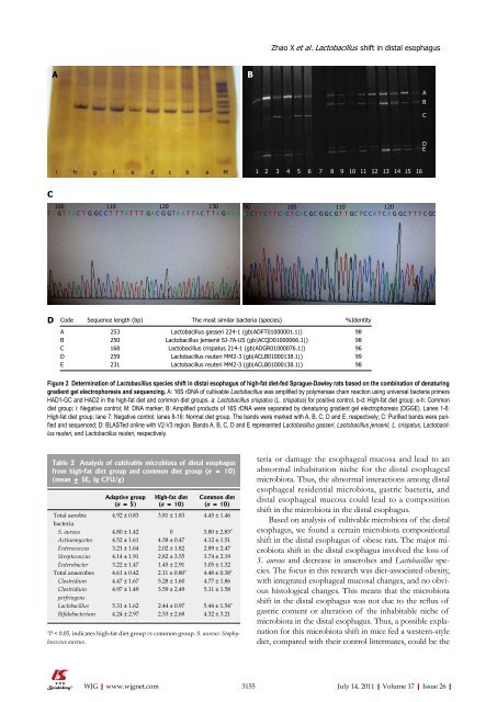
C<br />
A<br />
i h g f e d c b a M<br />
WJG|www.wjgnet.com<br />
B<br />
1 2 3 4 5 6 7 8 9 10 11 12 13 14 15 16<br />
100 110 120 130 90 100 110 120<br />
T A GT A A C T G GC C T T T A T T T GA C G GT A A T T A C T T A G A A A ACC T T C T T CA C T CAC GC G GC GT T GC T C C A T C A G GC T T T C GC<br />
D<br />
Code Sequence length (bp) The most similar bacteria (species) %Identity<br />
A 253 Lactobacillus gasseri 224-1 (gb|ADFT01000001.1|) 98<br />
B 250 Lactobacillus jensenii SJ-7A-US (gb|ACQD01000066.1|) 98<br />
C 168 Lactobacillus crispatus 214-1 (gb|ADGR01000076.1|) 96<br />
D 259 Lactobacillus reuteri MM2-3 (gb|ACLB01000138.1|) 99<br />
E 231 Lactobacillus reuteri MM2-3 (gb|ACLB01000138.1|) 98<br />
Figure 2 Determination <strong>of</strong> Lactobacillus species shift in distal esophagus <strong>of</strong> high-fat diet-fed Sprague-Dawley rats based on the combination <strong>of</strong> denaturing<br />
gradient gel electrophoresis and sequencing. A: 16S rDNA <strong>of</strong> cultivable Lactobacillus was amplified by polymerase chain reaction using universal bacteria primers<br />
HAD1-GC and HAD2 in the high-fat diet and common diet groups. a: Lactobacillus crispatus (L. crispatus) for positive control, b-d: High-fat diet group; e-h: Common<br />
diet group; i: Negative control; M: DNA marker; B: Amplified products <strong>of</strong> 16S rDNA were separated by denaturing gradient gel electrophoresis (DGGE). Lanes 1-6:<br />
High-fat diet group; lane 7: Negative control; lanes 8-16: Normal diet group. The bands were marked with A, B, C, D and E, respectively; C: Purified bands were purified<br />
and sequenced; D: BLASTed online with V2-V3 region. Bands A, B, C, D and E represented Lactobacillus gasseri, Lactobacillus jensenii, L. crispatus, Lactobacillus<br />
reuteri, and Lactobacillus reuteri, respectively.<br />
Table 2 Analysis <strong>of</strong> cultivable microbiota <strong>of</strong> distal esophagus<br />
from high-fat diet group and common diet group (n = 10)<br />
(mean ± SE, Ig CFU/g)<br />
Adaptive group<br />
(n = 5)<br />
High-fat diet<br />
(n = 10)<br />
Common diet<br />
(n = 10)<br />
Total aerobic<br />
bacteria<br />
4.92 ± 0.83 3.81 ± 1.83 4.45 ± 1.46<br />
S. aureus 4.80 ± 1.42 0 3.80 ± 2.83 a<br />
Actinomycetes 4.52 ± 1.61 4.58 ± 0.47 4.12 ± 1.51<br />
Enterococcus 3.21 ± 1.64 2.02 ± 1.82 2.89 ± 2.47<br />
Streptococcus 4.14 ± 1.91 2.82 ± 3.55 3.74 ± 2.19<br />
Enterobacter 3.22 ± 1.47 1.45 ± 2.91 3.05 ± 1.32<br />
Total anaerobes 4.61 ± 0.42 2.11 ± 0.80 a<br />
4.48 ± 0.38 a<br />
Clostridium 4.47 ± 1.67 5.28 ± 1.60 4.77 ± 1.86<br />
Clostridium<br />
perfringens<br />
4.97 ± 1.49 3.59 ± 2.49 5.11 ± 1.58<br />
Lactobacillus 5.31 ± 1.62 2.44 ± 0.97 5.44 ± 1.54 a<br />
Bifidobacterium 4.24 ± 2.97 2.53 ± 2.68 4.32 ± 3.21<br />
a P < 0.05, indicates high-fat diet group vs common group. S. aureus: Staphy-<br />
lococcus aureus.<br />
Zhao X et al . Lactobacillus shift in distal esophagus<br />
teria or damage the esophageal mucosa and lead to an<br />
abnormal inhabitation niche for the distal esophageal<br />
microbiota. Thus, the abnormal interactions among distal<br />
esophageal residential microbiota, gastric bacteria, and<br />
distal esophageal mucosa could lead to a composition<br />
shift in the microbiota in the distal esophagus.<br />
Based on analysis <strong>of</strong> cultivable microbiota <strong>of</strong> the distal<br />
esophagus, we found a certain microbiota compositional<br />
shift in the distal esophagus <strong>of</strong> obese rats. The major microbiota<br />
shift in the distal esophagus involved the loss <strong>of</strong><br />
S. aureus and decrease in anaerobes and Lactobacillus species.<br />
The focus in this research was diet-associated obesity,<br />
with integrated esophageal mucosal changes, and no obvious<br />
histological changes. This means that the microbiota<br />
shift in the distal esophagus was not due to the reflux <strong>of</strong><br />
gastric content or alteration <strong>of</strong> the inhabitable niche <strong>of</strong><br />
microbiota in the distal esophagus. Thus, a possible explanation<br />
for this microbiota shift in mice fed a western-style<br />
diet, compared with their control littermates, could be the<br />
3155 July 14, 2011|Volume 17|Issue <strong>26</strong>|<br />
A<br />
B<br />
C<br />
D<br />
E


