NEWSLETTER - ISRRT
NEWSLETTER - ISRRT
NEWSLETTER - ISRRT

You also want an ePaper? Increase the reach of your titles
YUMPU automatically turns print PDFs into web optimized ePapers that Google loves.
Introduction<br />
I was asked to spend a week in Fiji, based in Suva to do<br />
some training in Mammography and Breast Ultrasound.<br />
This was partly funded by the <strong>ISRRT</strong> and partly by the Fiji<br />
Government, Ministry of Health. Before I left for Fiji I tried<br />
to find what sort of training would be required. I also sent a<br />
questionnaire to be completed by those attending the training.<br />
I had also planned a rough program for the week, but<br />
could not make definite plans, as I did not know format of<br />
training, facilities that would be available, number of participants<br />
or their experience.<br />
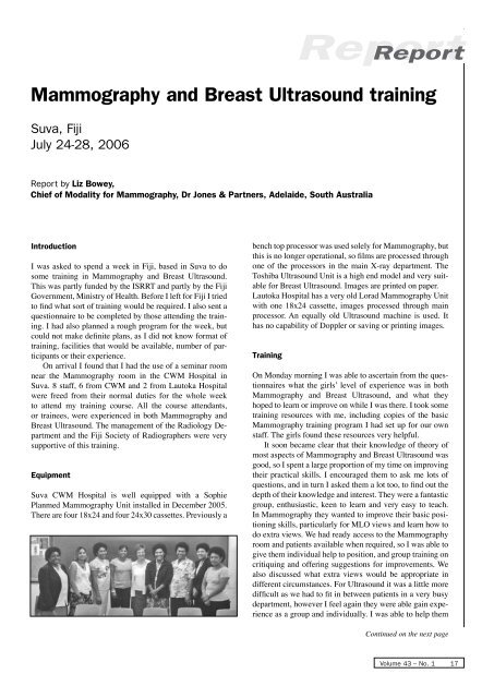
On arrival I found that I had the use of a seminar room<br />
near the Mammography room in the CWM Hospital in<br />
Suva. 8 staff, 6 from CWM and 2 from Lautoka Hospital<br />
were freed from their normal duties for the whole week<br />
to attend my training course. All the course attendants,<br />
or trainees, were experienced in both Mammography and<br />
Breast Ultrasound. The management of the Radiology Department<br />
and the Fiji Society of Radiographers were very<br />
supportive of this training.<br />
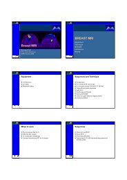
Equipment<br />
Suva CWM Hospital is well equipped with a Sophie<br />
Planmed Mammography Unit installed in December 2005.<br />
There are four 18x24 and four 24x30 cassettes. Previously a<br />
bench top processor was used solely for Mammography, but<br />
this is no longer operational, so films are processed through<br />
one of the processors in the main X-ray department. The<br />
Toshiba Ultrasound Unit is a high end model and very suitable<br />
for Breast Ultrasound. Images are printed on paper.<br />
Lautoka Hospital has a very old Lorad Mammography Unit<br />
with one 18x24 cassette, images processed through main<br />
processor. An equally old Ultrasound machine is used. It<br />
has no capability of Doppler or saving or printing images.<br />
Training<br />
Report Report<br />
Mammography and Breast Ultrasound training<br />
Suva, Fiji<br />
July 24-28, 2006<br />
Report by Liz Bowey,<br />
Chief of Modality for Mammography, Dr Jones & Partners, Adelaide, South Australia<br />
On Monday morning I was able to ascertain from the questionnaires<br />
what the girls’ level of experience was in both<br />
Mammography and Breast Ultrasound, and what they<br />
hoped to learn or improve on while I was there. I took some<br />
training resources with me, including copies of the basic<br />
Mammography training program I had set up for our own<br />
staff. The girls found these resources very helpful.<br />
It soon became clear that their knowledge of theory of<br />
most aspects of Mammography and Breast Ultrasound was<br />
good, so I spent a large proportion of my time on improving<br />
their practical skills. I encouraged them to ask me lots of<br />
questions, and in turn I asked them a lot too, to find out the<br />
depth of their knowledge and interest. They were a fantastic<br />
group, enthusiastic, keen to learn and very easy to teach.<br />
In Mammography they wanted to improve their basic positioning<br />
skills, particularly for MLO views and learn how to<br />
do extra views. We had ready access to the Mammography<br />
room and patients available when required, so I was able to<br />
give them individual help to position, and group training on<br />
critiquing and offering suggestions for improvements. We<br />
also discussed what extra views would be appropriate in<br />
different circumstances. For Ultrasound it was a little more<br />
difficult as we had to fit in between patients in a very busy<br />
department, however I feel again they were able gain experience<br />
as a group and individually. I was able to help them<br />
Continued on the next page<br />
Volume 43 – No. 1 17