Hormonas Tiroideas y Cerebro. Notas Sobre La Relación Bocio y ...
Hormonas Tiroideas y Cerebro. Notas Sobre La Relación Bocio y ...
Hormonas Tiroideas y Cerebro. Notas Sobre La Relación Bocio y ...

You also want an ePaper? Increase the reach of your titles
YUMPU automatically turns print PDFs into web optimized ePapers that Google loves.
T3<br />
T4<br />
D2<br />
T4<br />
D3<br />
T3<br />
D1<br />
rT3<br />
D3<br />
T2<br />
D2<br />
TR<br />
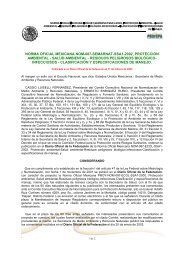
Figure. 1. Thyroid hormone transport, metabolism and action<br />
in a T3 target cell.<br />
TRE<br />
mRNA<br />
Protein<br />
RXR<br />
Nucleus<br />
Indeed much evidence has<br />
been published over the last three<br />
decades that cellular uptake and<br />
efflux of thyroid hormone do not take<br />
place by simple diffusion but are<br />
mediated by transporters (Fig. 1)<br />
(7). In vitro studies using isolated<br />
cells and perfusion of isolated tissue<br />
and in vivo studies in rats and<br />
humans have also strongly<br />
suggested that plasma membrane<br />
transport plays a rate-determining<br />
role in the hepatic metabolism of<br />
thyroid hormone, including the<br />
conversion of T4 to T3 (7).<br />
During the last few years thyroid hormone transporters have been characterized at the<br />
molecular level (7-11), including the Na + -taurocholate cotransporting polypeptide (NTCP), different<br />
members of the Na + -independent organic anion transporting polypeptide (OATP) family, the<br />
heterodimeric L-type amino acid transporters LAT1, LAT2, and the monocarboxylate transporters<br />
MCT8 and MCT10. Most of these transporters accept a variety of ligands, but OATP1C1, MCT8 and<br />
MCT10 show a high specificity towards iodothyronines (10, 12, 13).<br />
OATP1C1 is almost exclusively expressed in brain capillaries, and may be crucial for the<br />
transport of the prohormone T4 across the blood-brain barrier (13, 14). MCT8 is also expressed -<br />
among other tissues - in the brain, in particular in neurons (14, 15). MCT8 appears especially<br />
important for the uptake of the active hormone T3 into these cells, which is essential for optimal brain<br />
development. This T3 is produced from T4 by D2 in adjacent astrocytes. The neurons express D3<br />
which terminates T3 activity.<br />
The MCT8 gene is located on chromosome Xq13.2 and mutations in MCT8 have recently been<br />
associated with a syndrome combining severe X-linked psychomotor retardation, low serum T4 and<br />
strongly elevated T3 levels (16, 17). The mechanism of this disease involves a defect in the neuronal<br />
entry of T3, and thus in the action and metabolism of T3 in these cells, resulting in an impaired<br />
neurological development as well as a decrease in T3 clearance (16). This syndrome has now been<br />
documented in over 20 families and represents a novel mechanism of thyroid hormone resistance due<br />
to an impaired uptake of T3 in target cells.<br />
MCT8 null mice have been investigated by two groups with the same surprising results,<br />
showing exactly the same changes in thyroid hormone levels as affected patients but without any<br />
obvious neurological impairment (18, 19). MCT8 null mice show high liver and kidney D1, high brain<br />
and pituitary D2, and low brain D3 activities, changes which contribute to the abnormal serum thyroid<br />
parameters. MCT8 is importantly expressed in the hypothalamic area which explains the defect in<br />
negative feed-back action of thyroid hormone on TRH production and secretion (19, 20). Perhaps the<br />
most impressive demonstration of the function of MCT8 is the dramatic decrease in brain T3 uptake in<br />
MCT8 null versus wild-type mice (19).<br />
14