Evaluación cuantitativa del riesgo de cáncer de mama
Evaluación cuantitativa del riesgo de cáncer de mama
Evaluación cuantitativa del riesgo de cáncer de mama

You also want an ePaper? Increase the reach of your titles
YUMPU automatically turns print PDFs into web optimized ePapers that Google loves.
[Evaluación <strong>cuantitativa</strong> <strong><strong>de</strong>l</strong> <strong>riesgo</strong> <strong>de</strong> cáncer <strong>de</strong> <strong>mama</strong> - Dr. Fernando Gómez D.]El <strong>riesgo</strong> <strong>de</strong> un segundo cáncer <strong>de</strong> <strong>mama</strong>también está aumentado para mujerescon mutaciones. Las portadoras <strong>de</strong> mutaciónen el gen BRCA1 con diagnósticoprevio <strong>de</strong> cáncer <strong>de</strong> <strong>mama</strong> tienen un<strong>riesgo</strong> <strong>de</strong> un segundo primario <strong>de</strong> entreun 40 a 65% (90-92), mientras que paralas portadoras <strong><strong>de</strong>l</strong> gen BRCA2 el <strong>riesgo</strong>es <strong>de</strong> aproximadamente 50% (93). En lamujeres que han tenido cáncer <strong>de</strong> <strong>mama</strong>el <strong>riesgo</strong> <strong>de</strong> un cáncer <strong>de</strong> ovario es <strong>de</strong> 2 a3% (37), sin embargo para las que poseenla mutación en el gen BRCA1 aumentaentre un 19% (95% CI 14-24%) (91) y un44% (95% CI 28-56%) (94) para los 70años. En las portadoras <strong><strong>de</strong>l</strong> gen BRCA2el <strong>riesgo</strong> es <strong>de</strong> aproximadamente 15%(95% CI 8-23%) (93) para los 70 años.Los genes BRCA1 y 2 se han relacionadotambién con otros cánceres. El cáncer<strong>de</strong> próstata se ha visto relacionado conel gen BRCA2. Para los hombres que loportan, el <strong>riesgo</strong> relativo <strong>de</strong> cáncer <strong>de</strong>próstata es <strong>de</strong> 4.6 (95% CI 3.5-6.2), conun <strong>riesgo</strong> a los 80 años <strong>de</strong> 19.8% (95%CI 15.2-24.2%) (93). Otros cánceres sehan relacionado con baja o dudosa relacióna los genes BRCA1/2, como lo sonel cáncer <strong>de</strong> páncreas y el <strong>de</strong> colon, peroestos datos <strong>de</strong>berán confirmarse en el futuro(37).En conclusión, las pacientes con antece<strong>de</strong>ntes<strong>de</strong> cáncer <strong>de</strong> <strong>mama</strong> u ovario en lafamilia <strong>de</strong>ben consi<strong>de</strong>rarse para una evaluación<strong><strong>de</strong>l</strong> <strong>riesgo</strong>, <strong>de</strong>bido a que podríanser portadoras <strong>de</strong> mutaciones en los genesBRCA1/2 con el consiguiente <strong>riesgo</strong><strong>de</strong> <strong>de</strong>sarrollar un cáncer <strong>de</strong> <strong>mama</strong> u ovarioen el futuro. Por otra parte, quienes yahan tenido un cáncer <strong>de</strong> <strong>mama</strong> con mutacionesobjetivadas, <strong>de</strong>ben ser evaluadase informadas <strong>de</strong> su alto <strong>riesgo</strong> y <strong>de</strong>benconsi<strong>de</strong>rarse las medidas necesarias disponiblesque existen para disminuir laprobabilidad <strong>de</strong> <strong>de</strong>sarrollar un segundocáncer.Respecto a la indicación <strong>de</strong> pruebasgenéticas para estas mutaciones, la SociedadAmericana <strong>de</strong> Oncología Clínica(ASCO), recomienda lo siguiente pararealizar las pruebas <strong>de</strong> BRCA1/2 (95):(a) historia fuerte <strong>de</strong> cáncer en la familiao familiares con cánceres en eda<strong>de</strong>stempranas, (b) que el examen pueda serinterpretado a<strong>de</strong>cuadamente y (c) que elresultado pueda influir o cambiar el manejo<strong><strong>de</strong>l</strong> paciente o alguno <strong>de</strong> sus familiares.Por otro lado, hay que disponer <strong>de</strong> unlaboratorio confiable en el cual estén validadaslas técnicas metodológicas (14).c. Otros síndromes genéticosOtros síndromes relacionados a mutacionesgenéticas, son aquellos <strong>de</strong>nominadoscomo genes <strong>de</strong> baja penetrancia. Entreestos síndromes están el <strong>de</strong> Li-Fraumeni,la enfermedad <strong>de</strong> Cow<strong>de</strong>n, Peutz-Jeghers,Ataxia-Telangectasia y cáncer <strong>de</strong>colon no polipósico familiar. Es importantetener en cuenta estos síndromes almomento <strong>de</strong> reconstruir el pedigrí <strong>de</strong> laspacientes y en caso <strong>de</strong> i<strong>de</strong>ntificarlos <strong>de</strong>rivarlosal comité <strong>de</strong> alto <strong>riesgo</strong>.3. Lesiones precursoras previasSe sabe que existen diversas lesiones proliferativasbenignas <strong>de</strong> la <strong>mama</strong> que predisponena las mujeres a un alto <strong>riesgo</strong>.a. Hiperplasia epitelial con citologíaatípicaSe caracteriza por atipías celulares. Muchosautores han <strong>de</strong>mostrado un aumento<strong><strong>de</strong>l</strong> <strong>riesgo</strong> relativo <strong>de</strong> cáncer <strong>de</strong> <strong>mama</strong><strong>de</strong> alre<strong>de</strong>dor <strong>de</strong> dos veces (96,97). Estainformación es <strong>de</strong> gran importancia yse mencionará cuando se analicen lasnuevas técnicas <strong>de</strong> obtención <strong>de</strong> célulasepiteliales.b. Hiperplasias ductalesI. Hiperplasia ductal típica o usualSe caracteriza por un aumento <strong>de</strong> célulasen la capa epitelial pero sin característicasatípicas (98). Lo importante es queno es necesario adoptar ningún tipo <strong>de</strong>medida extra con este diagnóstico, <strong>de</strong>bidoa que no representa una lesión <strong>de</strong><strong>riesgo</strong> importante (37).III. Hiperplasia Ductal Atípica (HDA)Esta es una forma <strong>de</strong> hiperplasia ductalque morfológicamente simula a un carcinomaductal in situ (CDIS) (96,98).Esta lesión se consi<strong>de</strong>ra <strong>de</strong> mo<strong>de</strong>rado<strong>riesgo</strong> para cáncer <strong>de</strong> <strong>mama</strong>. El <strong>riesgo</strong>relativo se ha estimado entre 2.5 a 5 vecesen diversos estudios (96,99-102). El<strong>riesgo</strong> absoluto <strong>de</strong> <strong>de</strong>sarrollar un cáncer<strong>de</strong> <strong>mama</strong> <strong>de</strong>spués <strong><strong>de</strong>l</strong> diagnóstico <strong>de</strong> unaHDA <strong>de</strong>bidamente tratada es <strong>de</strong> 10% porun periodo <strong>de</strong> 10 a 15 años (99,103). El<strong>riesgo</strong> <strong>de</strong> cáncer invasor es mayor en lasmujeres peri-menopáusicas, llegando a6.1 veces (99). El <strong>riesgo</strong> no solo es ipsilateral,sino que también aumenta para la<strong>mama</strong> contralateral (99).Respecto al <strong>riesgo</strong> en las mujeres conantece<strong>de</strong>ntes familiares <strong>de</strong> cáncer <strong>de</strong><strong>mama</strong>, el <strong>riesgo</strong> relativo aumenta 10 vecesconjuntamente con HDA, comparadacon tres veces más para las que no tienenantece<strong>de</strong>ntes familiares (99,104).Es muy importante consi<strong>de</strong>rar a la HDAcomo antece<strong>de</strong>nte <strong>de</strong>bido al elevado<strong>riesgo</strong> que otorga a las pacientes en suevaluación. Este diagnóstico histológicose utiliza en la mayoría <strong>de</strong> los mo<strong><strong>de</strong>l</strong>osmatemáticos que calculan el <strong>riesgo</strong> <strong>de</strong> laspacientes.c. Lesiones lobularesLas dos lesiones características en estegrupo son la Hiperplasia Lobulillar Atípica(HLA) y el Carcinoma LobulillarIn Situ (CLIS). Se ha visto que el <strong>riesgo</strong>relativo <strong>de</strong> cáncer <strong>de</strong> <strong>mama</strong> invasor dadauna HLA (o neoplasia lobular grado 2) es<strong>de</strong> alre<strong>de</strong>dor <strong>de</strong> cuatro veces más que lasmujeres que no la tienen (99), similar a laADH. En el caso <strong><strong>de</strong>l</strong> CLIS (o neoplasialobular grado 3), el <strong>riesgo</strong> pue<strong>de</strong> llegarhasta 10 veces (105), similar a la ADHcon antece<strong>de</strong>ntes familiares. Por lo tantoestas lesiones también <strong>de</strong>ben ser consi<strong>de</strong>radasen la evaluación <strong><strong>de</strong>l</strong> <strong>riesgo</strong> <strong>de</strong> laspacientes que las portan.d. Otras lesiones <strong>de</strong> <strong>riesgo</strong>La cicatriz radiada es otro ejemplo <strong><strong>de</strong>l</strong>esión predisponerte <strong>de</strong> <strong>riesgo</strong>. Se haestimado que pacientes con esta lesión153149-163 153 5/12/06 05:10:38
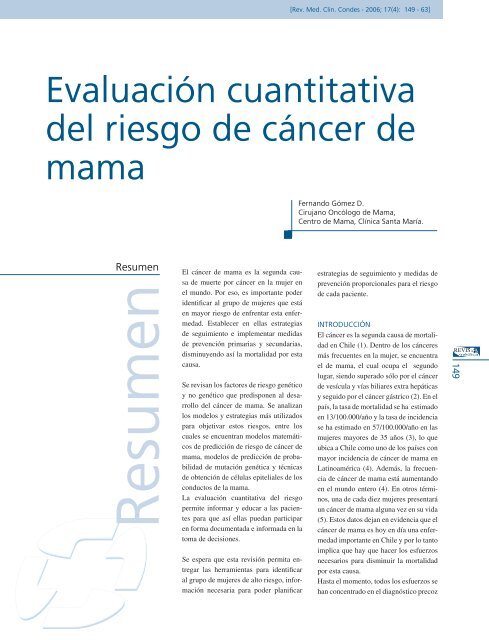
[Rev. Med. Clin. Con<strong>de</strong>s - 2006; 17(4): 149 - 63]154tienen dos a tres veces más <strong>riesgo</strong> que lasque no la tienen <strong>de</strong> presentar un cáncer<strong>de</strong> <strong>mama</strong> (106).Otra lesión <strong>de</strong> <strong>riesgo</strong> son los papilomasintraductales. Se ha calculado su <strong>riesgo</strong>en tres a cuatro veces más que las mujeresque no tienen esta lesión (96). Otrosautores reportan un <strong>riesgo</strong> extra <strong>de</strong> dosa tres veces y paradójicamente han observadoque los papilomas más pequeñosson los que tienen más <strong>riesgo</strong>, tendiendoa ser múltiples (107). Otras lesiones con<strong>riesgo</strong>s similares o menores son la hiperplasiaapocrina y la a<strong>de</strong>nosis esclerosante,que no aumentan en más <strong>de</strong> dos vecesel <strong>riesgo</strong> (14,37).Nuevamente, al igual que las lesionesductales y lobulares con atipías, estas lesionesaumentan el <strong>riesgo</strong> <strong>de</strong> las mujeres,por lo que hay que tenerlas presentes almomento <strong>de</strong> la evaluación final.2. OBJETIVANDO EL RIESGO1. Mo<strong><strong>de</strong>l</strong>os matemáticosEs muy común que las mujeres se presentenansiosas acerca <strong><strong>de</strong>l</strong> <strong>riesgo</strong> quetienen <strong>de</strong> <strong>de</strong>sarrollar un cáncer <strong>de</strong> <strong>mama</strong>y en general tien<strong>de</strong>n a sobre-estimarlo(14). Todos los datos relacionados alos factores <strong>de</strong> <strong>riesgo</strong> que existen en laliteratura y que han sido anteriormenteexpuestos, han permitido diseñar mo<strong><strong>de</strong>l</strong>osmatemáticos para la estimación <strong><strong>de</strong>l</strong><strong>riesgo</strong> en las pacientes. Estos mo<strong><strong>de</strong>l</strong>ospermiten cuantificar rápidamente el <strong>riesgo</strong><strong>de</strong> <strong>de</strong>sarrollar un cáncer <strong>de</strong> <strong>mama</strong> enun periodo o a una edad <strong>de</strong>terminada enlas pacientes. El po<strong>de</strong>r expresar este valoren términos objetivos y cuantitativosfacilita la educación <strong>de</strong> las pacientes, aligual que también permite diseñar racionalmenteuna estrategia para el manejo<strong><strong>de</strong>l</strong> <strong>riesgo</strong> y eventualmente la selección<strong>de</strong> pacientes para estudios clínicos <strong>de</strong>prevención (14).a. El mo<strong><strong>de</strong>l</strong>o <strong>de</strong> GailEn 1989, Gail y colaboradores <strong>de</strong>sarrollaronun mo<strong><strong>de</strong>l</strong>o para estimar el <strong>riesgo</strong>TABLA 1 / FACTORES DE RIESGORIESGO RELATIVO < 2 RIESGO RELATIVO 2-4 RIESGO RELATIVO > 4Menarquia tempranaMenopausia tardíaNuliparidad> 35 años 1° partoTHRObesidadAlcoholLesiones proliferativasBenignas<strong>de</strong> cáncer in situ e invasor <strong>de</strong> <strong>mama</strong> enmujeres participantes en un programa <strong>de</strong>screening llamado BCDDP que constaba<strong>de</strong> 284.780 mujeres (8). Este mo<strong><strong>de</strong>l</strong>oincluye variables como la edad, edad<strong>de</strong> menarquia y <strong><strong>de</strong>l</strong> primer embarazo <strong>de</strong>término, historia <strong>de</strong> familiares <strong>de</strong> primergrado con cáncer <strong>de</strong> <strong>mama</strong>, antece<strong>de</strong>ntes<strong>de</strong> biopsias <strong>mama</strong>rias previas y laraza. Una modificación <strong>de</strong> este mo<strong><strong>de</strong>l</strong>ofue propuesta por Costantino y colaboradoresel año 1999 (7), el cual calcula el<strong>riesgo</strong> <strong>de</strong> cáncer invasor solamente. Estemo<strong><strong>de</strong>l</strong>o se basó en los datos <strong><strong>de</strong>l</strong> estudio<strong>de</strong> prevención NSABP P-1 o BCPT11, eincluyó variables como tipo <strong>de</strong> histología<strong>de</strong> las biopsias previas respecto a lapresencia o no <strong>de</strong> atipías y variaciones enla raza <strong>de</strong> las pacientes.Ambos mo<strong><strong>de</strong>l</strong>os han sido validados pormúltiples estudios encontrándose unarelación entre los cánceres esperados ylos observados <strong>de</strong> 1.03, lo que es un muybuen resultado y habla <strong>de</strong> que el mo<strong><strong>de</strong>l</strong>ofunciona bastante bien (11,108-110).Existe un programa computacional quecalcula el <strong>riesgo</strong> según el mo<strong><strong>de</strong>l</strong>o <strong>de</strong> Gailmodificado por Costantino (versión 2.01en español).Si bien este mo<strong><strong>de</strong>l</strong>o ha <strong>de</strong>mostrado sereficiente, tiene sus limitaciones. Porejemplo, no consi<strong>de</strong>ra la edad <strong><strong>de</strong>l</strong> diagnóstico<strong>de</strong> los parientes con cáncer <strong>de</strong>Un familiar 1° gradoCa. MamaExposición a radiaciónCáncer <strong>de</strong> <strong>mama</strong> previoMamas <strong>de</strong>nsasDos familiares 1° gradoCa. MamaMutaciones genéticasCarcinoma Lobular InCarcinoma Ductal InHiperplasia DuctalAtípica (HDA)<strong>mama</strong>, ni tampoco a los familiares <strong>de</strong>segundo grado con cáncer <strong>de</strong> <strong>mama</strong>(no especifica si los cánceres <strong>de</strong> <strong>mama</strong>fueron bilaterales o no), ni tampoco serefiere a antece<strong>de</strong>ntes <strong>de</strong> cáncer <strong>de</strong> ovarioen la familia. Para pacientes que presentanestas condiciones el <strong>riesgo</strong> pue<strong>de</strong>ser también elevado por lo que <strong>de</strong>bieranser consi<strong>de</strong>radas como candidatas paraser <strong>de</strong>rivadas al consejo genético. Otralimitación <strong><strong>de</strong>l</strong> mo<strong><strong>de</strong>l</strong>o <strong>de</strong> Gail es que sobre-estimael <strong>riesgo</strong> <strong>de</strong> mujeres que noestán en un programa <strong>de</strong> screening enaproximadamente un 30% (109). Estoúltimo podría ser un problema <strong>de</strong>bido aque las mujeres podrían vivir con unaansiedad innecesaria o incluso podríanser sometidas a profilaxis innecesaria,como quimioprevención o incluso unamastectomía profiláctica (14). Por otrolado, el mo<strong><strong>de</strong>l</strong>o es bueno para calcular el<strong>riesgo</strong> <strong>de</strong> un grupo <strong>de</strong> mujeres con características<strong>de</strong>terminadas, pero no es tanbueno en pre<strong>de</strong>cir el <strong>riesgo</strong> individual <strong>de</strong>cada paciente. La precisión individual hasido estimada en 0.58 a cinco años (110).Utilizando como <strong>de</strong>finición <strong>de</strong> <strong>riesgo</strong> uníndice <strong>de</strong> Gail > 1.67 sólo un 44% <strong>de</strong>todos los cánceres que se diagnosticaronen estos programas tenían un <strong>riesgo</strong> estimadomayor a 1.67% a cinco años, enotras palabras, la mayoría <strong>de</strong> las pacientes(56%) que <strong>de</strong>sarrollaron un cáncer <strong>de</strong>ARCHIVO.indd 154 26/11/06 05:23:27
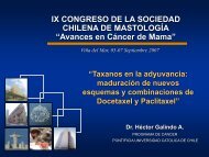
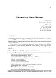
[Evaluación <strong>cuantitativa</strong> <strong><strong>de</strong>l</strong> <strong>riesgo</strong> <strong>de</strong> cáncer <strong>de</strong> <strong>mama</strong> - Dr. Fernando Gómez D.]<strong>mama</strong> tenían un <strong>riesgo</strong> estimado menora 1.67 a cinco años según el mo<strong><strong>de</strong>l</strong>o <strong>de</strong>Gail. Cuando se analizó cuales variableshacían que el <strong>riesgo</strong> estimado no fuerael correcto, se vio que el mo<strong><strong>de</strong>l</strong>o no funcionababien en mujeres menores <strong>de</strong> 35años, con historia familiar sugerente <strong>de</strong>mutaciones genéticas, historia previa <strong>de</strong>cáncer <strong>de</strong> <strong>mama</strong>, CLIS o CDIS, o paramujeres que no están en un programa <strong>de</strong>screening (37).En general, a pesar <strong>de</strong> todas las limitaciones<strong>de</strong> este mo<strong><strong>de</strong>l</strong>o, éste otorga enforma rápida la estimación <strong><strong>de</strong>l</strong> <strong>riesgo</strong> <strong>de</strong>cáncer <strong>de</strong> <strong>mama</strong> en las pacientes. Es unaherramienta útil y es el mo<strong><strong>de</strong>l</strong>o que hasido utilizado y que se está utilizando enlos gran<strong>de</strong>s estudios <strong>de</strong> prevención talescomo el NSABP P-1 y el NSABP P-2 oSTAR 11,(14).b. El mo<strong><strong>de</strong>l</strong>o <strong>de</strong> ClausEste mo<strong><strong>de</strong>l</strong>o fue publicado por Claus elaño 1994 (111) con los datos <strong><strong>de</strong>l</strong> estudioCASH y está diseñado para las mujerescon antece<strong>de</strong>ntes familiares <strong>de</strong> cáncer <strong>de</strong><strong>mama</strong>. A diferencia <strong><strong>de</strong>l</strong> mo<strong><strong>de</strong>l</strong>o <strong>de</strong> Gail,éste incluye antece<strong>de</strong>ntes familiares tanto<strong>de</strong> primer como <strong>de</strong> segundo grado, y laedad <strong>de</strong> diagnóstico <strong>de</strong> ellos. El mo<strong><strong>de</strong>l</strong>ocalcula la probabilidad <strong>de</strong> <strong>de</strong>sarrollar uncáncer <strong>de</strong> <strong>mama</strong> en un período <strong>de</strong> tiempo<strong>de</strong>terminado o a una edad <strong>de</strong>terminada.Es un buen complemento <strong><strong>de</strong>l</strong> mo<strong><strong>de</strong>l</strong>o <strong>de</strong>Gail en las mujeres con antece<strong>de</strong>ntes familiares,sobretodo <strong>de</strong> segundo grado.Es muy importante aclarar que tanto elmo<strong><strong>de</strong>l</strong>o <strong>de</strong> Gail modificado (por Costantino)como el mo<strong><strong>de</strong>l</strong>o <strong>de</strong> Claus, no <strong>de</strong>benser utilizados en pacientes con antece<strong>de</strong>ntespersonales <strong>de</strong> cáncer <strong>de</strong> <strong>mama</strong> oen portadoras conocidas <strong>de</strong> mutacionesgenéticas.c. Otros programas computacionalesSe han creado diversos softwares para laevaluación <strong>de</strong> las pacientes <strong>de</strong> alto <strong>riesgo</strong>.Uno <strong>de</strong> ellos es el creado por Parmigianiy colaboradores llamado el BRCAprogramo BRCA-PRO®, el cual fuediseñado para <strong>de</strong>terminar la probabilidadFIGURAS: Mo<strong><strong>de</strong>l</strong>o <strong>de</strong> Gail en españolque una mujer pue<strong>de</strong> tener <strong>de</strong> ser portadora<strong>de</strong> mutaciones en los genes BRCA1y/o BRCA2 (112,113). En sus trabajos,los autores <strong>de</strong>muestran que el programatiene muy buena correlación en la predicción<strong>de</strong> mutaciones genéticas <strong>de</strong> laspacientes. Es razonable referir al consejo<strong>de</strong> alto <strong>riesgo</strong> genético a pacientes queobtengan una probabilidad mayor al 5%<strong>de</strong> mutaciones para BRCA1/2, <strong>de</strong>bido aque estas pacientes tienen 50 veces más<strong>riesgo</strong> <strong>de</strong> ser portadoras que el 0.1% <strong>de</strong> lapoblación general (37). Cuando la probabilidad<strong>de</strong> mutación BRCA1 y/o BRCA2es mayor al 10%, hay que consi<strong>de</strong>rar larealización <strong><strong>de</strong>l</strong> test genético (37).Otro programa útil en la evaluación <strong><strong>de</strong>l</strong>as pacientes <strong>de</strong> alto <strong>riesgo</strong> es el Cancer-Gene®. Este fue diseñado para construirel árbol genealógico <strong>de</strong> las pacientes <strong>de</strong>155ARCHIVO.indd 155 26/11/06 05:23:32
[Rev. Med. Clin. Con<strong>de</strong>s - 2006; 17(4): 149 - 63]156FIGURA: Haciendo el pedigríFIGURA: Mo<strong><strong>de</strong>l</strong>o <strong>de</strong> ClausPRO®. Calcula los <strong>riesgo</strong>s y probabilida<strong>de</strong>sen función <strong>de</strong> los datos ingresadosen el pedigrí y en función <strong>de</strong> los datos ingresados<strong>de</strong> la paciente. A<strong>de</strong>más dispone<strong>de</strong> otros mo<strong><strong>de</strong>l</strong>os como el Myriad® I y IIy el Couch®.Como concepto general, es <strong>de</strong> gran utilidaddisponer <strong>de</strong> estas herramientas parael manejo <strong>de</strong> las pacientes <strong>de</strong> alto <strong>riesgo</strong><strong>de</strong>bido a que ahorran tiempo, estandarizanel <strong>riesgo</strong> en todas las pacientes y por-que son los utilizados por los principalesestudios internacionales multi-céntricosque nos han otorgado la información <strong><strong>de</strong>l</strong>os datos <strong>de</strong> prevención.2. Muestreo <strong>de</strong> epitelio ductalComo ya se mencionó antes, menos<strong><strong>de</strong>l</strong> 50% <strong>de</strong> las pacientes con cáncer <strong>de</strong><strong>mama</strong> pue<strong>de</strong>n ser i<strong>de</strong>ntificadas como <strong>de</strong>alto <strong>riesgo</strong> utilizando las variables clásicasantes analizadas (114), también semencionó que los mo<strong><strong>de</strong>l</strong>os <strong>de</strong> predicción<strong>de</strong> <strong>riesgo</strong> no son buenos para casos individuales(110).El fundamento para utilizar técnicas <strong>de</strong>muestreo epitelial se basa en que el cáncer<strong>de</strong> <strong>mama</strong> se genera a partir <strong>de</strong> cambiosen el epitelio. Estos primeros cambiosse <strong>de</strong>nominan hiperplasia o NeoplasiaIntra-Epitelial (NIE) (115). Una estimación<strong>de</strong> la frecuencia <strong>de</strong> estos cambiosepiteliales se han obtenido <strong>de</strong> estudiosque han analizado autopsias <strong>de</strong> mujerescon <strong>riesgo</strong> promedio, en los cuales se encontróentre un 12 a 25% <strong>de</strong> hiperplasiasen el tejido <strong>mama</strong>rio (116,117). Por otrolado, como ya se mencionó, se sabe quela hiperplasia atípica aumenta el <strong>riesgo</strong><strong>de</strong> cáncer <strong>de</strong> <strong>mama</strong> (118,119). Es poresto que estas técnicas pue<strong>de</strong>n ser <strong>de</strong>gran ayuda en la i<strong>de</strong>ntificación <strong><strong>de</strong>l</strong> <strong>riesgo</strong>individual <strong>de</strong> las mujeres con antece<strong>de</strong>ntes<strong>de</strong> <strong>riesgo</strong> que cuentan con un examenfísico e imagenológico normal. Por otrolado la documentación <strong>de</strong> cambios en elepitelio ductal ha <strong>de</strong>mostrado una mayoraceptación por parte <strong>de</strong> las pacientes paraingresar a estudios <strong>de</strong> quimioprevención(120,121).Los métodos que actualmente existenpara obtener muestras <strong><strong>de</strong>l</strong> epitelio ductalson: (a) Aspiración <strong>de</strong> fluido <strong><strong>de</strong>l</strong> pezón(AFP), (b) Punciones con aguja fina aleatorias(PAFA), (C) Lavado ductal (LD).La ductoscopía se utiliza como complementoa las técnicas anteriores.En esta revisión sólo se profundizará enlavado ductal, ya que los otros métodostienen más limitaciones.a. Lavado ductal (LD)El lavado ductal (LD) consiste en obtenercélulas <strong><strong>de</strong>l</strong> árbol ductal <strong>de</strong> la <strong>mama</strong>en búsqueda <strong>de</strong> atipías o malignidad enel grupo <strong>de</strong> mujeres <strong>de</strong> alto <strong>riesgo</strong> conel fin <strong>de</strong> objetivar el <strong>riesgo</strong> individual<strong>de</strong> cada paciente. Se ha reportado quees más sensible que las aspiraciones <strong><strong>de</strong>l</strong>fluido <strong><strong>de</strong>l</strong> pezón (AFP) y que a diferencia<strong>de</strong> ésta, el LD alcanza a obtener muestraincluso <strong>de</strong> los conductos terminales(122). El procedimiento consiste en:ARCHIVO.indd 156 26/11/06 05:23:51
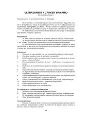
[Evaluación <strong>cuantitativa</strong> <strong><strong>de</strong>l</strong> <strong>riesgo</strong> <strong>de</strong> cáncer <strong>de</strong> <strong>mama</strong> - Dr. Fernando Gómez D.]FIGURA: Otros mo<strong><strong>de</strong>l</strong>os: Couch, Myriad I y II, etc.(a) aplicar masaje <strong>mama</strong>rio y vacío enel pezón, (b) canular conducto secretor,previa anestesia local, (c) lavar conductocon suero fisiológico para obtener lamuestra. Existen varios <strong>de</strong>talles paramejorar la técnica, como el uso <strong>de</strong> dilatadoreso nitroglicerina para evitar el espasmo<strong><strong>de</strong>l</strong> esfínter ductal (37). Una vezobtenida la muestra, ésta se centrifuga yse envía para evaluación citológica. Enel trabajo <strong>de</strong> Dooley y cols. (122) secomparó el lavado ductal con la AFP, obteniéndosemayor cantidad <strong>de</strong> muestrasina<strong>de</strong>cuadas en esta última (73 versus22%). A<strong>de</strong>más se vio que el número <strong>de</strong>células obtenida por conducto era significativamentemayor para el LD que paraAFP (13.000 versus 120). Otros trabajoshan publicado un promedio mayor <strong>de</strong>celularidad para AFP, pero la diferenciasigue siendo significativamente menor alcompararla con las células obtenidas porel LD (123,124), lo que le otorga al LDla posibilidad <strong>de</strong> realizar estudios biomolecularescon las muestras (37). Lasprincipales limitaciones <strong><strong>de</strong>l</strong> lavado ductalpublicadas por Dooley son: (a) en un10 a 15% <strong>de</strong> los casos no se logra obtenermuestra, (b) en un 2 a 5% no se logracanular el conducto y (c) un 15 a 20% <strong><strong>de</strong>l</strong>as muestras son acelulares. Esto quiere<strong>de</strong>cir que en suma el LD es exitoso en un60-65% <strong>de</strong> los casos.Los reportes citológicos se expresancomo; insuficiente, benigno, atipía mo<strong>de</strong>radao marcada atipía. Los diferentesestudios presentan rangos <strong>de</strong> 15-20%para citología insuficiente, 50-70% paracitología benigna, 17-25% para citologíacon mo<strong>de</strong>rada atipía y 2-7% para citologíacon marcada atipía (125-128).Actualmente no se sabe qué tanto varíael <strong>riesgo</strong> <strong>de</strong> pacientes portadoras <strong>de</strong> mutacionesen los genes BRCA1/2 cuandoexiste presencia <strong>de</strong> atipías, pero es muyprobable que aumente. Esto último lo<strong>de</strong>muestra Hoogerbrugge con 67 pacientes<strong>de</strong> muy alto <strong>riesgo</strong> (66% BRCA+),las cuales fueron a mastectomía profiláctica.De ellas 57% presentaron una omás lesiones patológicas <strong>de</strong> alto <strong>riesgo</strong>,se encontró HDA en un 37%, HLA enun 39%, CLIS en un 25% y CDIS en un15% (un caso <strong>de</strong> cáncer invasor) (129).En las pacientes mayores <strong>de</strong> 40 años seencontraban 6.6 veces más lesiones queen las menores <strong>de</strong> 40 (p= 0.01) y en laspacientes con ooforectomía previa seencontraban cinco veces menos lesiones.Esto hace pensar que el LD pue<strong>de</strong>encontrar atipías ocultas y ayudar a estasmujeres a tomar una <strong>de</strong>cisión frente a lasalternativas <strong>de</strong> prevención, incluso cirugíaprofiláctica.Otra ventaja <strong><strong>de</strong>l</strong> LD es que se pue<strong>de</strong> realizarseguimiento <strong>de</strong> los mismos conductosevaluados previamente, esto cobravital importancia en la evaluación <strong>de</strong> loscambios epiteliales con el uso <strong>de</strong> quimioprevención,ya que como se mencionóanteriormente es posible realizar estudiosbiomoleculares e incluso genéticos(130,131).Para tratar <strong>de</strong> ver cual era la sensibilidad<strong><strong>de</strong>l</strong> LD frente a un cáncer <strong>de</strong> <strong>mama</strong>, elCentro <strong>de</strong> Mama <strong>de</strong> Lynn Sage <strong>de</strong> la NorthWesternUniversity <strong>de</strong> Chicago realizóLD a pacientes que iban a ser sometidasa una mastectomía momentos antes <strong>de</strong> lacirugía en el pabellón (132). Los resultadosno fueron muy alentadores, dado quela sensibilidad fue sólo <strong><strong>de</strong>l</strong> 40%, aunqueFIGURA: Mo<strong><strong>de</strong>l</strong>os <strong>de</strong> <strong>riesgo</strong> para PDAs.157ARCHIVO.indd 157 26/11/06 05:23:59
[Rev. Med. Clin. Con<strong>de</strong>s - 2006; 17(4): 149 - 63]abb. DuctoscopíaConsiste en el uso <strong>de</strong> una fibra ópticaque canaliza los conductos <strong>mama</strong>rios<strong><strong>de</strong>l</strong> pezón con el fin <strong>de</strong> visualizar endoscópicamenteel árbol ductal. Se havisto que la ductoscopía es posible <strong>de</strong>realizar, ya que otorga una buena visualizacióny permite realizar lavado ductal(133-136). Debido a las propieda<strong>de</strong>s <strong>de</strong>visualización que tiene la ductoscopía,incluso en conductos distales al pezón ya que es un procedimiento mínimamenteinvasivo, se cree que la ductoscopíapue<strong>de</strong> servir como segundo paso en elmanejo <strong>de</strong> las pacientes <strong>de</strong> alto <strong>riesgo</strong>que presentan una citología alterada enel lavado ductal (37,127).En resumen, la ductoscopía parece seruna herramienta útil en el seguimiento<strong>de</strong> pacientes <strong>de</strong> alto <strong>riesgo</strong>, sin embargose requieren más estudios para establecersu verda<strong>de</strong>ra utilidad.cdFIGURAS: Lavado ductal. (Figuras extraídas <strong><strong>de</strong>l</strong> libro <strong><strong>de</strong>l</strong> libro “Managing Breast Cancer Risk”37)158TABLA 2 / ESTUDIOS DE LAVADO DUCTAL EN MUJERES DE ALTO RIESGOSERIESNúmero%con AFP%Insuficiente%Benigno% AtipiaMo<strong>de</strong>rada% AtipiaMarcadaDooley et al 1500842254177Francesca ttiy Woods 2122921657.124.12.6Woods 2 yEkbom 3160?1972.426.32Khan et al 4129852053234la especificidad fue cercana al 100%, vale<strong>de</strong>cir el hecho <strong>de</strong> que el LD sea negativono implica que no exista un cáncer,pero el hecho <strong>de</strong> que muestre atipías si secorrelacionaba con la presencia <strong>de</strong> éste.En resumen, el lavado ductal pue<strong>de</strong>ofrecer una serie <strong>de</strong> ventajas en la evaluación<strong>de</strong> las pacientes <strong>de</strong> alto <strong>riesgo</strong>,sin embargo aún queda mucho por investigar.FIGURA: Ductoscopía, lesión resultó ser unCDIS (Figuras extraídas <strong><strong>de</strong>l</strong> libro <strong><strong>de</strong>l</strong> libro“Managing Breast Cancer Risk”37)ARCHIVO.indd 158 26/11/06 05:24:06
[Evaluación <strong>cuantitativa</strong> <strong><strong>de</strong>l</strong> <strong>riesgo</strong> <strong>de</strong> cáncer <strong>de</strong> <strong>mama</strong> - Dr. Fernando Gómez D.]RESUMENEn la práctica clínica diaria un 10% <strong><strong>de</strong>l</strong>as mujeres que asisten a una consultason <strong>de</strong> alto <strong>riesgo</strong>. Es importante i<strong>de</strong>ntificarlasy <strong>de</strong>rivarlas al especialista.El manejo <strong>de</strong> mujeres <strong>de</strong> alto <strong>riesgo</strong> compren<strong>de</strong>tres pilares:• Pilar 1: Evaluación <strong>cuantitativa</strong> <strong><strong>de</strong>l</strong><strong>riesgo</strong>• Pilar 2: Estrategias <strong>de</strong> seguimiento• Pilar 3: Medidas <strong>de</strong> prevenciónA continuación se presentan algoritmos <strong>de</strong> acción:DERIVACIÓN PROGRAMA DE ALTO RIESGO• Familiares <strong>de</strong> primera línea o ≥ 2 familiares <strong>de</strong> segunda línea condiagnóstico <strong>de</strong> cáncer <strong>de</strong> <strong>mama</strong> antes <strong>de</strong> los 50 años.• Lesiones precursoras: HDA, HLA, CLIS.• Gail alterado (menarquia precoz, paridad tardía, edad, raza, etc.).• Radiación torácica previa.• Historia personal <strong>de</strong> cáncer <strong>de</strong> <strong>mama</strong> uni lateral antes <strong>de</strong> los 50 años o bilateral acualquier edad, o a<strong>de</strong>más cáncer <strong>de</strong> ovario.• Cáncer <strong>de</strong> <strong>mama</strong> en hombre.• Sospecha clínica <strong>de</strong> alto <strong>riesgo</strong>.Para objetivar el <strong>riesgo</strong> es necesario conocerlos factores <strong>de</strong> <strong>riesgo</strong> y aplicar losmo<strong><strong>de</strong>l</strong>os matemáticos existentes. Paracalcular el <strong>riesgo</strong> <strong>de</strong> cáncer <strong>de</strong> <strong>mama</strong> hayque utilizar el mo<strong><strong>de</strong>l</strong>o <strong>de</strong> Gail y Claus.Para la probabilidad <strong>de</strong> mutación genéticahay que utilizar el BRCA-PRO (incluidoen Cancergene).Es posible individualizar el <strong>riesgo</strong> conmuestras <strong>de</strong> epitelio ductal, ya que en un20 a 25% <strong>de</strong> las mujeres <strong>de</strong> alto <strong>riesgo</strong> seobtienen células atípicas.PROGRAMA ALTO RIESGOESPECIALISTA ALTO RIESGO• Historia familiar <strong>de</strong>tallada y otros fact. <strong>riesgo</strong>• Construcción <strong>de</strong> pedigrí si es indicado• Utilización <strong>de</strong> Gail y otros mo<strong><strong>de</strong>l</strong>os matemáticosESTRATIFICACIÓN DEL RIESGOBIBLIOGRAFÍA1> INE: Causas <strong>de</strong> Mortalidad segúncertificado <strong>de</strong> <strong>de</strong>función, Anuario <strong>de</strong> Demografía.Instituto Nacional <strong>de</strong> Estadísticas,1999.2> MINSAL: Estadísticas <strong>de</strong> Natalidad yMortalidad en Chile. Ministerio <strong>de</strong> Salud<strong>de</strong> Chile, 2000.3> Servicios <strong>de</strong> Salud M: Inci<strong>de</strong>ncia <strong><strong>de</strong>l</strong>Cáncer <strong>de</strong> Mama, Programa <strong>de</strong> Cáncer<strong>de</strong> Mama. Unidad <strong>de</strong> Patologías Mamarias.2000.4> Harris J: Diseases of the Breast, 2ndEd. Lippincott W&W, Epi<strong>de</strong>miologic andAssessing and Managing Risk. 1999.5> Parker SL, Tong T, Bol<strong>de</strong>n S, et al:Cancer statistics, 1997. CA Cancer J Clin47:5-27, 1997.6> Tabar L, Fagerberg CJ, Gad A, etal: Reduction in mortality from breastcancer after mass screening with mammography.Randomised trial from theRIESGO POBL. GRALGail y otros mo<strong><strong>de</strong>l</strong>osno muestran aumento<strong><strong>de</strong>l</strong> <strong>riesgo</strong>.A<strong>de</strong>más no haysospecha clínica.RIESGO POBL. GRAL RIESGO ELEVADO RIESGO MUY ALTOSCREENINGIngresa al programa<strong>de</strong> screening habitualRIESGO ELEVADO• Gail > 1.66 y < 4 a 5a• RR 2 a 4• Riesgo vida > 15%• BRCA-PRO < 10%• Criterio clínico <strong>de</strong><strong>riesgo</strong> mo<strong>de</strong>radoCOMITÉ ALTO RIESGORIESGO MUY ALTO• Gail ≥4% a 5a• RR > 4• Riesgo vida > 30%• BRCA-PRO ≥ 10%• Criterio clínico <strong>de</strong><strong>riesgo</strong> elevado92% 8%159ARCHIVO.indd 159 26/11/06 05:24:19
[Rev. Med. Clin. Con<strong>de</strong>s - 2006; 17(4): 149 - 63]160Breast Cancer Screening Working Groupof the Swedish National Board of Healthand Welfare. Lancet 1:829-32, 1985.7> Costantino JP, Gail MH, Pee D, etal: Validation studies for mo<strong><strong>de</strong>l</strong>s projectingthe risk of invasive and total breastcancer inci<strong>de</strong>nce. J Natl Cancer Inst91:1541-8, 1999.8> Gail MH, Brinton LA, Byar DP, et al:Projecting individualized probabilities of<strong>de</strong>veloping breast cancer for white femaleswho are being examined annually. JNatl Cancer Inst 81:1879-86, 1989.9> Burke W, Daly M, Garber J, et al:Recommendations for follow-up care ofindividuals with an inherited predispositionto cancer. II. BRCA1 and BRCA2.Cancer Genetics Studies Consortium.Jama 277:997-1003, 1997.10> Sakorafas GH: The management ofwomen at high risk for the <strong>de</strong>velopmentof breast cancer: risk estimation and preventativestrategies. Cancer Treat Rev29:79-89, 2003.11> Fisher B, Costantino JP, WickerhamDL, et al: Tamoxifen for prevention ofbreast cancer: report of the National SurgicalAdjuvant Breast and Bowel ProjectP-1 Study. J Natl Cancer Inst 90:1371-88, 1998.12> Veronesi U, Maisonneuve P, RotmenszN, et al: Italian randomized trialamong women with hysterectomy: tamoxifenand hormone-<strong>de</strong>pen<strong>de</strong>nt breastcancer in high-risk women. J Natl CancerInst 95:160-5, 2003.13> Colditz GA, Rosner BA, Speizer FE:Risk factors for breast cancer accordingto family history of breast cancer.For theNurses’ Health Study Research Group. JNatl Cancer Inst 88:365-71, 1996.14> Vogel VG: Management of the highriskpatient. Surg Clin North Am 83:733-51, 2003.15> Ries LAG, Kosary, C.L., Hankey,B.F., Miller, B.A., and Edwards, B.K.:SEER Cancer Statistics Review, 1973-1995. National Cancer Institute Bethesda,MD, 1998.16> Vogel VG: High-risk populationsas targets for breast cancer preventiontrials. Prev Med 20:86-100, 1991.17> Eidson M, Becker TM, Wiggins CL,et al: Breast cancer among Hispanics,American Indians and non-Hispanicwhites in New Mexico. Int J Epi<strong>de</strong>miol23:231-7, 1994.18> Kelsey JL, Gammon MD, John EM:Reproductive factors and breast cancer.Epi<strong>de</strong>miol Rev 15:36-47, 1993.19> MacMahon B, Trichopoulos D,Brown J, et al: Age at menarche, urineestrogens and breast cancer risk. Int JCancer 30:427-31, 1982.20> Bruzzi P, Negri E, La Vecchia C, etal: Short term increase in risk of breastcancer after full term pregnancy. Bmj297:1096-8, 1988.21> Newcomb PA, Storer BE, LongneckerMP, et al: Lactation and a reducedrisk of premenopausal breast cancer. NEngl J Med 330:81-7, 1994.22> Romieu I, Hernan<strong>de</strong>z-Avila M, LazcanoE, et al: Breast cancer and lactationhistory in Mexican women. Am J Epi<strong>de</strong>miol143:543-52, 1996.23> Trichopoulos D, MacMahon B, ColeP: Menopause and breast cancer risk. JNatl Cancer Inst 48:605-13, 1972.24> Parazzini F, Braga C, La Vecchia C,et al: Hysterectomy, oophorectomy inpremenopause, and risk of breast cancer.Obstet Gynecol 90:453-6, 1997.25> Breast cancer and hormonal contraceptives:collaborative reanalysisof individual data on 53 297 womenwith breast cancer and 100 239 womenwithout breast cancer from 54 epi<strong>de</strong>miologicalstudies. Collaborative Group onHormonal Factors in Breast Cancer. Lancet347:1713-27, 1996.26> Bernstein: Exogenous Hormones.Cancer Epi<strong>de</strong>miology and Prevention,2nd Ed. New York: WB Saun<strong>de</strong>rs, Co,p462, 1996.27> Marchbanks PA, McDonald JA,Wilson HG, et al: Oral contraceptivesand the risk of breast cancer. N Engl JMed 346:2025-32, 2002.28> Ross RK, Paganini-Hill A, Wan PC,et al: Effect of hormone replacementtherapy on breast cancer risk: estrogenversus estrogen plus progestin. J NatlCancer Inst 92:328-32, 2000.29> Steinberg KK, Thacker SB, Smith SJ,et al: A meta-analysis of the effect of estrogenreplacement therapy on the risk ofbreast cancer. Jama 265:1985-90, 1991.30> Breast cancer and hormone replacementtherapy: collaborative reanalysisof data from 51 epi<strong>de</strong>miological studiesof 52,705 women with breast cancer and108,411 women without breast cancer.Collaborative Group on Hormonal Factorsin Breast Cancer. Lancet 350:1047-59, 1997.31> Magnusson C, Baron JA, CorreiaN, et al: Breast-cancer risk followinglong-term oestrogen- and oestrogen-progestin-replacementtherapy. Int J Cancer81:339-44, 1999.32> Chen CL, Weiss NS, Newcomb P,et al: Hormone replacement therapy inrelation to breast cancer. Jama 287:734-41, 2002.33> Daling JR, Malone KE, Doody DR,et al: Relation of regimens of combinedhormone replacement therapy to lobular,ductal, and other histologic types ofbreast carcinoma. Cancer 95:2455-64,2002.34> Pilote L, Hlatky MA: Attitu<strong>de</strong>s ofwomen toward hormone therapy andprevention of heart disease. Am Heart J129:1237-8, 1995.35> Ettinger B: Hormone replacementtherapy and coronary heart disease. ObstetGynecol Clin North Am 17:741-57,1990.36> Rossouw JE, An<strong>de</strong>rson GL, PrenticeRL, et al: Risks and benefits of estrogenplus progestin in healthy postmenopausalwomen: principal results From theWomen’s Health Initiative randomizedcontrolled trial. Jama 288:321-33, 200237> Morrow M, Jordan: Managing BreastCancer Risk. BC Decker Inc., 2003.38> Wyn<strong>de</strong>r EL, Cohen LA, Muscat JE,et al: Breast cancer: weighing the evi<strong>de</strong>ncefor a promoting role of dietary fat.J Natl Cancer Inst 89:766-75, 1997.39> Armstrong B, Doll R: Environmentalfactors and cancer inci<strong>de</strong>nce and mor-ARCHIVO.indd 160 26/11/06 05:24:27
[Evaluación <strong>cuantitativa</strong> <strong><strong>de</strong>l</strong> <strong>riesgo</strong> <strong>de</strong> cáncer <strong>de</strong> <strong>mama</strong> - Dr. Fernando Gómez D.]tality in different countries, with specialreference to dietary practices. Int J Cancer15:617-31, 1975.40> Howe GR, Hirohata T, Hislop TG, etal: Dietary factors and risk of breast cancer:combined analysis of 12 case-controlstudies. J Natl Cancer Inst 82:561-9,1990.41> Hunter DJ, Spiegelman D, AdamiHO, et al: Cohort studies of fat intake andthe risk of breast cancer--a pooled analysis.N Engl J Med 334:356-61, 1996.42> Smith-Warner SA, Spiegelman D,Adami HO, et al: Types of dietary fat andbreast cancer: a pooled analysis of cohortstudies. Int J Cancer 92:767-74, 2001.43> Gla<strong>de</strong> MJ: Food, nutrition, and theprevention of cancer: a global perspective.American Institute for Cancer Research/WorldCancer Research Fund,American Institute for Cancer Research,1997. Nutrition 15:523-6, 1999.44> Sato R, Helzlsouer KJ, Alberg AJ,et al: Prospective study of carotenoids,tocopherols, and retinoid concentrationsand the risk of breast cancer. Cancer Epi<strong>de</strong>miolBiomarkers Prev 11:451-7, 2002.45> Toniolo P, Van Kappel AL, AkhmedkhanovA, et al: Serum carotenoids andbreast cancer. Am J Epi<strong>de</strong>miol 153:1142-7, 2001.46> Dorgan JF, Sowell A, Swanson CA,et al: Relationships of serum carotenoids,retinol, alpha-tocopherol, and seleniumwith breast cancer risk: results from aprospective study in Columbia, Missouri(United States). Cancer Causes Control9:89-97, 1998.47> Longnecker MP, Berlin JA, OrzaMJ, et al: A meta-analysis of alcoholconsumption in relation to risk of breastcancer. Jama 260:652-6, 1988.48> Gapstur SM, Potter JD, Sellers TA,et al: Increased risk of breast cancer withalcohol consumption in postmenopausalwomen. Am J Epi<strong>de</strong>miol 136:1221-31,1992.49> Willett WC, Hunter DJ, StampferMJ, et al: Dietary fat and fiber in relationto risk of breast cancer. An 8-yearfollow-up. Jama 268:2037-44, 1992.50> Folsom AR, McKenzie DR, BisgardKM, et al: No association between caffeineintake and postmenopausal breastcancer inci<strong>de</strong>nce in the Iowa Women’sHealth Study. Am J Epi<strong>de</strong>miol 138:380-3, 1993.51> Vatten LJ, Solvoll K, Loken EB: Coffeeconsumption and the risk of breastcancer. A prospective study of 14,593Norwegian women. Br J Cancer 62:267-70, 1990.52> Toniolo P, Riboli E, Shore RE, et al:Consumption of meat, animal products,protein, and fat and risk of breast cancer:a prospective cohort study in New York.Epi<strong>de</strong>miology 5:391-7, 199453> Sellers TA, Kushi LH, Cerhan JR, etal: Dietary folate intake, alcohol, and riskof breast cancer in a prospective study ofpostmenopausal women. Epi<strong>de</strong>miology12:420-8, 2001.54> Frie<strong>de</strong>nreich CM: Review of anthropometricfactors and breast cancer risk.Eur J Cancer Prev 10:15-32, 2001.55> van <strong>de</strong>n Brandt PA, Spiegelman D,Yaun SS, et al: Pooled analysis of prospectivecohort studies on height, weight,and breast cancer risk. Am J Epi<strong>de</strong>miol152:514-27, 2000.56> Barnes-Josiah D, Potter JD, SellersTA, et al: Early body size and subsequentweight gain as predictors of breast cancerinci<strong>de</strong>nce (Iowa, United States). CancerCauses Control 6:112-8, 1995.57> Huang Z, Hankinson SE, ColditzGA, et al: Dual effects of weight andweight gain on breast cancer risk. Jama278:1407-11, 1997.58> Trentham-Dietz A, Newcomb PA,Egan KM, et al: Weight change and riskof postmenopausal breast cancer (UnitedStates). Cancer Causes Control 11:533-42, 2000.59> Frie<strong>de</strong>nreich CM, Courneya KS,Bryant HE: Case-control study of anthropometricmeasures and breast cancerrisk. Int J Cancer 99:445-52, 2002.60> Frie<strong>de</strong>nreich CM, Thune I, BrintonLA, et al: Epi<strong>de</strong>miologic issues relatedto the association between physical activityand breast cancer. Cancer 83:600-10, 1998.61> Gilliland FD, Li YF, BaumgartnerK, et al: Physical activity and breastcancer risk in hispanic and non-hispanicwhite women. Am J Epi<strong>de</strong>miol 154:442-50, 2001.62> Verloop J, Rookus MA, van <strong>de</strong>rKooy K, et al: Physical activity and breastcancer risk in women aged 20-54 years. JNatl Cancer Inst 92:128-35, 2000.63> Lee IM, Cook NR, Rexro<strong>de</strong> KM,et al: Lifetime physical activity and riskof breast cancer. Br J Cancer 85:962-5,2001.64> Luoto R, Latikka P, Pukkala E, et al:The effect of physical activity on breastcancer risk: a cohort study of 30,548 women.Eur J Epi<strong>de</strong>miol 16:973-80, 2000.65> Rockhill B, Willett WC, Hunter DJ,et al: Physical activity and breast cancerrisk in a cohort of young women. J NatlCancer Inst 90:1155-60, 1998.66> Byers T, Nestle M, McTiernan A,et al: American Cancer Society gui<strong><strong>de</strong>l</strong>ineson nutrition and physical activityfor cancer prevention: Reducing therisk of cancer with healthy food choicesand physical activity. CA Cancer J Clin52:92-119, 2002.67> Tokunaga M, Land CE, Tokuoka S,et al: Inci<strong>de</strong>nce of female breast canceramong atomic bomb survivors, 1950-1985. Radiat Res 138:209-23, 1994.68> Miller AB, Howe GR, Sherman GJ,et al: Mortality from breast cancer afterirradiation during fluoroscopic examinationsin patients being treated for tuberculosis.N Engl J Med 321:1285-9,1989.69> Hancock SL, Tucker MA, HoppeRT: Breast cancer after treatment ofHodgkin’s disease. J Natl Cancer Inst85:25-31, 1993.70> Haffty BG: Radiation therapy andthe risk of contralateral breast cancer.Int J Radiat Oncol Biol Phys 56:920-1,2003.71> Obedian E, Fischer DB, Haffty BG:Second malignancies after treatment ofearly-stage breast cancer: lumpectomyand radiation therapy versus mastec-161ARCHIVO.indd 161 26/11/06 05:24:34
[Rev. Med. Clin. Con<strong>de</strong>s - 2006; 17(4): 149 - 63]162tomy. J Clin Oncol 18:2406-12, 2000.72> Fowble B, Hanlon A, Freedman G,et al: Second cancers after conservativesurgery and radiation for stages I-IIbreast cancer: i<strong>de</strong>ntifying a subset of womenat increased risk. Int J Radiat OncolBiol Phys 51:679-90, 2001.73> La<strong>de</strong>n F, Hunter DJ: Environmentalrisk factors and female breast cancer. AnnuRev Public Health 19:101-23, 1998.74> Terry PD, Rohan TE: Cigarette smokingand the risk of breast cancer in women:a review of the literature. CancerEpi<strong>de</strong>miol Biomarkers Prev 11:953-71,2002.75> Ambrosone C: Impact of genesis onthe relation-ship between smoking andbreast cancer risk. J Womens Cancer3:17-22, 2001.76> Slattery ML, Kerber RA: A comprehensiveevaluation of family history andbreast cancer risk. The Utah PopulationDatabase. Jama 270:1563-8, 1993.77> Claus EB, Risch NJ, ThompsonWD: Age at onset as an indicator of familialrisk of breast cancer. Am J Epi<strong>de</strong>miol131:961-72, 1990.78> Claus EB, Schildkraut JM, ThompsonWD, et al: The genetic attributablerisk of breast and ovarian cancer. Cancer77:2318-24, 1996.79> Struewing JP, Hartge P, Wachol<strong>de</strong>rS, et al: The risk of cancer associatedwith specific mutations of BRCA1 andBRCA2 among Ashkenazi Jews. N EnglJ Med 336:1401-8, 1997.80> Ford D, Easton DF, Peto J: Estimatesof the gene frequency of BRCA1 and itscontribution to breast and ovarian cancerinci<strong>de</strong>nce. Am J Hum Genet 57:1457-62,1995.81> Peto J, Collins N, Barfoot R, et al:Prevalence of BRCA1 and BRCA2 genemutations in patients with early-onsetbreast cancer. J Natl Cancer Inst 91:943-9, 1999.82> Satagopan JM, Offit K, Foulkes W,et al: The lifetime risks of breast cancerin Ashkenazi Jewish carriers of BRCA1and BRCA2 mutations. Cancer Epi<strong>de</strong>miolBiomarkers Prev 10:467-73, 2001.83> Hall JM, Lee MK, Newman B, etal: Linkage of early-onset familial breastcancer to chromosome 17q21. Science250:1684-9, 1990.84> Newman B, Mu H, Butler LM, et al:Frequency of breast cancer attributableto BRCA1 in a population-based seriesof American women. Jama 279:915-21,1998.85> Whittemore AS, Gong G, Itnyre J:Prevalence and contribution of BRCA1mutations in breast cancer and ovariancancer: results from three U.S. population-basedcase-control studies of ovariancancer. Am J Hum Genet 60:496-504, 1997.86> Couch FJ, DeShano ML, BlackwoodMA, et al: BRCA1 mutations in womenattending clinics that evaluate the risk ofbreast cancer. N Engl J Med 336:1409-15, 1997.87> Shattuck-Ei<strong>de</strong>ns D, Oliphant A, Mc-Clure M, et al: BRCA1 sequence analysisin women at high risk for susceptibilitymutations. Risk factor analysis andimplications for genetic testing. Jama278:1242-50, 1997.88> Wooster R, Neuhausen SL, MangionJ, et al: Localization of a breast cancersusceptibility gene, BRCA2, to chromosome13q12-13. Science 265:2088-90,1994.89> Ford D, Easton DF, Stratton M, etal: Genetic heterogeneity and penetranceanalysis of the BRCA1 and BRCA2 genesin breast cancer families. The BreastCancer Linkage Consortium. Am J HumGenet 62:676-89, 1998.90> Easton DF, Ford D, Bishop DT:Breast and ovarian cancer inci<strong>de</strong>nce inBRCA1-mutation carriers. Breast CancerLinkage Consortium. Am J Hum Genet56:265-71, 1995.91> Brose MS, Rebbeck TR, CalzoneKA, et al: Cancer risk estimates forBRCA1 mutation carriers i<strong>de</strong>ntified in arisk evaluation program. J Natl CancerInst 94:1365-72, 2002.92> Marcus JN, Watson P, Page DL, etal: Hereditary breast cancer: pathobiology,prognosis, and BRCA1 and BRCA2gene linkage. Cancer 77:697-709, 199693> Cancer risks in BRCA2 mutation carriers.TheBreast Cancer Linkage Consortium.J Natl Cancer Inst 91:1310-6, 1999.94> Ford D, Easton DF, Bishop DT, etal: Risks of cancer in BRCA1-mutationcarriers. Breast Cancer Linkage Consortium.Lancet 343:692-5, 1994.95> Statement of the American Societyof Clinical Oncology: genetic testing forcancer susceptibility, Adopted on February20, 1996. J Clin Oncol 14:1730-6;discussion 1737-40, 1996.96> Krieger N, Hiatt RA: Risk of breastcancer after benign breast diseases. Variationby histologic type, <strong>de</strong>gree of atypia,age at biopsy, and length of followup.Am J Epi<strong>de</strong>miol 135:619-31, 1992.97> Mommers EC, Page DL, DupontWD, et al: Prognostic value of morphometryin patients with normal breasttissue or usual ductal hyperplasia of thebreast. Int J Cancer 95:282-5, 2001.98> Bodian CA, Perzin KH, Lattes R,et al: Prognostic significance of benignproliferative breast disease. Cancer71:3896-907, 1993.99> Page DL, Dupont WD, Rogers LW,et al: Atypical hyperplastic lesions of thefemale breast. A long-term follow-upstudy. Cancer 55:2698-708, 1985.100> Tavassoli FA, Norris HJ: A comparisonof the results of long-term followupfor atypical intraductal hyperplasiaand intraductal hyperplasia of the breast.Cancer 65:518-29, 1990.101> Moskowitz M, Gartsi<strong>de</strong> P, WirmanJA, et al: Proliferative disor<strong>de</strong>rs of thebreast as risk factors for breast cancerin a self-selected screened population:pathologic markers. Radiology 134:289-91, 1980.102> Marshall LM, Hunter DJ, ConnollyJL, et al: Risk of breast cancer associatedwith atypical hyperplasia of lobular andductal types. Cancer Epi<strong>de</strong>miol BiomarkersPrev 6:297-301, 1997.103> Dupont WD, Page DL: Relativerisk of breast cancer varies with timesince diagnosis of atypical hyperplasia.Hum Pathol 20:723-5, 1989.ARCHIVO.indd 162 26/11/06 05:24:39
[Evaluación <strong>cuantitativa</strong> <strong><strong>de</strong>l</strong> <strong>riesgo</strong> <strong>de</strong> cáncer <strong>de</strong> <strong>mama</strong> - Dr. Fernando Gómez D.]104> Webb PM, Byrne C, Schnitt SJ, etal: Family history of breast cancer, ageand benign breast disease. Int J Cancer100:375-8, 2002.105> Connolly JL, Schnitt SJ: Benignbreast disease. Resolved and unresolvedissues. Cancer 71:1187-9, 1993.106> Jacobs TW, Byrne C, Colditz G, etal: Radial scars in benign breast-biopsyspecimens and the risk of breast cancer.N Engl J Med 340:430-6, 1999.107> Page DL, Salhany KE, Jensen RA,et al: Subsequent breast carcinoma riskafter biopsy with atypia in a breast papilloma.Cancer 78:258-66, 1996.108> Bondy ML, Lustba<strong>de</strong>r ED, HalabiS, et al: Validation of a breast cancer riskassessment mo<strong><strong>de</strong>l</strong> in women with a positivefamily history. J Natl Cancer Inst86:620-5, 1994.109> Spiegelman D, Colditz GA, HunterD, et al: Validation of the Gail et al.mo<strong><strong>de</strong>l</strong> for predicting individual breastcancer risk. J Natl Cancer Inst 86:600-7,1994.110> Rockhill B, Spiegelman D, ByrneC, et al: Validation of the Gail et al. mo<strong><strong>de</strong>l</strong>of breast cancer risk prediction andimplications for chemoprevention. J NatlCancer Inst 93:358-66, 2001.111> Claus EB, Risch N, Thompson WD:Autosomal dominant inheritance of earlyonsetbreast cancer. Implications for riskprediction. Cancer 73:643-51, 1994.112> Parmigiani G, Berry D, AguilarO: Determining carrier probabilitiesfor breast cancer-susceptibility genesBRCA1 and BRCA2. Am J Hum Genet62:145-58, 1998.113> Berry DA, Parmigiani G, SanchezJ, et al: Probability of carrying amutation of breast-ovarian cancer geneBRCA1 based on family history. J NatlCancer Inst 89:227-38, 1997.114> Seidman H, Stellman SD, MushinskiMH: A different perspective on breastcancer risk factors: some implicationsof the nonattributable risk. CA Cancer JClin 32:301-13, 1982.115> O’Shaughnessy JA, Kelloff GJ, GordonGB, et al: Treatment and preventionof intraepithelial neoplasia: an importanttarget for accelerated new agent <strong>de</strong>velopment.Clin Cancer Res 8:314-46, 2002.116> Bhathal PS, Brown RW, LesueurGC, et al: Frequency of benign and malignantbreast lesions in 207 consecutiveautopsies in Australian women. Br JCancer 51:271-8, 1985.117> Nielsen M, Thomsen JL, PrimdahlS, et al: Breast cancer and atypia amongyoung and middle-aged women: a studyof 110 medicolegal autopsies. Br J Cancer56:814-9, 1987.118> Dupont WD, Page DL: Risk factorsfor breast cancer in women with proliferativebreast disease. N Engl J Med312:146-51, 1985.119> Brown TA, Wall JW, ChristensenED, et al: Atypical hyperplasia in the eraof stereotactic core needle biopsy. J SurgOncol 67:168-73, 1998.120> Vogel VG, Costantino JP, WickerhamDL, et al: Re: tamoxifen for preventionof breast cancer: report of theNational Surgical Adjuvant Breast andBowel Project P-1 Study. J Natl CancerInst 94:1504, 2002.121> Tchou J HN, Ra<strong>de</strong>maker A et al.:Patient acceptance of tamoxifen as chemoprevention.ProcAm Soc Clin Oncol22:551, 2003.122> Dooley WC, Ljung BM, VeronesiU, et al: Ductal lavage for <strong>de</strong>tection ofcellular atypia in women at high riskfor breast cancer. J Natl Cancer Inst93:1624-32, 2001.123> Papanicolaou GN, Holmquist DG,Ba<strong>de</strong>r GM, et al: Exioliative cytology ofthe human mammary gland and its valuein the diagnosis of cancer and other diseasesof the breast. Cancer 11:377-409,1958.124> Buehring GC: Screening for breastatypias using exfoliative cytology. Cancer43:1788-99, 1979.125> Petrakis NL, Ernster VL, Sacks ST,et al: Epi<strong>de</strong>miology of breast fluid secretion:association with breast cancer riskfactors and cerumen type. J Natl CancerInst 67:277-84, 1981.126> Francescatti D WJ: Ductal Lavage:the learning curve. Proceeding of theFourth Annual Lynn Sage Symposium,2002; Chicago, IL. B6, 2002.127> Woods J, Ekbom GA: Managinghigh risk patients with ductal lavage.Proceeding of the Fourth Annual LynnSage Symposium, 2002; Chicago, IL.B5, 2002.128> Khan SA BC, Ramakrishnan R, etal: Proportion of cytologically evaluablesamples from duct lavage procedures.Proceeding of the American Associationof Cancer Research Frontiers in CancerResearch, 2002.129> Hoogerbrugge N, Bult P, <strong>de</strong> Widt-Levert LM, et al: High prevalence ofpremalignant lesions in prophylacticallyremoved breasts from women at hereditaryrisk for breast cancer. J Clin Oncol21:41-5, 2003.130> Evron E, Dooley WC, UmbrichtCB, et al: Detection of breast cancer cellsin ductal lavage fluid by methylationspecificPCR. Lancet 357:1335-6, 2001131> King BL, Crisi GM, Tsai SC, et al:Immunocytochemical analysis of breastcells obtained by ductal lavage. Cancer96:244-9, 2002.132> Khan SA RN, Baird C, et al: DuctalLavage finding in women with knownbreast cancer un<strong>de</strong>rgoing mastectomy.Proceeding of the San Antonio BreastCancer Symposium, 2002.133> Makita M, Sakamoto G, AkiyamaF, et al: Duct endoscopy and endoscopicbiopsy in the evaluation of nipple discharge.Breast Cancer Res Treat 18:179-87, 1991.134> Okazaki A, Okazaki M, Asaishi K,et al: Fiberoptic ductoscopy of the breast:a new diagnostic procedure for nippledischarge. Jpn J Clin Oncol 21:188-93,1991.135> Shen KW, Wu J, Lu JS, et al: Fiberopticductoscopy for patients with nippledischarge. Cancer 89:1512-9, 2000136> Dietz JR, Crowe JP, Grundfest S, etal: Directed duct excision by using mammaryductoscopy in patients with pathologicnipple discharge. Surgery 132:582-7; discussion 587-8, 2002.163ARCHIVO.indd 163 26/11/06 05:24:49