Impiego delle tecniche di imaging nelle demenze - Istituto Superiore ...
Impiego delle tecniche di imaging nelle demenze - Istituto Superiore ...
Impiego delle tecniche di imaging nelle demenze - Istituto Superiore ...

Create successful ePaper yourself
Turn your PDF publications into a flip-book with our unique Google optimized e-Paper software.
<strong>Impiego</strong> <strong>delle</strong> <strong>tecniche</strong> <strong>di</strong> <strong>imaging</strong> <strong>nelle</strong> <strong>demenze</strong><br />
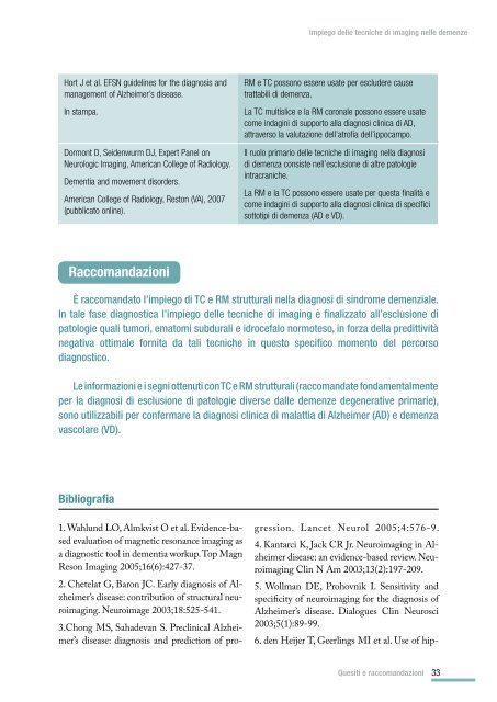
Hort J et al. EFSN guidelines for the <strong>di</strong>agnosis and<br />
management of Alzheimer’s <strong>di</strong>sease.<br />
In stampa.<br />
Dormont D, Seidenwurm DJ, Expert Panel on<br />
Neurologic Imaging, American College of Ra<strong>di</strong>ology.<br />
Dementia and movement <strong>di</strong>sorders.<br />
American College of Ra<strong>di</strong>ology, Reston (VA), 2007<br />
(pubblicato online).<br />
RM e TC possono essere usate per escludere cause<br />
trattabili <strong>di</strong> demenza.<br />
La TC multislice e la RM coronale possono essere usate<br />
come indagini <strong>di</strong> supporto alla <strong>di</strong>agnosi clinica <strong>di</strong> AD,<br />
attraverso la valutazione dell’atrofia dell’ippocampo.<br />
Il ruolo primario <strong>delle</strong> <strong>tecniche</strong> <strong>di</strong> <strong>imaging</strong> nella <strong>di</strong>agnosi<br />
<strong>di</strong> demenza consiste nell’esclusione <strong>di</strong> altre patologie<br />
intracraniche.<br />
La RM e la TC possono essere usate per questa finalità e<br />
come indagini <strong>di</strong> supporto alla <strong>di</strong>agnosi clinica <strong>di</strong> specifici<br />
sottotipi <strong>di</strong> demenza (AD e VD).<br />
Raccomandazioni<br />
È raccomandato l’impiego <strong>di</strong> TC e RM strutturali nella <strong>di</strong>agnosi <strong>di</strong> sindrome demenziale.<br />
In tale fase <strong>di</strong>agnostica l’impiego <strong>delle</strong> <strong>tecniche</strong> <strong>di</strong> <strong>imaging</strong> è finalizzato all’esclusione <strong>di</strong><br />
patologie quali tumori, ematomi subdurali e idrocefalo normoteso, in forza della pre<strong>di</strong>ttività<br />
negativa ottimale fornita da tali <strong>tecniche</strong> in questo specifico momento del percorso<br />
<strong>di</strong>agnostico.<br />
Le informazioni e i segni ottenuti con TC e RM strutturali (raccomandate fondamentalmente<br />
per la <strong>di</strong>agnosi <strong>di</strong> esclusione <strong>di</strong> patologie <strong>di</strong>verse dalle <strong>demenze</strong> degenerative primarie),<br />
sono utilizzabili per confermare la <strong>di</strong>agnosi clinica <strong>di</strong> malattia <strong>di</strong> Alzheimer (AD) e demenza<br />
vascolare (VD).<br />
Bibliografia<br />
1. Wahlund LO, Almkvist O et al. Evidence-based<br />
evaluation of magnetic resonance <strong>imaging</strong> as<br />
a <strong>di</strong>agnostic tool in dementia workup. Top Magn<br />
Reson Imaging 2005;16(6):427-37.<br />
2. Chetelat G, Baron JC. Early <strong>di</strong>agnosis of Alzheimer’s<br />
<strong>di</strong>sease: contribution of structural neuro<strong>imaging</strong>.<br />
Neuroimage 2003;18:525-541.<br />
3.Chong MS, Sahadevan S. Preclinical Alzheimer’s<br />
<strong>di</strong>sease: <strong>di</strong>agnosis and pre<strong>di</strong>ction of progression.<br />
Lancet Neurol 2005;4:576-9.<br />
4. Kantarci K, Jack CR Jr. Neuro<strong>imaging</strong> in Alzheimer<br />
<strong>di</strong>sease: an evidence-based review. Neuro<strong>imaging</strong><br />
Clin N Am 2003;13(2):197-209.<br />
5. Wollman DE, Prohovnik I. Sensitivity and<br />
specificity of neuro<strong>imaging</strong> for the <strong>di</strong>agnosis of<br />
Alzheimer’s <strong>di</strong>sease. Dialogues Clin Neurosci<br />
2003;5(1):89-99.<br />
6. den Heijer T, Geerlings MI et al. Use of hip-<br />
Quesiti e raccomandazioni<br />
33