Ana Paula Dias Ribeiro - Faculdade de Odontologia - Unesp
Ana Paula Dias Ribeiro - Faculdade de Odontologia - Unesp
Ana Paula Dias Ribeiro - Faculdade de Odontologia - Unesp
- No tags were found...
You also want an ePaper? Increase the reach of your titles
YUMPU automatically turns print PDFs into web optimized ePapers that Google loves.
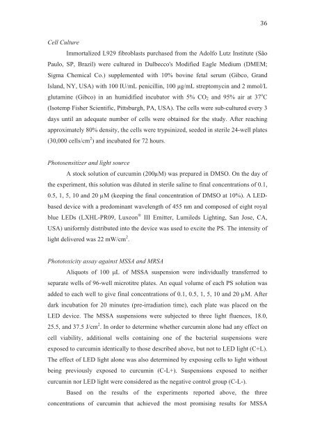
Cell CultureImmortalized L929 fibroblasts purchased from the Adolfo Lutz Institute (SãoPaulo, SP, Brazil) were cultured in Dulbecco's Modified Eagle Medium (DMEM;Sigma Chemical Co.) supplemented with 10% bovine fetal serum (Gibco, GrandIsland, NY, USA) with 100 IU/mL penicillin, 100 μg/mL streptomycin and 2 mmol/Lglutamine (Gibco) in an humidified incubator with 5% CO 2 and 95% air at 37 o C(Isotemp Fisher Scientific, Pittsburgh, PA, USA). The cells were sub-cultured every 3days until an a<strong>de</strong>quate number of cells were obtained for the study. After reachingapproximately 80% <strong>de</strong>nsity, the cells were trypsinized, see<strong>de</strong>d in sterile 24-well plates(30,000 cells/cm 2 ) and incubated for 72 hours.Photosensitizer and light sourceA stock solution of curcumin (200µM) was prepared in DMSO. On the day ofthe experiment, this solution was diluted in sterile saline to final concentrations of 0.1,0.5, 1, 5, 10 and 20 µM (keeping the final concentration of DMSO at 10%). A LEDbased<strong>de</strong>vice with a predominant wavelength of 455 nm and composed of eight royalblue LEDs (LXHL-PR09, Luxeon ® III Emitter, Lumileds Lighting, San Jose, CA,USA) uniformly distributed into the <strong>de</strong>vice was used to excite the PS. The intensity oflight <strong>de</strong>livered was 22 mW/cm 2 .Phototoxicity assay against MSSA and MRSAAliquots of 100 μL of MSSA suspension were individually transferred toseparate wells of 96-well microtitre plates. An equal volume of each PS solution wasad<strong>de</strong>d to each well to give final concentrations of 0.1, 0.5, 1, 5, 10 and 20 µM. Afterdark incubation for 20 minutes (pre-irradiation time), each plate was placed on theLED <strong>de</strong>vice. The MSSA suspensions were subjected to three light fluences, 18.0,25.5, and 37.5 J/cm 2 . In or<strong>de</strong>r to <strong>de</strong>termine whether curcumin alone had any effect oncell viability, additional wells containing one of the bacterial suspensions wereexposed to curcumin i<strong>de</strong>ntically to those <strong>de</strong>scribed above, but not to LED light (C+L).The effect of LED light alone was also <strong>de</strong>termined by exposing cells to light withoutbeing previously exposed to curcumin (C-L+). Suspensions exposed to neithercurcumin nor LED light were consi<strong>de</strong>red as the negative control group (C-L-).Based on the results of the experiments reported above, the threeconcentrations of curcumin that achieved the most promising results for MSSA


